. 2016 Dec 16;91(1):e02075-16.
doi: 10.1128/JVI.02075-16. Print 2017 Jan 1.

Transcriptional Silencing of Moloney Murine Leukemia Virus in Human Embryonic Carcinoma Cells

Affiliations

Transcriptional Silencing of Moloney Murine Leukemia Virus in Human Embryonic Carcinoma Cells

Gary Z Wang et al. J Virol. .

Abstract

Embryonic carcinoma (EC) cells are malignant counterparts of embryonic stem (ES) cells and serve as useful models for investigating cellular differentiation and human embryogenesis. Though the susceptibility of murine EC cells to retroviral infection has been extensively analyzed, few studies of retrovirus infection of human EC cells have been performed. We tested the susceptibility of human EC cells to transduction by retroviral vectors derived from three different retroviral genera. We show that human EC cells efficiently express reporter genes delivered by vectors based on human immunodeficiency virus type 1 (HIV-1) and Mason-Pfizer monkey virus (M-PMV) but not Moloney murine leukemia virus (MLV). In human EC cells, MLV integration occurs normally, but no viral gene expression is observed. The block to MLV expression of MLV genomes is relieved upon cellular differentiation. The lack of gene expression is correlated with transcriptional silencing of the MLV promoter through the deposition of repressive histone marks as well as DNA methylation. Moreover, depletion of SETDB1, a histone methyltransferase, resulted in a loss of transcriptional silencing and upregulation of MLV gene expression. Finally, we provide evidence showing that the lack of MLV gene expression may be attributed in part to the lack of MLV enhancer function in human EC cells.

Importance: Human embryonic carcinoma (EC) cells are shown to restrict the expression of murine leukemia virus genomes but not retroviral genomes of the lentiviral or betaretroviral families. The block occurs at the level of transcription and is accompanied by the deposition of repressive histone marks and methylation of the integrated proviral DNA. The host machinery required for silencing in human EC cells is distinct from that in murine EC cell lines: the histone methyltransferase SETDB1 is required, but the widely utilized corepressor TRIM28/Kap1 is not. A transcriptional enhancer element from the Mason-Pfizer monkey virus can override the silencing and promote transcription of chimeric proviral DNAs. The findings reveal novel features of human EC gene regulation not present in their murine counterparts.

Keywords: DNA methylation; chromatin immunoprecipitation; enhancer; histones; repressor.


Figures

FIG 1
FIG 1
Characterizing retroviral transduction of human embryonic carcinoma cells. (A) Flow cytometry analysis of HeLa or human EC cells (NTERA2 and 2102Ep) infected with VSV-G-pseudotyped single-round MLV-GFP, M-PMV–GFP, or HIV-1–mCherry reporter viruses. Data shown are from day 2 postinfection. The y axis shows side scatter (SSC); the x axis shows GFP or mCherry intensity. One representative experiment of three independent experiments is shown. (B) Quantification of the results from panel A from multiple experiments. The logarithmic graph shows the percentage of GFP+ or mCherry+ cells 2 days postinfection. Results shown are means ± standard errors of the mean (SEM) from three independent experiments. (C) Determination of viral DNA copy number. Cell lines shown were infected with VSV-G-pseudotyped MLV-GFP reporter viruses and propagated for 14 days in culture. Viral integration was determined by performing real-time quantitative PCR using primers specific for GFP and Tert on genomic DNA isolated from infected cells. The extent of viral integration (relative viral copy number) was quantified using the 2−ΔΔCT method by normalizing the GFP signal to the Tert gene. Results shown are means ± SEM from three independent experiments performed in duplicate. (D) Characterizing MLV LTR activity in HeLa and human EC cells. Individual cell lines were cotransfected with LTR firefly luciferase reporter constructs and a Renilla luciferase control plasmid for 36 h. Relative luciferase expression was calculated by dividing the firefly luciferase signal by the Renilla luciferase signal, and the resulting ratio was then normalized to the signal obtained from the same cells transfected with the M-PMV LTR luciferase construct (set to 1). Results shown are means ± SEMs from three independent experiments.
FIG 2
FIG 2
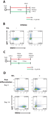
The block to MLV infection is relieved by differentiation of human EC cells (A) Scheme of experimental setup. (B) Flow cytometry analysis of undifferentiated and retinoic acid-differentiated NTERA2 cells infected with VSV-G-pseudotyped MLV-GFP reporter viruses. Results shown are at day 2 postinfection. The x axis shows SSEA4 staining (a marker of pluripotency); the y axis shows GFP intensity. One representative experiment of three independent experiments is shown. (C) Scheme of experimental setup. (D) NTERA2 cells were infected with VSV-G-pseudotyped MLV-GFP reporter viruses. At 1 day postinfection, cells were cultured with or without retinoic acid to induce differentiation. Flow cytometry analysis was carried out at the indicated times postdifferentiation to monitor MLV gene expression. The x axis shows SSEA4 staining (a marker of pluripotency); the y axis shows GFP intensity. One representative experiment of two independent experiments is shown. RA, retinoic acid; FACS, fluorescence-activated cell sorting.
FIG 3
FIG 3
MLV silencing in human EC cells is associated with deposition of repressive chromatin marks. (A) Undifferentiated NTERA2 cells or retinoic acid (RA)-induced differentiated NTERA2 cells infected with MLV-GFP for 3 days were subjected to ChIP analysis using the indicated antibodies. ChIP data are presented as the percentage of input DNA. Results shown are means ± standard deviations (SDs) from two independent experiments performed in duplicate. (B) 2102Ep cells infected with MLV-GFP for 5 days were subjected to ChIP analysis using the indicated antibodies. ChIP data are presented as the percentage of input DNA. Results shown are means ± SDs from three independent experiments performed in duplicate. (C) Experiment similar to that in panel A but performed in F9 mouse EC cells. (D) Experiment similar to that in panel A but performed in HeLa cells. (E) 2102Ep cells infected with M-PMV–GFP for 5 days were subjected to ChIP analysis as described above. qPCR, quantitative PCR.
FIG 4
FIG 4
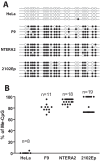
MLV silencing in human EC cells is associated with DNA methylation of the viral promoter. (A) At 14 days postinfection, bisulfite sequencing analysis of the 5′ LTR of the MLV proviral genomic DNA. Representative clones of proviral DNA genome are shown for each infected cell line. Open circles represent unmethylated CpG, filled circles represent methylated CpG (Me-CpG). (B) Quantification of the results from panel A from multiple MLV proviral genomic DNA clones for each cell line.
FIG 5
FIG 5
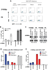
Depletion of SETDB1 but not TRIM28 relieves MLV transcriptional repression in human EC cells. (A) Flow cytometry analysis of indicated 2102Ep or F9 cells infected with VSV-G-pseudotyped MLV-GFP reporter viruses; this is at 2 days postinfection. The y axis shows side scatter (SSC); the x axis shows GFP intensity. Results of one representative experiment of three independent experiments are shown. (B) Quantification of the results from panel A from multiple experiments. Results shown are means ± SEM from three independent experiments. (C) Lysates prepared from 2102Ep or F9 cells stably transduced with either scrambled shRNA or TRIM28- or SETDB1-specific shRNAs were subjected to Western blotting as indicated. (D) 2102Ep cells infected with MLV-GFP for 3 days were subjected to ChIP analysis using antibodies against H3K9me3. ChIP data are presented as the percentage of input DNA. Results shown are means ± SDs from two independent experiments performed in duplicate. (E) Experiments similar to those in panel D but using antibodies against acetylated histone H3 (H3Ac).
FIG 6
FIG 6
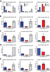
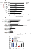
MLV gene expression in human EC cells is rescued by strong transcriptional enhancer sequences derived from M-PMV. (A) Functional analysis of MLV LTR sequences using mutant LTR reporter constructs. HeLa or 2102Ep cells were cotransfected with LTR firefly luciferase reporter constructs and a Renilla luciferase control plasmid for 36 h. Relative luciferase expression was calculated by dividing the firefly luciferase signal by the Renilla luciferase signal, and the resulting ratio was then normalized to the signal obtained from the same cell transfected with the M-PMV LTR luciferase construct (set to 1). Results shown are means ± SEMs from at least three independent experiments. NCR, negative-control region in MLV LTR; DR, direct repeat in MLV LTR (i.e., MLV transcriptional enhancer). Student's t test was used for statistical analysis. **, P < 0.01; *, P < 0.05 (compared to either 2102Ep or HeLa cells transfected with M-PMV LTR luciferase). (B) Assay similar to that outlined in for panel A but using chimeric LTR reporter constructs in which DNA sequences spanning the DR region of the MLV LTR were replaced with sequences from the U3 region of the M-PMV LTR. Student's t test was used for statistical analysis. **, P < 0.01; *, P < 0.05 (compared to either 2102Ep or HeLa cells transfected with M-PMV LTR luciferase). (C) Functional analysis of MLV LTR sequences using mutant LTR reporter constructs. Undifferentiated or retinoic acid (RA)-induced differentiated NTERA2 cells were cotransfected with LTR firefly luciferase reporter constructs and a Renilla luciferase control plasmid for 36 h. Relative luciferase expression was calculated as outlined above. Results shown are means ± SDs from two independent experiments. DR, direct repeat in MLV LTR (i.e., MLV transcriptional enhancer).

Similar articles

Cited by

References

    1. Andrews PW. 2002. From teratocarcinomas to embryonic stem cells. Philos Trans R Soc Lond B Biol Sci 357:405–417. doi:10.1098/rstb.2002.1058. - DOI - PMC - PubMed
    1. Andrews PW, Damjanov I, Simon D, Banting GS, Carlin C, Dracopoli NC, Fogh J. 1984. Pluripotent embryonal carcinoma clones derived from the human teratocarcinoma cell line Tera-2. Differentiation in vivo and in vitro. Lab Invest 50:147–162. - PubMed
    1. Pera MF, Cooper S, Mills J, Parrington JM. 1989. Isolation and characterization of a multipotent clone of human embryonal carcinoma cells. Differentiation 42:10–23. doi:10.1111/j.1432-0436.1989.tb00602.x. - DOI - PubMed
    1. Shevinsky LH, Knowles BB, Damjanov I, Solter D. 1982. Monoclonal antibody to murine embryos defines a stage-specific embryonic antigen expressed on mouse embryos and human teratocarcinoma cells. Cell 30:697–705. doi:10.1016/0092-8674(82)90274-4. - DOI - PubMed
    1. Kannagi R, Cochran NA, Ishigami F, Hakomori S, Andrews PW, Knowles BB, Solter D. 1983. Stage-specific embryonic antigens (SSEA-3 and -4) are epitopes of a unique globo-series ganglioside isolated from human teratocarcinoma cells. EMBO J 2:2355–2361. - PMC - PubMed

MeSH terms

LinkOut - more resources