. 2016 Oct 25;14(10):e1002571.
doi: 10.1371/journal.pbio.1002571. eCollection 2016 Oct.

snRNA 3' End Processing by a CPSF73-Containing Complex Essential for Development in Arabidopsis

Affiliations

snRNA 3' End Processing by a CPSF73-Containing Complex Essential for Development in Arabidopsis

Yunfeng Liu et al. PLoS Biol. .

Abstract

Uridine-rich small nuclear RNAs (snRNAs) are the basal components of the spliceosome and play essential roles in splicing. The biogenesis of the majority of snRNAs involves 3' end endonucleolytic cleavage of the nascent transcript from the elongating DNA-dependent RNA ploymerase II. However, the protein factors responsible for this process remain elusive in plants. Here, we show that DEFECTIVE in snRNA PROCESSING 1 (DSP1) is an essential protein for snRNA 3' end maturation in Arabidopsis. A hypomorphic dsp1-1 mutation causes pleiotropic developmental defects, impairs the 3' end processing of snRNAs, increases the levels of snRNA primary transcripts (pre-snRNAs), and alters the occupancy of Pol II at snRNA loci. In addition, DSP1 binds snRNA loci and interacts with Pol-II in a DNA/RNA-dependent manner. We further show that DSP1 forms a conserved complex, which contains at least four additional proteins, to catalyze snRNA 3' end maturation in Arabidopsis. The catalytic component of this complex is likely the cleavage and polyadenylation specificity factor 73 kDa-I (CSPF73-I), which is the nuclease cleaving the pre-mRNA 3' end. However, the DSP1 complex does not affect pre-mRNA 3' end cleavage, suggesting that plants may use different CPSF73-I-containing complexes to process snRNAs and pre-mRNAs. This study identifies a complex responsible for the snRNA 3' end maturation in plants and uncovers a previously unknown function of CPSF73 in snRNA maturation.


Conflict of interest statement

The authors have declared that no competing interests exist.

Figures

Fig 1
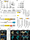
Fig 1. dsp1-1 increases the abundance of pre-U2.3 snRNAs and causes pleiotropic developmental defects.
(A) The accumulation of pre-U2.3 snRNAs detected by quantitative RT-PCR (qRT-PCR). dsp1-1+DSP1: dsp1-1 harboring a WT copy of DSP1; Col: WT. The levels of pre-U2.3 snRNAs in dsp1-1 were normalized to those of UBIQUITIN 5 (UBQ5) and compared with Col. Error bars indicate standard deviation (SD) of three technical replicates (**p < 0.01). Similar results were obtained from three biological replicates. Black arrowheads: primers used for RT-PCR; Black arrow: cleavage sites. (B) and (C) Morphological phenotypes of various genotypes. (D) Dissected green siliques of Col and DSP1/dsp1-2. (E) Embryo development of Col (top row) and dsp1-1 (bottom row). Embryos from siliques that were pollinated and harvested at the same time are shown in one column for comparison (Col versus dsp1-1). Embryo stages: preglobular (P), globular (G), transition (T), heart (H), torpedo (To), walking-stick (W), and mature embryo (M). (F) Pollen viability of Col and dsp1-1. Viable pollens are purple and round, whereas defective pollens are less stained. Bars = 100 μm (E) and 20 μm (F).
Fig 2
Fig 2. DSP1 is required for snRNA 3′ end maturation.
(A) The accumulation of various pre-snRNAs detected by qRT-PCR. The levels of pre-snRNAs in dsp1-1 were normalized to those of UBQ5 and compared with Col. Error bars indicate SD of three technical replications (**p < 0.01). Black arrowheads: primers used for RT-PCR; Black arrow: cleavage sites. (B) The accumulation of mature U1 and U2 snRNAs detected by northern Blot. UBQ5 was blotted as a loading control. (C) In vitro processing of pre-U2.3 snRNAs by nuclear protein extracts from Col (WT) and dsp1-1. Arrow indicates mature snRNAs. (D) Quantification of mature U2.3 RNA production in dsp1-1 relative to Col. The reaction stopped at 90 min was used for quantification analysis. The radioactive signal of mature U2.3 RNAs was quantified with Quantity One and then normalized to input. The relative levels of mature U2 snRNAs produced by dsp1-1 were then compared with those produced by Col. The value of Col was set to 1. The value represents the mean of three repeats. *p < 0.05 (t test). (E) Diagram of the pU2::pre-U2-GUS and pU2::pre-U2m-GUS transgenes. GUS+: GUS protein was produced. GUS-: no GUS protein was produced. (F) GUS levels detected by histochemical staining in various tissues of transgenic plants harboring pU2::pre-U2-GUS (Top row) or pU2::pre-U2m-GUS (Bottom row). Twenty plants were analyzed. A representative image is shown in each case. DSP+: DSP1/DSP1 or DSP1/dsp1-1. (G) The expression of pre-U2-GUS RNAs and pre-U2m-GUS RNAs in DSP+ and dsp1-1 detected by RT-PCR. Black arrowheads: primers used for PCR. The levels of pre-U2-GUS/pre-U2m-GUS were normalized to UBQ5 and compared with those in DSP+ (value set to 1). RNAs extracted from inflorescences were used for qRT-PCR analyses.
Fig 3
Fig 3. DSP1 binds the snRNA loci and affects the occupancy of Pol II at the snRNA loci.
(A) Subcellular localization of DSP1-GFP in tobacco leaf epidermal cells. The nuclei were visualized by DAPI staining of DNA. Scale bars: 20 μm. (B) Detection of DSP1-GFP in the nuclear and cytoplasmic protein fractions. T: Total proteins; N: Nuclear fraction; C: Cytoplasmic fraction. PEPC: the cytoplasm-localized phosphoenolpyruvate carboxylase. (C) Diagram showing the regions within the U2.3 and U1a loci detected by ChIP. (D–F) The occupancy of DSP1 at various loci in the cotyledons of plants harboring DSP1-GFP or GFP. Actin2-P: the promoter region of ACTIN2; Actin2-T: the coding region of ACTIN2. DNAs co-purified with DSP1-GFP or GFP were analyzed using quantitative PCR (qPCR). Means and SDs of three technical repeats are presented. Three biological replicates gave similar results. **p < 0.01, * p < 0.05 (t test). (G) and (H) Co-immunoprecipitation (Co-IP) between GFP-DSP1 and Pol II. (I) and (J) Co-IP between GFP-DSP1 and Pol II requires both DNA and RNA. Transgenic plants or nontransgenic plants (No) were indicated on Top. Proteins detected by western blot are labeled on the left. In: Input; MNase: Micrococcal Nuclease. (K–M) The occupancy of Pol II at various DNA loci in Col and dsp1-1. Actin2-P: the promoter region of ACTIN2; Actin2-T: the coding region of ACTIN2. DNAs co-purified with Pol II were analyzed using qPCR. Pol II C1 is used as a negative control for Pol II occupancy. Means and SDs of three technical repeats are presented. Similar results were obtained from three biological replicates. **p < 0.01, *p < 0.05 (t test).
Fig 4
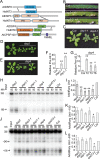
Fig 4. CPSF73-I is required for pre-snRNA processing.
(A) Diagram showing protein alignments. The grey lines define the homologous regions of two proteins. Protein domains are indicated in color. ARM: armadillo-like fold; β-Casp: After metallo‐β‐lactamase‐associated CPSF Artemis SNM1/PSO2; RMMBL: RNA-metabolizing metallo-beta-lactamase. (B–D) The effect of amiRCPSF73-I on plant development and the transcript levels of CPSF73-I and pre-U2.3 snRNA. cpsf73-I: amiRCPSF73-I. (E) Expression of an amiRCPSF73-I resistance CPSF73-I transgene recovers the levels of pre-U2.3 in the amiRCPSF73-I transgenic line. The levels of pre-U2.3 snRNAs and CPSF73-I were normalized to those of UBQ5 and compared with Col. Error bars indicate SD of three technical replicates (**p < 0.01). (F) The occupancy of CFPSF73-I at the U2.3 locus detected by ChIP. DNA co-purified with CPSF73-I was analyzed using qPCR. ChIP was performed on N. benthamiana leaves harboring the GFP-CPSF73-I and pU2::pre-U2-GUS transgenes. (G) CPSF73-I does not interact with Pol II. IP was performed using Arabidopsis harboring the 35S::GFP-CPSF73-I transgene.
Fig 5
Fig 5. Identification of other components involved in snRNA maturation.
(A) Schemes showing DSP2, DSP3, DSP4, and their human homologs. The grey lines define the homologous regions of two proteins. Protein domains are indicated in color. DUF2356: Domain of unknown function 2356; β-Casp: After metallo‐β‐lactamase‐associated CPSF Artemis SNM1/PSO2; RMMBL: RNA-metabolizing metallo-beta-lactamase. (B) Dissected green siliques of Col, DSP2/dsp2, and DSP3/dsp3-2. (C–E) Three-week-old seedlings of various genotypes. dsp2: amiRDSP2; dsp4: amiRDSP4. (F) and (G) The accumulation of pre-U2.3 snRNAs in various genotypes. Transcript levels of DSP2, DSP4, and pre-U2.3 snRNA in various mutants were normalized to UBQ and compared with those in Col (value set as 1). **p < 0.01, *p < 0.05 (t test). (H) In vitro processing of pre-U2.3 snRNAs in nuclear protein extracts from various genotypes. (I) Quantification of mature U2.3 snRNA production in various mutants relative to their levels in Col. dsp2: amiRDSP2 (T2); dsp4: amiRDSP4 (T4); cpsf73-I: amiRCPSF73-I (T3). (J) In vitro processing of the 3′-UTR of the Rubisco small subunit gene (RSB-3) in nuclear protein extracts from various genotypes. (K) and (L) Quantification of ~190 and 240 nt cleavage products generated in various mutants relative to their levels in Col. dsp2: amiRDSP2 (T2); dsp4: amiRDSP4 (T4); cpsf73-I: amiRCPSF73-I (T3). The radioactive signal of cleavage products from U2.3 snRNAs or RSB-3 was quantified with Quantity One and then normalized to input. The levels of cleavage products generated by dsp1-1, dsp2, dsp3-1, dsp4, or cpsf73-I were then compared with those of Col, respectively. The value of Col was set to 1. The value represents mean of three repeats. **p < 0.01; *p < 0.05 (t test).
Fig 6
Fig 6. DSP1, DSP2, DSP3, DSP4, and CPSF73-I form a complex.
(A–D) The interactions of DSP1 with DSP2, DSP3, DSP4, or CPSF73-I. (E–G) The interactions of DSP2 with DSP3, DSP4, or CPSF73-I. (H) The interaction of DSP4 with DSP3. (I) and (J) The interactions of CPSF73-I with DSP3-MYC or MYC-DSP4. GFP tagged proteins (1) or GFP (2) were co-expressed with various proteins as indicated on the left of the top row in N. benthamiana, except for DSP3-MYC. In vitro–translated DSP3-MYC was added to protein extracts containing GFP-tagged proteins (1) or GFP (2) as indicated on the left of the top row. Proteins detected by western blot are indicated on the left. (K) and (L) The interactions of DSP1 with CPSF73-I or DSP4 in Arabidopsis. (M) The interactions of DSP4 with CPSF73-I in Arabidopsis. IP was performed on protein extracts from Arabidopsis harboring GFP-DSP1/MYC-CPSF73-I, GFP-DSP1/MYC-DSP4, or GFP-DSP4/MYC-CPSF73-I with anti-GFP antibodies. Proteins detected by western blot are indicated on the left. (N) DSP1 pulls down DSP3 in the presence of CPSF73-I. GFP-DSP1 (1) or GFP (2) were co-expressed with HA-CPSF73-I. Protein extracts were mixed with in vitro–translated DSP3-MYC. Proteins detected by western blot are indicated on the left. (O) DSP4 pulls down DSP1, DSP2, and CPSF73-I. GFP-DSP4 (1) or GFP (2) were co-expressed with MYC-DSP1, HA-DSP2, and MYC-CPSF73-I in N. benthamiana. Proteins detected by western blot are indicated on the left. (P) CPSF73-I pulls down DSP1, DSP2, and DSP4. GFP-CPSF73-I (1) or GFP (2) were co-expressed with MYC-DSP1, HA-DSP2, and MYC.
Fig 7
Fig 7. Models for the 3′ end processing of snRNAs and mRNAs in Arabidopsis.
DSP1, DSP2, DSP3, DSP4, and CPSF73-I form a complex to co-transcriptionally cleave the pre-snRNAs upstream of the 3′ box. This complex may contain additional proteins. In contrast, a complex composed of CPSF100, CPSF73-I, and other proteins acts specifically on the 3′ end of pre-mRNAs. However, whether these two complexes have overlapping functions on some substrates remains to be investigated. FUE: far-upstream element; NUE: near-upstream element; CE: Cleavage element; PAP: Poly (A) polymerase; Fip1: Factor interacting with poly (A) polymerase 1; CstF: Cleavage stimulatory factor; CFIm: Mammalian cleavage factor I. The dashed lines indicate potential physical interaction. The dashed arrow indicates potential connection between the DSP1 complex and the CPSF complex.

Similar articles

Cited by

References

    1. Guthrie C, Patterson B. Spliceosomal snRNAs. Annual review of genetics. 1988;22:387–419. 10.1146/annurev.ge.22.120188.002131 . - DOI - PubMed
    1. Wahl MC, Will CL, Luhrmann R. The spliceosome: design principles of a dynamic RNP machine. Cell. 2009;136(4):701–18. 10.1016/j.cell.2009.02.009 . - DOI - PubMed
    1. Matera AG, Terns RM, Terns MP. Non-coding RNAs: lessons from the small nuclear and small nucleolar RNAs. Nat Rev Mol Cell Biol. 2007;8(3):209–20. 10.1038/nrm2124 . - DOI - PubMed
    1. Hernandez N. Small nuclear RNA genes: a model system to study fundamental mechanisms of transcription. J Biol Chem. 2001;276(29):26733–6. 10.1074/jbc.R100032200 . - DOI - PubMed
    1. Carbon P, Murgo S, Ebel JP, Krol A, Tebb G, Mattaj LW. A common octamer motif binding protein is involved in the transcription of U6 snRNA by RNA polymerase III and U2 snRNA by RNA polymerase II. Cell. 1987;51(1):71–9. . - PubMed

MeSH terms

Substances

Grants and funding

This work is supported by a National Science Foundation Grant MCB-1121193 (to BY). The funders had no role in study design, data collection and analysis, decision to publish, or preparation of the manuscript.