. 2016 Oct 24;12(10):e1006361.
doi: 10.1371/journal.pgen.1006361. eCollection 2016 Oct.

The Skp1 Homologs SKR-1/2 Are Required for the Caenorhabditis elegans SKN-1 Antioxidant/Detoxification Response Independently of p38 MAPK

Affiliations

The Skp1 Homologs SKR-1/2 Are Required for the Caenorhabditis elegans SKN-1 Antioxidant/Detoxification Response Independently of p38 MAPK

Cheng-Wei Wu et al. PLoS Genet. .

Abstract

SKN-1/Nrf are the primary antioxidant/detoxification response transcription factors in animals and they promote health and longevity in many contexts. SKN-1/Nrf are activated by a remarkably broad-range of natural and synthetic compounds and physiological conditions. Defining the signaling mechanisms that regulate SKN-1/Nrf activation provides insights into how cells coordinate responses to stress. Nrf2 in mammals is regulated in part by the redox sensor repressor protein named Keap1. In C. elegans, the p38 MAPK cascade in the intestine activates SKN-1 during oxidative stress by promoting its nuclear accumulation. Interestingly, we find variation in the kinetics of p38 MAPK activation and tissues with SKN-1 nuclear accumulation among different pro-oxidants that all trigger strong induction of SKN-1 target genes. Using genome-wide RNAi screening, we identify new genes that are required for activation of the core SKN-1 target gene gst-4 during exposure to the natural pro-oxidant juglone. Among 10 putative activators identified in this screen was skr-1/2, highly conserved homologs of yeast and mammalian Skp1, which function to assemble protein complexes. Silencing of skr-1/2 inhibits induction of SKN-1 dependent detoxification genes and reduces resistance to pro-oxidants without decreasing p38 MAPK activation. Global transcriptomics revealed strong correlation between genes that are regulated by SKR-1/2 and SKN-1 indicating a high degree of specificity. We also show that SKR-1/2 functions upstream of the WD40 repeat protein WDR-23, which binds to and inhibits SKN-1. Together, these results identify a novel p38 MAPK independent signaling mechanism that activates SKN-1 via SKR-1/2 and involves WDR-23.


Conflict of interest statement

The authors have declared that no competing interests exist.

Figures
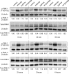
Fig 1
Fig 1. Kinetics of PMK-1 activation.
Relative level of PMK-1 phosphorylation at the kinase activating residues (Y180/182) and total PMK-1 protein levels in L4/YA worms treated with 5 mM sodium arsenite, 38 μM juglone, or 7 mM acrylamide for up to four hours. Band intensities relative to β-tubulin are given below each pair of blots with control arbitrarily set to 1.
Fig 2
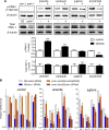
Fig 2. PMK-1 activation and its requirement for SKN-1 transcriptional responses.
(A) Relative level of PMK-1 phosphorylation at the kinase activating residues (Y180/182) and total PMK-1 protein levels in L4/YA worms treated with 5 mM sodium arsenite for 1 h, 35 mM paraquat for 2 h, 38 μM juglone for 3 h, or 7 mM acrylamide for 4 h. PMK-1 phosphorylation and protein levels were normalized to β-tubulin, which was detected on the same blot after stripping; histograms represent mean plus standard error of densitometry from n = 4 replicates of ~1,000 worms. ***P<0.001 compared to corresponding control as determined by Student’s T-test. Representative Western blot images are shown. (B) Fold changes in gst-4, gst-10, gst-12, gst-30, and gcs-1 mRNA levels relative to N2 control in L4/YA N2 wildtype and pmk-1(km25) mutant worms treated with 5 mM sodium arsenite or 38 μM juglone for 1 h, worms were fed either control RNAi or skn-1 RNAi from L1. Histograms represent mean plus standard error of n = 4 replicates of 200–300 worms. All genes were induced significantly by arsenite or juglone (P<0.001); *P<0.05, *** P<0.001 compared to N2;control(RNAi), ‡ P<0.001 compared to pmk-1(km25);control(RNAi).
Fig 3
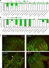
Fig 3. SKN-1 is localized to multiple tissues in a stress dependent manner.
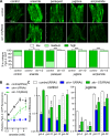
Animals integrated with a SKN-1b/c::GFP transgene were treated with 5 mM sodium arsenite, 5 mM sodium azide, 35 mM paraquat, 38 μM juglone, or 7 mM acrylamide. Accumulation of intestinal (A) and head (B) SKN-1b/c::GFP were scored separately. n = 60–100 worms from 3 independent trials. ***P<0.001 from corresponding controls as determined by Chi-Square tests. For intestinal SKN-1b/c::GFP, low refers to little to no SKN-1b/c::GFP, medium refers to SKN-1b/c::GFP observed only at the anterior or posterior of the intestine, and high refers to SKN-1b/c::GFP observed throughout the intestine. For head SKN-1b/c::GFP, negative refers to no observation of GFP signals in the head region, SKN-1b/c::GFP positive refers to GFP signals observed throughout the head region. (C) Representative fluorescence micrographs are shown for arsenite and juglone. Arrows mark head GFP and arrowheads mark intestinal nuclei.
Fig 4
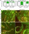
Fig 4. SKN-1b/c::GFP can accumulate in the head without pmk-1.
(A) Scoring of SKN-1b/c::GFP was conducted as in Fig 2. **P<0.01 and ***P<0.001 relative to the corresponding controls without arsenite as determined by Chi-Square tests, †P<0.05 and ††P<0.01 relative to the arsenite exposed wildtype worms as determined by Chi-Square tests, n = 51–92. (B) Representative images of SKN-1b/c::GFP. Arrows mark head GFP and arrowheads mark intestinal nuclei.
Fig 5
Fig 5. New gst-4 regulators identified through genome wide RNAi screening function upstream of wdr-23 and skn-1.
Animals integrated with Pgst-4::GFP were fed bacteria producing dsRNA to positive hits obtained from the RNAi screen and exposed to juglone. (A) Pgst-4::GFP fluorescence was scored and representative images are shown. For Pgst-4::GFP scoring, low refers to little to no GFP signals observed throughout the worm, medium refers to GFP signals observed at the anterior and posterior ends of the worm, and high refers to GFP signals observed throughout the body. n = 50–118 worms. (B) Pgst-4::GFP reporter scoring in either a wdr-23 loss of function (tm1817) or a skn-1 gain of function allele (k1023) mutant background. n = 54–75 worms. *P<0.05, **P<0.01 and ***P<0.001 relative to corresponding control as determined by Chi-Square tests; note that pad-1(RNAi) in skn-1(k1023) could not be tested for significance because it had zero worms with low or medium fluorescence.
Fig 6
Fig 6. skr-1/2 is required for induction of SKN-1 dependent detoxification genes.
(A) Pgst-4::GFP fluorescence scoring and representative fluorescence micrographs of worms fed with control or skr-1/2 dsRNA after exposure to 5 mM sodium arsenite for 1 h (recovered for 3 h on NGM agar to induce GFP), 35 mM paraquat for 2 h (recovered for 2 h), 38 μM juglone for 3 h (recovered for 1 h), or 7 mM acrylamide for 4 h (no recovery). n = 70–89 worms from 3 independent trials, *P<0.05, ***P<0.001 as determined by Chi-Square test. (B) Relative Pgst-4::GFP fluorescence measured by a plate reader after a 6 h exposure to a range of arsenite concentrations; all values are normalized to control(RNAi) with no arsenite. n = 4–8 wells of worms in a 384 well plate; P<0.001 for skr-1/2(RNAi) and skn-1(RNAi) versus control(RNAi) at all concentrations except for the lowest. (C) Fold changes in mRNA of gst-4, gst-10, gst-12, and gst-30 relative to control (no stressors) in worms with control, skn-1, or skr-1/2(RNAi) after exposure to 38 μM juglone for 3 h. mRNA levels were normalized to rpl-2; values are means plus standard error of n = 4 replicates of 200–400 worms. All genes were induced significantly by juglone (P<0.001); ***P<0.001 compared to control (RNAi).
Fig 7
Fig 7. Whole transcriptome profiling reveals correlation between loss of skn-1 and skr-1/2.
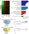
(A) Heat map of log2 fold-change of 174 genes found to be significantly up-regulated by juglone (38 μM for 3 h) with their corresponding fold-changes in worms fed with skn-1, skr-1/2, or daf-16 dsRNA compared to control dsRNA. Fold-change analysis for RNA-seq data were carried out using the CuffDiff application in Galaxy with q-value <0.05 determined by a false discovery rate of <5%. n = 3 replicates of 1,000–2,000 worms. Coefficients of correlation are listed below the map and 95% confidence intervals are as follows: skn-1 and skr-1/2 (0.693–0.818), skn-1 and daf-16 (0.533–0.712), and skr-1/2 and daf-16 (0.090–0.370). (B) Venn diagrams showing numbers of genes overlapping. (C) DAVID functional enrichment analysis of skr-1/2(RNAi) down-regulated genes. (D) Linear regression analysis of all genes down-regulated by either skn-1 or daf-16(RNAi) plotted against their fold change with skr-1/2(RNAi). ***P<0.001 as determined by linear regression F-test.
Fig 8
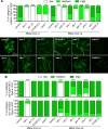
Fig 8. skr-1/2 is required for juglone and arsenite resistance but not for SKN-1 accumulation or PMK-1 phosphorylation.
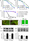
(A) Survival of eri-1 worms fed control, skn-1, or skr-1/2 RNAi on 125 μM juglone after a 2 h pre-treatment at 38 μM. ***P<0.001 compared to control RNAi as determined by Log-rank (Mantel-Cox) test. n = 98–109 worms from a single trial; statistics and results from two other trials are shown in S9 Fig. (B) Survival of N2 worms fed control, skn-1, or skr-1/2 RNAi on 10 mM arsenite. ***P<0.001 compared to control RNAi as determined by Log-rank (Mantel-Cox). n = 140–170 worms from a single trial; statistics and results from two other trials are shown in S9 Fig. (C) Survival of worms with enhanced SKN-1 activity exposed to a range of juglone concentrations for 16 h. skr-1/2(RNAi) did not significantly decrease survival at any concentration in any strain. n = 4 independent trials of 12–78 worms per condition and trial. (D) Animals with integrated SKN-1b/c::GFP were fed with either control or skr-1/2(RNAi) and treated with 38 μM juglone for 3 h and accumulation of head SKN-1b/c::GFP was scored. Representative fluorescence micrographs of head SKN-1b/c::GFP are shown with arrows marking GFP. n = 45 to 54 worms. (E) Phosphorylation of PMK-1 was measured in worms fed control or skr-1/2 dsRNA and exposed to 5 mM sodium arsenite for 1 h. PMK-1 phosphorylation levels were normalized to β-tubulin and then to control (without stressor); β-tubulin was detected on the same blots after stripping. Total PMK-1 was measured on the same lysates in a different blot and also normalized to β-tubulin. Values are mean plus standard error for densitometry of protein bands from n = 3 replicates of >1,000 worms. Representative immunoblot bands are shown, ***P<0.001 relative to control. There was no significant effect of skr-1/2(RNAi).
Fig 9
Fig 9. SKR-1 is expressed broadly and interacts with WDR-23.
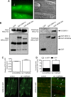
(A) Fluorescence and differential interference contrast micrographs showing cytoplasmic and nuclear (arrow) expression of SKR-1::GFP in the intestine (QV254). (B) SKR-1 interacts with WDR-23. HEK293 cells were co-transfected with V5-SKR-1 fusion protein along with either GST only vector (pDEST27), GST-SKN-1c fusion protein, or GST-WDR23a fusion protein. Complexes were captured with GSH beads and interactions with SKR-1 were determined by immunoblotting with anti-V5 mAb. Co-pulldown of V5-SKR-1 with GST-WDR-23a was also detected in a separate independent trial. (C-D) Total WDR-23::GFP fluorescence (normalized to RFP) and percentage of worms with visible nuclear WDR-23::GFP fluorescence with and without skr-1/2(RNAi). *P<0.05 and **P<0.01. Arrows point to hypodermal nuclei and asterisks mark neuronal cells.
Fig 10
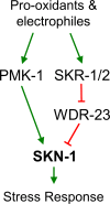
Fig 10. Model of SKR-1 activation by pro-oxidants and reactive small molecules.
In response to pro-oxidants, PMK-1 is activated and contributes to SKN-1 activation. Additional parallel or compensatory mechanisms likely exist given the ability of detoxification genes to be induced strongly without PMK-1 (Fig 2B). In response to diverse pro-oxidants and electrophiles, SKN-1 dependent gene expression is induced by a mechanism that requires SKR-1/2 (Figs 5 and 6). SKR-1/2 is not required for phosphorylation of p38 MAPK (Fig 8) and likely functions independently. SKR-1 acts upstream of WDR-23 (Fig 5), is able to interact with WDR-23 (Fig 9), and influences its nuclear levels suggesting a mechanism through this direct SKN-1 repressor.

Similar articles

Cited by

References

    1. Finkel T (2011) Signal transduction by reactive oxygen species. J Cell Biol 194: 7–15. 10.1083/jcb.201102095 - DOI - PMC - PubMed
    1. Finkel T, Holbrook NJ (2000) Oxidants, oxidative stress and the biology of ageing. Nature 408: 239–247. 10.1038/35041687 - DOI - PubMed
    1. Waris G, Ahsan H (2006) Reactive oxygen species: role in the development of cancer and various chronic conditions. J Carcinog 5: 14 10.1186/1477-3163-5-14 - DOI - PMC - PubMed
    1. Uttara B, Singh AV, Zamboni P, Mahajan RT (2009) Oxidative stress and neurodegenerative diseases: a review of upstream and downstream antioxidant therapeutic options. Curr Neuropharmacol 7: 65–74. 10.2174/157015909787602823 - DOI - PMC - PubMed
    1. Blackwell TK, Steinbaugh MJ, Hourihan JM, Ewald CY, Isik M (2015) SKN-1/Nrf, stress responses, and aging in Caenorhabditis elegans. Free Radic Biol Med 88: 290–301. 10.1016/j.freeradbiomed.2015.06.008 - DOI - PMC - PubMed

MeSH terms

Substances