. 2016 Oct 21:6:35399.
doi: 10.1038/srep35399.

The serologically defined colon cancer antigen-3 (SDCCAG3) is involved in the regulation of ciliogenesis

Affiliations

The serologically defined colon cancer antigen-3 (SDCCAG3) is involved in the regulation of ciliogenesis

Fangyan Yu et al. Sci Rep. .

Abstract

A primary cilium is present on most eukaryotic cells and represents a specialized organelle dedicated to signal transduction and mechanosensing. Defects in cilia function are the cause for several human diseases called ciliopathies. The serologically defined colon cancer antigen-3 (SDCCAG3) is a recently described novel endosomal protein mainly localized at early and recycling endosomes and interacting with several components of membrane trafficking pathways. Here we describe localization of SDCCAG3 to the basal body of primary cilia. Furthermore, we demonstrate that decreased expression levels of SDCCAG3 correlate with decreased ciliary length and a reduced percentage of ciliated cells. We show that SDCCAG3 interacts with the intraflagellar transport protein 88 (IFT88), a crucial component of ciliogenesis and intraciliary transport. Mapping experiments revealed that the N-terminus of SDCCAG3 mediates this interaction by binding to a region within IFT88 comprising several tetratricopeptide (TRP) repeats. Finally, we demonstrate that SDCCAG3 is important for ciliary localization of the membrane protein Polycystin-2, a protein playing an important role in the formation of polycystic kidney disease, but not for Rab8 another ciliary protein. Together these data suggest a novel role for SDCCAG3 in ciliogenesis and in localization of cargo to primary cilia.


Figures

Figure 1
Figure 1. SDCCAG3 localizes to the cilia basal body.
RPE1 cells stably expressing ARL13b-EGFP were serum-starved for 48 h to induce primary cilia, and then stained with anti-SDCCAG3 antibody (Ab2), anti-pericentrin antibody (PCNT), and DAPI. Images were obtained by fluorescence microscopy. Scale Bar represents 5 μm. Inset, scale bar represents 2 μm.
Figure 2
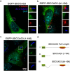
Figure 2. Localization of transfected EGFP-SDCCAG3 to cilia in IMCD3 cells.
IMCD3 cells were transfected with the indicated EGFP-SDCCAG3 expression constructs (green) and stained for the cilia axoneme marker acetylated tubulin (blue) and the basal body marker gamma-tubulin (red). (a) Full-length EGFP-SDCCAG3 co-localized with gamma-tubulin at the basal body. (b) A deletion construct of SDCCAG3 lacking amino acids 1-100 (Δ1-100) did not co-localize with gamma-tubulin at the basal body. (c) A truncated version of SDCCAG3 comprising only amino acids 1-100 was sufficient for basal body localization of SDCCAG3. Images are representative of three different experiments. Scale bar represents 5μm. (d) A schematic diagram of the transfected EGFP-SDCCAG3 expression constructs showing the full length protein (I), the construct comprising only amino acids 1-100 (II) and SDCCAG3 lacking amino acids 1-100 (III).
Figure 3
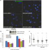
Figure 3. SDCCAG3 depletion impairs ciliogenesis.
(a) Representative figures of ciliated RPE1 cells transfected with SDCCAG3 or control siRNAs. RPE1 cells were transfected with the indicated siRNA. After 24 h, cells were incubated in low serum for 48 h and immunostained with anti-acetylated-α-tubulin antibody. Scale bar represents 5 μm. (b) Upper panel, the knockdown efficiency of three different SDCCAG3 siRNAs is demonstrated by western-blotting. Cropped blots are shown, full-length blots are presented in supplementary figure S6. Lower panel, RPE1 cells were transfected with the indicated siRNA. 24 h post transfection, cells were starved for an additional 24 h or 48 h. Cilia were labelled with anti-acetylated-α-tubulin antibody and the percentages of ciliated cells were quantified. (n = the number of cilia, n > 300; N = number of independent experiments, N = 3; unpaired t-test). (c) The length of cilia at 48 hours post serum starvation was measured (n > 100, N = 3, F-test). *p < 0.05, **p < 0.01. ***p < 0.001. Error bars represent s.e.m.
Figure 4
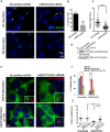
Figure 4. Impaired ciliogenesis observed after transfection of mouse IMCD3 cells with mSDCCAG3 siRNA can be rescued by overexpression of human SDCCAG3.
(a) IMCD3 cells were transfected with scrambled or siRNA targeting mouse SDCCAG3 for 48 hours and then cultured without serum for 0 and 16h and stained with acetylated tubulin (green) or DAPI (blue). Arrows represent nascent cilia. Scale bar represents 5μm. (b) Quantification of the percentage of ciliated cells from three independent experiments; unpaired t-test, error bar represents ± s.e.m **P < 0.01. (c) Cells were treated as described in (a) and ciliary length was measured 16 hours post-serum starvation (N = 3, F-test,***p < 0.001). (d) Immunoblot analysis of knockdown of SDCCAG3 upon treatment with mSDCCAG3 siRNA. Cropped blots are shown, full-length blots are presented in supplementary figure S6. (e) Localization of transfected human EGFP-SDCCAG3 or EGFP-SDCCAG3 Δ1-100 in IMCD3 cells treated with scrambled or mSDCCAG3 siRNA and stained with acetylated tubulin (red) and DAPI (blue). Scale bar represents 5μm. (f) Quantification of the percentage of ciliated cells showing rescue of the ciliogenesis defect in mSDCCAG3 siRNA treated cells upon expression of full-length EGFP-SDCCAG3 but not upon EGFP alone or EGFP-SDCCAG3 Δ1-100 expression. N = 3, unpaired t-test,*p < 0.05, ***p < 0.001, error bars represent ± s.e.m. (g) Quantification of ciliary length in scrambled or mSDCCAG3 siRNA treated cells after 16 hours of serum-starvation and upon expression of full-length EGFP-SDCCAG3 or upon expression of EGFP alone. (N = 3, F-test, ***p < 0.001, ns-not significant).
Figure 5
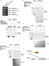
Figure 5. SDCCAG3 interacts with the intraflagellar transport protein IFT88.
(a) Yeast two-hybrid analysis of the interaction between amino acids 1-100 of SDCCAG3 with IFT88. An expression construct for IFT88 (full-length) was co-transformed into yeast cells with expression vectors for SDCCAG3 (amino acids 1-100), or the indicated negative control plasmids (FRMPD4 = Ferm and PDZ domain containing protein 4, PTPN13 = Protein typrosine phosphatase 13, pGBT9 = empty bait expression vector). Growth on media deficient for tryptophan (Trp), leucine (Leu) and histidine (His) indicates interaction of the corresponding proteins. (b) GST-SDCCAG3 interacts with full-length IFT88. Lysate of Cos7 cells transiently transfected with EGFP or EGFP-IFT88 expression constructs were incubated with purified GST, GST-SDCCAG3 and GST-SDCCAG3 1-100aa beads respectively. The pulldown result was analyzed by western blotting using an anti-GFP antibody. (c) GFP-trap pulldown of EGFP-IFT88 and Myc-SDCCAG3. Cos7 cells were transiently transfected with expression plasmids for myc-SDCCAG3 or the indicated GFP fusion proteins. Corresponding cell lysates were mixed as indicated, and subsequently subjected to GFP-Trap®_A pulldown. Bound proteins were detected via western blot with anti-myc and anti-GFP antibody. Cropped blots are shown, full-length blots are presented in supplementary figure S6. (d) Amino acids 400-550 of IFT88 are sufficient and necessary for interaction with SDCCAG3. HEK293 cells were transfected with expression constructs for EGFP, EGFP-IFT88 aa400-550 or EGFP-IFT88Δaa400-550. After 48 h, cell lysates were incubated with purified GST, GST-SDCCAG3 1-100aa or GST-SDCCAG3 glutathione agarose beads; protein complexes were separated by SDS–PAGE and detected with anti-GFP antibody. The arrow indicated the position of EGFP-IFT88 aa400-550. (e) Amino acids 1-100 of SDCCAG3 are required for interaction with IFT88. The indicated SDCCCAG3 GST-fusion proteins were incubated with lysate of EGFP or EGFP-IFT88 transfected HeLa cells. Bound proteins were separated by SDS-PAGE and detected with anti-GFP antibody. (f) Schematic diagram of the interaction of SDCCAG3 with IFT88. Ovals indicate coiled coil domains; the grey rectangles represent tetratricopeptide repeats (TPR).
Figure 6
Figure 6. Co-localization of SDCCAG3 and IFT88 in cells.
(a) Co-localization of SDCCAG3 and EGFP-IFT88 at centrosome. HeLa cells were grown on coverslip and transiently transfected with an expression construct for EGFP-IFT88. After 20 hours, cells were fixed and immunostained for SDCCAG3 and pericentrin (PCNT). Arrows indicate the co-localization at centrosomes. Scale bar represents 5 μm. (b) Localization of SDCCAG3 to the basal body is independent of IFT88. IMCD3 cells treated with scrambled or mouse IFT88 siRNA and transfected with EGFP-SDCCAG3 were stained with gamma-tubulin antibody. Scale bar represents 5 μm. (c) Quantification of co-localization of EGFP-SDCCAG3 with gamma-tubulin in cells treated with the indicated siRNA; n = 3, unpaired t-test, error bars ± s.e.m, ns = non-significant. (d) Immunoblot analysis validating knockdown of IFT88 in mIFT88 siRNA treated IMCD3 cells. Cropped blots are shown, full-length blots are presented in supplementary figure S6.
Figure 7
Figure 7. SDCCAG3 depletion impairs Polycystin-2 localization to primary cilia.
(a) IMCD3 cells transfected with control siRNA or mSDCCAG3 siRNA for 48 h were serum starved overnight (16 hours). Cells were then fixed and immunostained with anti-acetylated-α-tubulin antibody (red), anti-polycystin-2 (green) antibody and DAPI (blue). Scale bar represents 5 μm. (b) Quantification of Polycystin-2 localization to cilia after SDCCAG3 knockdown. The number of Polycystin-2 positive cilia of siRNA transfected cells were quantified. (c) Rescue of ciliary Polycystin-2 upon overexpression of human EGFP-SDCCAG3 in mSDCCAG3 siRNA treated IMCD3 cells. IMCD3 cells were transfected with siRNA targeting mouse SDCCAG3, after 24 hours cells were also transfected with expression plasmids for EGFP or EGFP-SDCCAG3 for another 48 hours. Transfected cells were subsequently serum starved for 16 hours and stained with anti-acetylated-α-tubulin (blue) and anti-Polycystin-2 (red) antibodies. The number of cilia positive for Polycystin-2 was quantified as above; n = 3, unpaired t-test, **p < 0.05,error bars ± s.e.m. (d) Depletion of SDCCAG3 did not alter localization of Rab8 to cilia in IMCD3 cells transfected with mSDCCAG3 or scrambled siRNA. Cells were treated with the indicated siRNAs as described above and stained for Rab8, acetylated-α-tubulin and DAPI. Images were taken by confocal microscopy. Scale bar represents 5 μm. (e) Quantification of the percentage of Rab8 positive cilia in SDCCAG3 siRNA and control siRNA treated cells; n = 3, unpaired t-test, error bars ± s.e.m, ns = non-significant.

Similar articles

Cited by

References

    1. Wheatley D. N., Wang A. M. & Strugnell G. E. Expression of primary cilia in mammalian cells. Cell Biol Int. 20, 73–81 (1996). - PubMed
    1. Bukoreshtliev N. V., Haase K. & Pelling A. E. Mechanical cues in cellular signalling and communication. Cell Tissue Res. 352, 77–94 (2013). - PubMed
    1. Huangfu D. & Anderson K. V. Cilia and Hedgehog responsiveness in the mouse. Proc Natl Acad Sci USA 102, 11325–11330 (2005). - PMC - PubMed
    1. Waters A. M. & Beales P. L. Ciliopathies: an expanding disease spectrum. Pediatr Nephrol. 26, 1039–1056 (2011). - PMC - PubMed
    1. Hildebrandt F., Benzing T. & Katsanis N. Ciliopathies. N Engl J Med. 364, 1533–1543 (2011). - PMC - PubMed

Publication types

MeSH terms

Substances