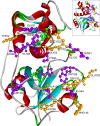
Structural Dynamics Investigation of Human Family 1 & 2 Cystatin-Cathepsin L1 Interaction: A Comparison of Binding Modes
- PMID: 27764212
- PMCID: PMC5072729
- DOI: 10.1371/journal.pone.0164970
Structural Dynamics Investigation of Human Family 1 & 2 Cystatin-Cathepsin L1 Interaction: A Comparison of Binding Modes
Abstract
Cystatin superfamily is a large group of evolutionarily related proteins involved in numerous physiological activities through their inhibitory activity towards cysteine proteases. Despite sharing the same cystatin fold, and inhibiting cysteine proteases through the same tripartite edge involving highly conserved N-terminal region, L1 and L2 loop; cystatins differ widely in their inhibitory affinity towards C1 family of cysteine proteases and molecular details of these interactions are still elusive. In this study, inhibitory interactions of human family 1 & 2 cystatins with cathepsin L1 are predicted and their stability and viability are verified through protein docking & comparative molecular dynamics. An overall stabilization effect is observed in all cystatins on complex formation. Complexes are mostly dominated by van der Waals interaction but the relative participation of the conserved regions varied extensively. While van der Waals contacts prevail in L1 and L2 loop, N-terminal segment chiefly acts as electrostatic interaction site. In fact the comparative dynamics study points towards the instrumental role of L1 loop in directing the total interaction profile of the complex either towards electrostatic or van der Waals contacts. The key amino acid residues surfaced via interaction energy, hydrogen bonding and solvent accessible surface area analysis for each cystatin-cathepsin L1 complex influence the mode of binding and thus control the diverse inhibitory affinity of cystatins towards cysteine proteases.
Conflict of interest statement
The authors have declared that no competing interests exist.
Figures


Similar articles
-
Modelling family 2 cystatins and their interaction with papain.J Biomol Struct Dyn. 2013;31(6):649-64. doi: 10.1080/07391102.2012.706403. Epub 2012 Aug 13. J Biomol Struct Dyn. 2013. PMID: 22881286
-
Studies on the interactions of SAP-1 (an N-terminal truncated form of cystatin S) with its binding partners by CD-spectroscopic and molecular docking methods.J Biomol Struct Dyn. 2015;33(1):147-57. doi: 10.1080/07391102.2013.855882. Epub 2013 Nov 21. J Biomol Struct Dyn. 2015. PMID: 24261636
-
Grafting of features of cystatins C or B into the N-terminal region or second binding loop of cystatin A (stefin A) substantially enhances inhibition of cysteine proteinases.Biochemistry. 2003 Sep 30;42(38):11326-33. doi: 10.1021/bi030119v. Biochemistry. 2003. PMID: 14503883
-
Cloning and characterisation of novel cystatins from elapid snake venom glands.Biochimie. 2011 Apr;93(4):659-68. doi: 10.1016/j.biochi.2010.12.008. Epub 2010 Dec 21. Biochimie. 2011. PMID: 21172403
-
Differential effect toward inhibition of papain and cathepsin C by recombinant human salivary cystatin SN and its variants produced by a baculovirus system.Arch Biochem Biophys. 2000 Aug 1;380(1):133-40. doi: 10.1006/abbi.2000.1909. Arch Biochem Biophys. 2000. PMID: 10900142
Cited by
-
Type 2 Cystatins and Their Roles in the Regulation of Human Immune Response and Cancer Progression.Cancers (Basel). 2023 Nov 10;15(22):5363. doi: 10.3390/cancers15225363. Cancers (Basel). 2023. PMID: 38001623 Free PMC article. Review.
-
Protease-bound structure of Ricistatin provides insights into the mechanism of action of tick salivary cystatins in the vertebrate host.Cell Mol Life Sci. 2023 Oct 28;80(11):339. doi: 10.1007/s00018-023-04993-4. Cell Mol Life Sci. 2023. PMID: 37898573 Free PMC article.
-
The Human Salivary Antimicrobial Peptide Profile according to the Oral Microbiota in Health, Periodontitis and Smoking.J Innate Immun. 2019;11(5):432-444. doi: 10.1159/000494146. Epub 2018 Nov 28. J Innate Immun. 2019. PMID: 30485856 Free PMC article.
-
Predicting Diagnostic Potential of Cathepsin in Epithelial Ovarian Cancer: A Design Validated by Computational, Biophysical and Electrochemical Data.Biomolecules. 2021 Dec 30;12(1):53. doi: 10.3390/biom12010053. Biomolecules. 2021. PMID: 35053201 Free PMC article.
-
Mialostatin, a Novel Midgut Cystatin from Ixodes ricinus Ticks: Crystal Structure and Regulation of Host Blood Digestion.Int J Mol Sci. 2021 May 20;22(10):5371. doi: 10.3390/ijms22105371. Int J Mol Sci. 2021. PMID: 34065290 Free PMC article.
References
-
- Turk V, Stoka V, Turk D. Cystatins: biochemical and structural properties, and medical relevance. Front Biosci. 2008; 13: 5406–5420. - PubMed
Publication types
MeSH terms
Substances
Grants and funding
LinkOut - more resources
Full Text Sources
Other Literature Sources
Molecular Biology Databases

