. 2016 Oct 13;7(10):e2408.
doi: 10.1038/cddis.2016.249.

Loss of Rictor with aging in osteoblasts promotes age-related bone loss

Affiliations

Loss of Rictor with aging in osteoblasts promotes age-related bone loss

Pinling Lai et al. Cell Death Dis. .

Abstract

Osteoblast dysfunction is a major cause of age-related bone loss, but the mechanisms underlying changes in osteoblast function with aging are poorly understood. This study demonstrates that osteoblasts in aged mice exhibit markedly impaired adhesion to the bone formation surface and reduced mineralization in vivo and in vitro. Rictor, a specific component of the mechanistic target of rapamycin complex 2 (mTORC2) that controls cytoskeletal organization and cell survival, is downregulated with aging in osteoblasts. Mechanistically, we found that an increased level of reactive oxygen species with aging stimulates the expression of miR-218, which directly targets Rictor and reduces osteoblast bone surface adhesion and survival, resulting in a decreased number of functional osteoblasts and accelerated bone loss in aged mice. Our findings reveal a novel functional pathway important for age-related bone loss and support for miR-218 and Rictor as potential targets for therapeutic intervention for age-related osteoporosis treatment.


Figures

Figure 1
Figure 1
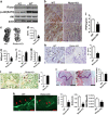
Osteoblast adhesion and survival are markedly reduced and Rictor expression is downregulated with aging in vitro and in vivo. (a) Representative immunohistochemical (IHC) staining for osteocalcin (OCN) from 3-month-old and 16-month-old mice (n=6). Arrow, osteoblast; BS, bone surface; scale bar, 50 μm; *P<0.01 versus 3M. (b) SEM image of femora trabecular bones from 3- and 16-month-old mice. Upper panel showed less and thinner trabecula bone in 16-month-old mice. Black arrow, osteoblasts or osteocytes, up panel scale bar, 500 μm; lower panel scale bar, 50 μm. (c) Photomicrographs of fluorescent calcein staining of 3- and 16-month-old mice and quantification of mineralizing surface/bone surface (MS/BS), mineral apposition rate (MAR) and MAR/osteoblast. Arrow: bone formation surface; scale bar, 20 μm (n=3 *P<0.01 versus 3M). (d) Cell adhesion analyses of calvarial osteoblast cultures from 3- and 16-month-old mice with (lower panel) or without (upper panel) matrigel in the plate (n=3 *P<0.01 versus 3M). (e) Representative photomicrographs of bone nodule formation in MSC cultures from 3- and 16-month-old mice. Scale bar, 400 μm. (f) Western blot analysis of cleaved-poly ADP-ribose polymerase (PARP) in lysates of trabecular bone dissected from femora of 3- and 16-month-old mice. (g) Western blot analysis of Rictor and P-Akt(S473) expression in lysates of trabecular bone from 3- and 16-month-old mice. (h) Western blot analysis of mTOR, Rictor, Raptor and P-Akt(S473) expression in lysates of lung, spleen and kidney from 3- and 16-month-old mice. (i) Western blot analysis of Rictor, mTOR, Raptor, osteocalcin (OCN) and P-Akt(S473) expression in primary calvarial osteoblasts from 3- and 16-month-old mice
Figure 2
Figure 2
Deletion of Rictor in osteoblasts impairs bone formation and stimulates aged-related bone loss. (a) Western blot analysis of Rictor and P-Akt(S473) expression in primary cultured calvarial osteoblasts from OBRictorKO and wild-type mice. (b) Representative IHC staining for p-Akt(S473) of distal femora from OBRictorKO and wild-type mice; scale bar, 100 μm. (c) Representative micro-CT scans of vertebrae from 6-month-old OBRictorKO and wild-type mice. (dg) Changes in vertebral trabecular BV/TV, BMD, Tb.Sp and Tb.N with age in OBRictorKO and wild-type mice (n=6, *P<0.01). (h) Representative IHC staining for OCN in distal femora from 6-month-old OBRictorKO and wild-type mice (n=6). Arrow, osteoblast; scale bar, 50 μm; *P<0.01 between genotypes. (i) Representative TRAP staining for osteoclasts of distal femora from 6-month-old OBRictorKO and wild-type mice (n=6). NS, no significant difference between genotypes; scale bar, 100 μm. (j) Enzyme-linked immunosorbnent assay (ELISA) analysis of serum Procollagen Type I N-terminal propeptide (PINP) levels in 6-month-old OBRictorKO and wild-type mice (n=6).*P<0.01 versus WT. (k) Representative calcein staining and quantification of MS/BS, MAR and MAR/osteoblast in distal femora from 6-month-old OBRictorKO and wild-type mice (n=3 *P<0.01 versus OBRictorKO). (l) Representative HE staining of distal femora from OBRictorKO and wild-type mice, adipocytes were counted (n=6). NS, no significant difference between genotypes; scale bar, 1 mm
Figure 3
Figure 3
Rictor is essential for osteoblast adhesion, mineralization and survival. (a) Photomicrographs of calcium nodule in MSCs isolated from wild-type and OBRictorKO mice after osteognenic induction for 21 days. (b) Western blot analysis of Rictor and OCN expression in MSCs from OBRictorKO and wild-type mice that underwent osteogenic induction for 14 days. (c) Photomicrographs of bone nodule formation in osteoblast cultures from OBRictorKO and wild-type mice. (d) Cell adhesion analysis of primary osteoblast cultures from OBRictorKO and wild-type mice with (lower panel) or without (upper panel) matrigel in the plate (n=3). *P<0.01 compared with wild-type mice. (e) Western blot analysis of cleaved-PARP expression in trabecular bone lysates from 6-month-old OBRictorKO and wild-type mice. (f) Representative photomicrographs of F-actin (red fluorescence) and p-Paxillin (Y118 green fluorescence) expression in primary osteoblasts from OBRictorKO and wild-type mice. Arrow, p-Paxillin (Y118); scare bar, 5 μm. (g) SEM analysis of trabecular bone from 6-month-old OBRictorKO and wild-type mice. Upper panel showed less and thinner trabecula bone in OBRictorKO mice. Black arrow, osteoblast or osteocyte; upper panel bar, 500 μm; lower panel bar, 50 μm
Figure 4
Figure 4
Rictor is a target of miR-218 in osteoblasts. (a) Real-time PCR analysis of 13 miRNAs expression in trabecular bone from 3- and 16-month-old mice (n=6). *P<0.01 versus 3M. (b) Western blot analysis of Rictor, p-Paxillin(Y118) and P-Akt(S473) expression in osteoblasts transfected with miR-218 mimics and miR-218 inhibitors. (c) The putative miR-218 binding site in the Rictor 3′ UTR. (d) Luciferase activity in MC3T3-E1 cells co-transfected with miR-218 mimics or negative control (NC) and the indicated 3′ UTR-driven reporter constructs (n=3). *P<0.01 versus NC; **P<0.05 versus NC
Figure 5
Figure 5
miR-218 represses osteoblast adhesion and survival by targeting Rictor. (a) Adhesion analysis of MC3T3-E1 cells transfected with miR-218 mimics, miR-218 inhibitors and NCs (n=3). *P<0.01, **P<0.05. (b) AnnexinV analysis of apoptosis in MC3T3-E1 cells transfected with miR-218 mimics or miR-218 inhibitor and serum-starved for 48 h (n=3). *P<0.01, **P<0.05. (c) Western blot analysis of Rictor, p-Paxillin(Y118), Runx2 and p-Akt(S473) expression in MC3T3-E1 cells transfected with miR-218 mimics. (d) Western blot analysis of Rictor, p-Paxillin(Y118), Runx2 and p-Akt(S473) expression in MC3T3-E1 cells transfected with miR-218 inhibitor or NCs. (e) Western blot analysis of Rictor, p-Paxillin(Y118) and p-Akt(S473) expression in MC3T3-E1 cells transfected with miR-218 mimics and/or Rictor-expressing vectors. (f) Adhesion analysis of MC3T3-E1 cells transfected with miR-218 mimics and/or Rictor-expressing vector (n=3). *P<0.01, **P<0.05. (g) AnnexinV analysis of apoptosis in MC3T3-E1 cells transfected with miR-218 mimics and/or Rictor-expressing vector (n=3); *P<0.01, **P<0.05
Figure 6
Figure 6
ROS stimulates miR-218 to downregulate Rictor in aged mice osteoblast. (a) ROS levels in trabecular bones of 3- and 16-month-old mice (n=6); *P<0.01 compared with 3-month-old mice. (b) Effect of H2O2 (1μM) on miR-218 expression in MC3T3-E1 cells (n=3). *P<0.01 versus controls. (c) Western blot analysis of Rictor, mTOR and Raptor expression in MC3T3-E1 cells treated with H2O2 (5, 10 μM) for 48 h. (d) Effect of H2O2 (1 μM for 24 h) on MC3T3-E1 cell adhesion (n=3). *P<0.01 versus controls. (e) Effect of H2O2 (10 μM for 36 h) and on MC3T3-E1 cell apoptosis(n=3). *P<0.01 versus controls
Figure 7
Figure 7
ROS scavenger reduces miR-218 and Rictor expression and aged-related bone loss in mice. (a) Western blot analysis of Rictor, Raptor and P-Akt(S473) expression in trabecular bone of 3-, 9- and 16-month-old mice. (b) Trabecular bone ROS levels in 9-month-old mice treated with N-acetyl-l-cysteine (NAC; 2 mg/ml) or vehicle for 7 months (n=10). *P<0.01 versus controls. (c) Representative micro-CT scans of vertebrae from mice described in (b). (dg) Trabecular bone BV/TV, BMD, Tb.Sp and Tb.N in mice described in (c) (n=10); *P<0.01 versus controls. (h,i) Oestocalcin IHC (h) or TRAP (i) staining of distal femora from mice described in (b). Scale bar, 50 μm; *P<0.01 versus controls. (j) Real-time PCR analysis of miR-218 expression in trabecular bones of mice described in (b)(n=10). *P<0.01 versus controls. (k) Westem bolt analysis of c-PARP expression in trabecular bones of mice described in (b). (l) Western blot analysis of Rictor, Raptor and P-Akt(S473) expression in trabecular bones of mice described in (b). (m) A schematic model depicting that the upregulation of miR-218 and loss of Rictor with aging induces the pathogenesis of age-related bone loss

Similar articles

Cited by

References

    1. Rachner TD, Khosla S, Hofbauer LC. Osteoporosis: now and the future. Lancet 2011; 377: 1276–1287. - PMC - PubMed
    1. Raisz LG. Pathogenesis of osteoporosis: concepts, conflicts, and prospects. J Clin Invest 2005; 115: 3318–3325. - PMC - PubMed
    1. Canalis E, Giustina A, Bilezikian JP. Mechanisms of anabolic therapies for osteoporosis. N Engl J Med 2007; 357: 905–916. - PubMed
    1. Khosla S. Pathogenesis of age-related bone loss in humans. J Gerontol Ser A-Biol Sci Med Sci 2013; 68: 1226–1235. - PMC - PubMed
    1. Duque G, Troen BR. Understanding the mechanisms of senile osteoporosis: new facts for a major geriatric syndrome. J Am Geriatr Soc. 2008; 56: 935–941. - PubMed

Publication types

MeSH terms