. 2016 Oct 6;1(16):e89371.
doi: 10.1172/jci.insight.89371.

Circumventing furin enhances factor VIII biological activity and ameliorates bleeding phenotypes in hemophilia models

Affiliations

Circumventing furin enhances factor VIII biological activity and ameliorates bleeding phenotypes in hemophilia models

Joshua I Siner et al. JCI Insight. .

Abstract

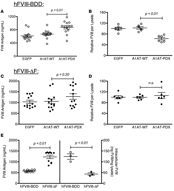
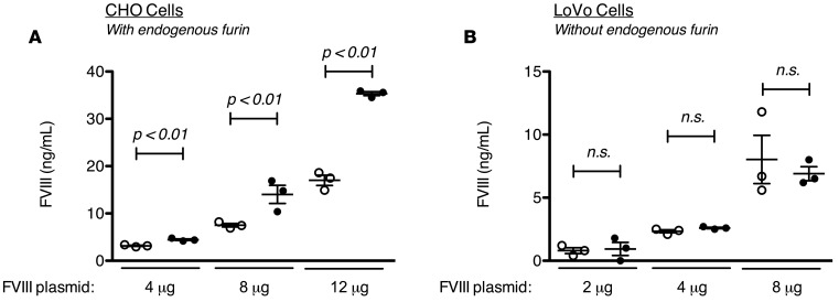
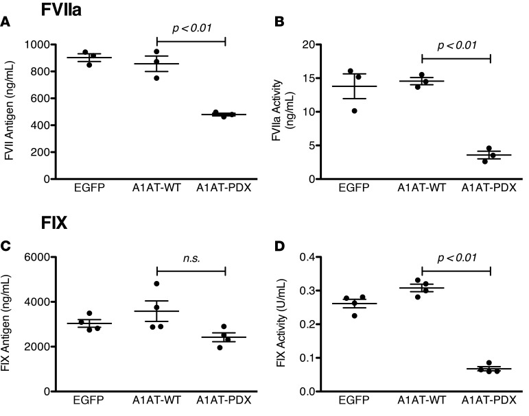
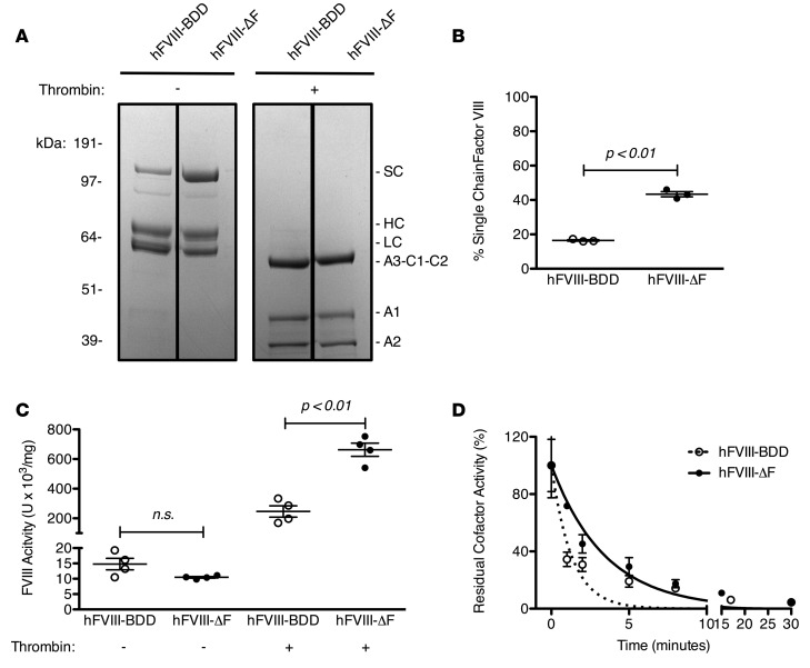
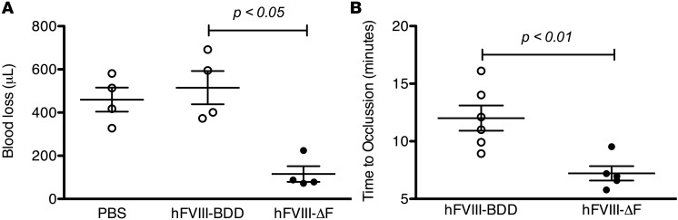
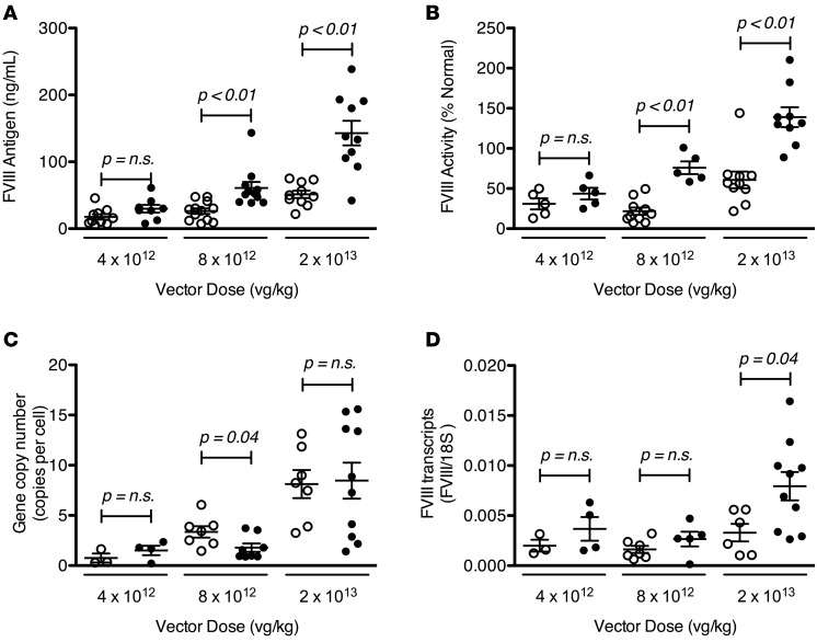
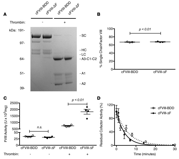
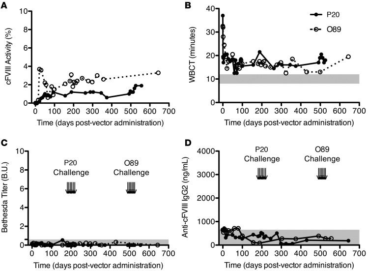
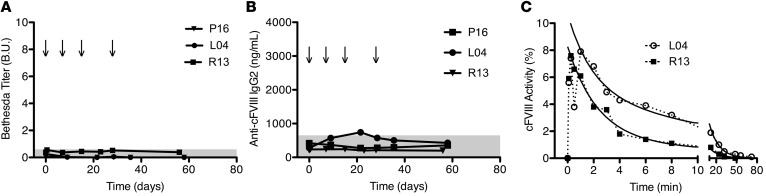
Processing by the proprotein convertase furin is believed to be critical for the biological activity of multiple proteins involved in hemostasis, including coagulation factor VIII (FVIII). This belief prompted the retention of the furin recognition motif (amino acids 1645-1648) in the design of B-domain-deleted FVIII (FVIII-BDD) products in current clinical use and in the drug development pipeline, as well as in experimental FVIII gene therapy strategies. Here, we report that processing by furin is in fact deleterious to FVIII-BDD secretion and procoagulant activity. Inhibition of furin increases the secretion and decreases the intracellular retention of FVIII-BDD protein in mammalian cells. Our new variant (FVIII-ΔF), in which this recognition motif is removed, efficiently circumvents furin. FVIII-ΔF demonstrates increased recombinant protein yields, enhanced clotting activity, and higher circulating FVIII levels after adeno-associated viral vector-based liver gene therapy in a murine model of severe hemophilia A (HA) compared with FVIII-BDD. Moreover, we observed an amelioration of the bleeding phenotype in severe HA dogs with sustained therapeutic FVIII levels after FVIII-ΔF gene therapy at a lower vector dose than previously employed in this model. The immunogenicity of FVIII-ΔF did not differ from that of FVIII-BDD as a protein or a gene therapeutic. Thus, contrary to previous suppositions, FVIII variants that can avoid furin processing are likely to have enhanced translational potential for HA therapy.


Figures

Figure 1
Figure 1. Effect of furin inhibition on the secretion and intracellular retention of human factor VIII (hFVIII) variants.
The amount of hFVIII secreted (A and C) or intracellularly retained (B and D) from baby hamster kidney (BHK) cells stably expressing either hFVIII-BDD (A and B) or hFVIII-ΔF (C and D) after transduction with vectors containing either the furin-inhibiting α1-antitrypsin Portland (A1AT-PDX) or a negative control (A1AT-wild type [A1AT-WT] or EGFP). Intracellular FVIII is reported as the relative amount of FVIII after transduction compared with the mean of the EGFP-transduced group. (E) Direct comparison of the amount of secreted (left) and intracellularly retained (right) hFVIII in BHK cells expressing either hFVIII-BDD or hFVIII-ΔF. All FVIII levels were determined by ELISA in duplicate. Each data point represents a distinct transduction and results are aggregates of 2–4 distinct experiments. Means were compared by 2-tailed Student’s t test. P values greater than 0.05 were considered not significant (n.s.). Horizontal markers in whisker plots represent the mean and 1 SEM.
Figure 2
Figure 2. Comparative secretion advantage of human factor VIII-ΔF (hFVIII-ΔF) over hFVIII-BDD requires furin.
Transient transfection with Lipofectamine 2000 of (A) CHO (furin-expressing) and (B) LoVo (furin-deficient) cells with hFVIII-BDD– (open circles) or hFVIII-ΔF–containing (closed circles) plasmids at 3 different plasmid amounts, as indicated. FVIII antigen levels were determined 48 hours after transfection and normalized for transfection efficiency. Each data point represents a distinct transfection. Means were compared by 2-tailed Student’s t test. P values greater than 0.05 were considered not significant (n.s.). Horizontal markers in whisker plots represent the mean and 1 SEM.
Figure 3
Figure 3. Effect of furin inhibition on the secretion and clotting activity of human factor VIIa (hFVIIa) and hFIX.
The amount of factor secreted from either FVIIa- (A) or FIX-producing (C) HEK293 cells after transduction with vectors containing either the furin-inhibiting α1-antitrypsin Portland (A1AT-PDX) or the negative control, A1AT-wild type (A1AT-WT). The amount of FVIIa activity (B) or FIX activity (D) from the same conditioned media. Each data point represents a distinct transduction. Means were compared by 2-tailed Student’s t test. P values greater than 0.05 were considered not significant (n.s.). Horizontal markers in whisker plots represent the mean and 1 SEM.
Figure 4
Figure 4. Biochemical characterization of recombinant human factor VIII-ΔF (hFVIII-ΔF) compared with hFVIII-BDD.
(A) SDS-PAGE analysis of 3 μg of purified recombinant hFVIII-BDD and hFVIII-ΔF staining with Coomassie blue before (−) or after (+) activation with 20 nM thrombin. Identified protein species are single chain (SC), heavy chain (HC), and light chain (LC). (B) Quantification of percentage single-chain of each hFVIII variant by densitometric analysis of SDS-PAGE. Each data point represents a distinct measurement. Similar results were obtained with at least 2 distinct protein preparations. (C) hFVIII clotting activity was determined by 1-stage or 2-stage clotting assay. Each data point represents a distinct dilution. Similar results were obtained with at least 2 distinct protein preparations. (D) Decay of activated hFVIII variants following thrombin activation. Residual activity was determined by 2-stage clotting assay and normalized by the 0 time point. Error bars represent SEM of at least 3 separate dilutions. Lines are single-exponential fittings. The half-lives of activated hFVIII-ΔF and hFVIII-BDD are 2.4 ± 0.3 minutes (R2 = 0.96) and 0.91 ± 0.2 minutes (R2 = 0.91), respectively. Means were compared by 2-tailed Student’s t test. P values greater than 0.05 were considered not significant (n.s.). Horizontal markers in whisker plots represent the mean and 1 SEM.
Figure 5
Figure 5. Hemostatic effect of recombinant human factor VIII (hFVIII) variants in severe hemophilia A mice.
Mice received PBS or 10 μg/kg recombinant hFVIII protein immediately prior to injury. (A) In the tail-clip challenge, blood loss over 10 minutes was determined after complete tail transection at 3-mm diameter. Median blood loss for mice that received PBS, hFVIII-BDD, and hFVIII-ΔF was 460 ± 60, 520 ± 80, and 120 ± 40 μL, respectively. FVIII antigen levels after infusion were similar, as shown in Supplemental Figure 2. (B) In the FeCl3 challenge, time to occlusion of the carotid artery was measured after 7.5% FeCl3–induced injury. Median time to occlusion for mice that received hFVIII-BDD and hFVIII-ΔF was 12 ± 1 and 7 ± 1 minutes, respectively. Each data point represents an individual animal. Horizontal markers in whisker plots represent the mean and 1 SEM.
Figure 6
Figure 6. Expression of human factor VIII (hFVIII) variants following adeno-associated viral serotype 8 (AAV8)–mediated liver gene transfer in severe hemophilia A (HA) mice.
Three dosing cohorts of mice received either AAV8-hFVIII-ΔF (closed circles) or AAV8-hFVIII-BDD (open circles) with a liver-specific promoter at the indicated vector doses. Circulating FVIII antigen (A) and activity (B), as determined by chromogenic assay, was measured 4–8 weeks after vector infusion. Liver sections were harvested at least 4 weeks after injection and used to determine vector DNA content (C) and mRNA level (D). Each data point represents a single animal. Horizontal markers in whisker plots represent the mean and 1 SEM.
Figure 7
Figure 7. Biochemical characterization of recombinant canine factor VIII-ΔF (cFVIII-ΔF ) compared with cFVIII-BDD.
(A) SDS-PAGE analysis of 3 μg of cFVIII-BDD and cFVIII-ΔF staining with Coomassie blue before (−) or after (+) activation with thrombin. Identified protein species are single chain (SC), heavy chain (HC), and light chain (LC). (B) Quantification of percentage of SC of each cFVIII variant by densitometric analysis of SDS-PAGE. Each data point represents a distinct measurement. (C) cFVIII clotting activity was determined by 1-stage or 2-stage clotting assay. Each data point represents a distinct dilution. (D) Decay of cFVIII variants following thrombin activation. Error bars represent SEM of at least 3 separate dilutions. Lines are single-exponential fittings. The half-lives of activated cFVIII-ΔF and cFVIII-BDD are 2.2 ± 0.2 and 4.7 ± 0.4 minutes, respectively; R2 = 0.99 and 0.99, respectively. Means were compared by 2-tailed Student’s t test. P values greater than 0.05 considered not significant (n.s.). Horizontal markers in whisker plots represent the mean and 1 SEM.
Figure 8
Figure 8. Adeno-associated viral serotype 8 (AAV8)–mediated canine factor VIII-ΔF (cFVIII-ΔF) gene therapy in severe hemophilia A dogs.
Two dogs, P20 (closed circles) and O89 (open circles), received 6 × 10¹² vector genome/kg (vg/kg) of AAV8-cFVIII-ΔF. Time course of cFVIII activity level (A) and whole-blood clotting time (WBCT) (B) for both dogs. Gray bar represents WBCT of dogs without hemophilia. (C) Time course of Bethesda titers for inhibitory antibodies for both dogs. Gray bar represents 0.6 Bethesda unit (B.U.) threshold. (D) Time course of anti-cFVIII IgG2 levels for both dogs. Gray bar represents baseline levels. Arrows in (C) and (D) indicate protein challenges with 2 μg/kg cFVIII-BDD recombinant protein. Each time point run in at least duplicate.
Figure 9
Figure 9. Canine factor VIII-ΔF (cFVIII-ΔF) protein therapy in naive severe hemophilia A dogs.
(A) Bethesda titers for inhibitory antibodies while receiving 2 μg/kg cFVIII-ΔF protein infusions, as indicated by arrows. Gray bar represents 0.6 Bethesda unit (B.U.) threshold. (B) Anti-cFVIII IgG2 levels of the same dogs. Gray bar represents baseline levels. (C) Time course of cFVIII activity after infusion of 2 μg/kg cFVIII-ΔF protein. Each time point run in at least duplicate. Solid lines are the bi-exponential fitting (R2 = 0.99 and 0.98) used to calculate the terminal (β) half-lives of 10 and 14 hours.

Similar articles

Cited by

References

    1. Nathwani AC, et al. Long-term safety and efficacy of factor IX gene therapy in hemophilia B. N Engl J Med. 2014;371(21):1994–2004. doi: 10.1056/NEJMoa1407309. - DOI - PMC - PubMed
    1. Nathwani AC, et al. Adenovirus-associated virus vector-mediated gene transfer in hemophilia B. N Engl J Med. 2011;365(25):2357–2365. doi: 10.1056/NEJMoa1108046. - DOI - PMC - PubMed
    1. Arruda VR, Samelson-Jones BJ. Obstacles and future of gene therapy for hemophilia. Expert Opin Orphan Drugs. 2015;3(9):997–1010. doi: 10.1517/21678707.2015.1069179. - DOI - PMC - PubMed
    1. Jiang H, et al. Multiyear therapeutic benefit of AAV serotypes 2, 6, and 8 delivering factor VIII to hemophilia A mice and dogs. Blood. 2006;108(1):107–115. doi: 10.1182/blood-2005-12-5115. - DOI - PubMed
    1. Sabatino DE, et al. Efficacy and safety of long-term prophylaxis in severe hemophilia A dogs following liver gene therapy using AAV vectors. Mol Ther. 2011;19(3):442–449. doi: 10.1038/mt.2010.240. - DOI - PMC - PubMed

Publication types