. 2016 Oct 12;11(10):e0164544.
doi: 10.1371/journal.pone.0164544. eCollection 2016.

Tomosyn Negatively Regulates Arginine Vasopressin Secretion in Embryonic Stem Cell-Derived Neurons

Affiliations

Tomosyn Negatively Regulates Arginine Vasopressin Secretion in Embryonic Stem Cell-Derived Neurons

Seiji Takeuchi et al. PLoS One. .

Abstract

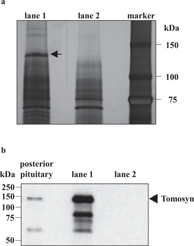
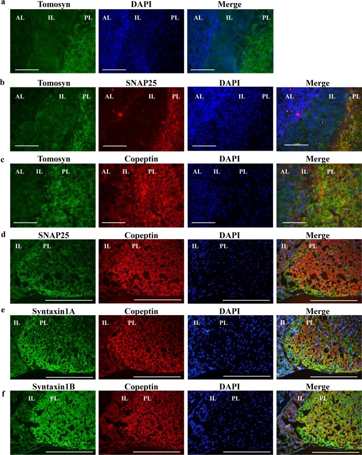
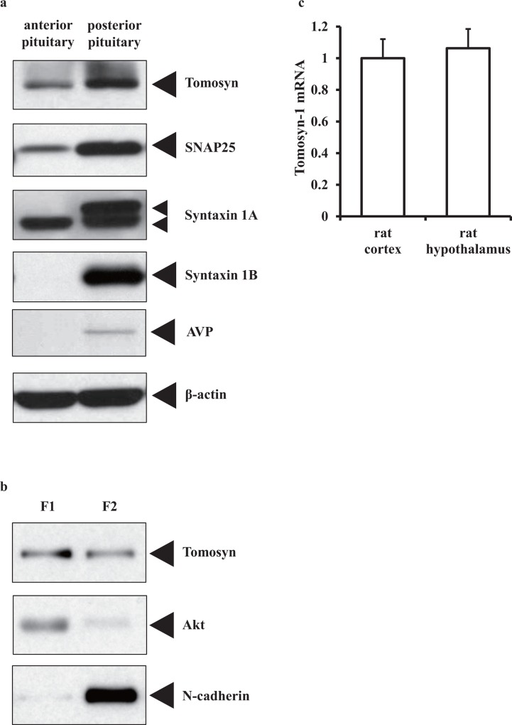
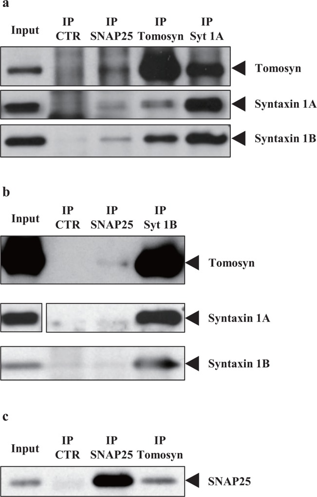
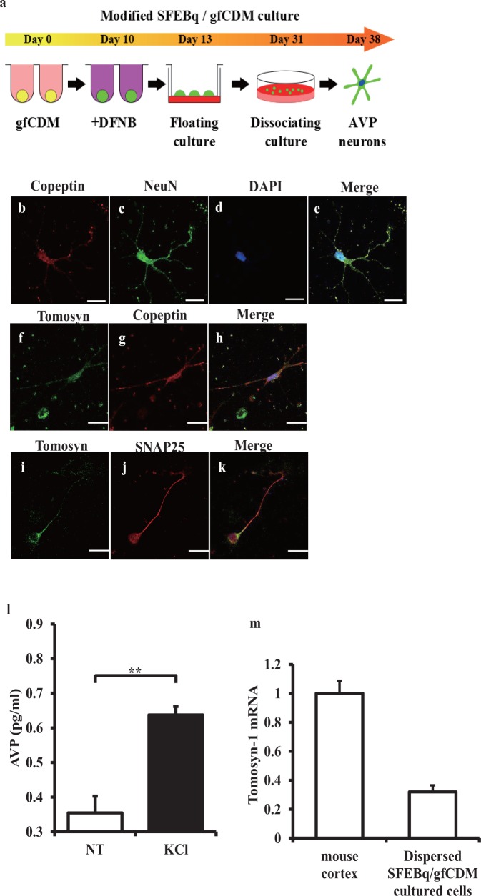
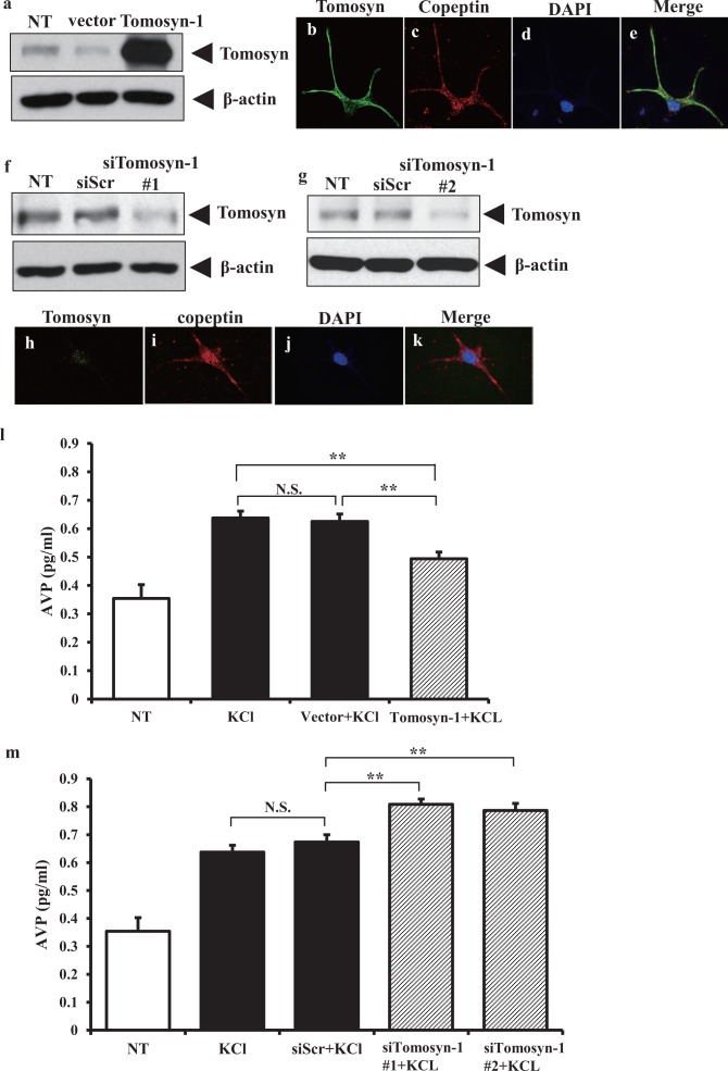
Arginine vasopressin (AVP) is secreted via exocytosis; however, the precise molecular mechanism underlying the exocytosis of AVP remains to be elucidated. To better understand the mechanisms of AVP secretion, in our study we have identified proteins that bind with a 25 kDa synaptosomal-associated protein (SNAP25). SNAP25 plays a crucial role in exocytosis, in the posterior pituitary. Embryonic stem (ES) cell-derived AVP neurons were established to investigate the functions of the identified proteins. Using glutathione S-transferase (GST)-pulldown assays and proteomic analyses, we identified tomosyn-1 (syntaxin-binding protein 5) as a SNAP25-binding protein in the posterior pituitary. Coimmunoprecipitation assays indicated that tomosyn formed N-ethylmaleimide-sensitive factor attachment protein receptor (SNARE) complexes with SNAP25 and syntaxin1. Immunohistochemistry showed that tomosyn localized to the posterior pituitary. Mouse ES cells self-differentiated into AVP neurons (mES-AVP) that expressed tomosyn and two transmembrane SNARE proteins, including SNAP25 and syntaxin1. KCl increased AVP secretion in mES-AVP, and overexpression of tomosyn-1 reduced KCl-stimulated AVP secretion. Downregulation of tomosyn-1 with siRNA increased KCl-stimulated AVP secretion. These results suggested that tomosyn-1 negatively regulated AVP secretion in mES-AVP and further suggest the possibility of using mES-AVP culture systems to evaluate the role of synaptic proteins from AVP neurons.


Conflict of interest statement

The authors have declared that no competing interests exist.

Figures

Fig 1
Fig 1. Tomosyn is a SNAP25 binding protein in the posterior pituitary.
(a) In vitro binding assay. Rat posterior pituitary lysates was incubated with GST-SNAP25 and glutathione (GSH) Sepharose. Eluted proteins bound to the beads were separated on SDS-PAGE followed by silver staining of proteins. The arrow indicates the band of interest. (b) Western blotting of pull-down samples. The arrowhead indicates the location of the immune reactive band reacting with anti-tomosyn antibody (Santa Cruz Biotechnology #sc-136105). Lane 1 = GST-SNAP25 with rat posterior pituitary lysates; lane 2 = GST-SNAP25 without rat posterior pituitary lysates (for both a, and b); Marker = molecular weight markers.
Fig 2
Fig 2. Tomosyn localizes in posterior pituitary.
(a) The cryosections of rat posterior pituitary were single stained with anti-tomosyn or DAPI (labelled on the top). The merged image is show at the right. (b–f) Double stained images are shown and the antibodies used are shown on top each image. Merged images are shown at right. Scale bars indicate 100 μm (white bars). AL, anterior lobe of the pituitary; IL, intermediate lobe; and PL, posterior lobe.
Fig 3
Fig 3. Tomosyn is expressed in the rat hypothalamo-posterior pituitary axis.
(a) Presence of tomosyn in the rat anterior and posterior pituitary as indicated by western blotting. Pituitary lysates were separated on SDS-PAGE followed by western blotting using anti-tomosyn, anti-SNAP25, anti-syntaxin 1A, anti-syntaxin 1B, anti-AVP, and anti-β-actin antibodies (as a loading control). (b) Subcellular fractionation of rat posterior pituitary samples. Fraction 1 (cytosolic proteins, F1) and Fraction 2 (membranes and membrane organelles, F2) were subjected to SDS-PAGE followed by western blotting using anti-tomosyn antibody. Five μg of proteins was loaded onto each lane. The subcellular fractionation was confirmed by western blotting with anti-Akt (cytosolic marker) and anti-N-cadherin (membrane fraction marker) antibodies. Tomosyn was present in both fractions. (c) Expression levels of tomosyn-1 mRNA in rat cortex and hypothalamus are similar. The amount of mRNA was determined using quantitative RT-PCR. The values are normalized to β-actin mRNA and are expressed as the mean ± SEM.
Fig 4
Fig 4. Tomosyn forms SNARE complexes with SNAP25 and syntaxin 1 in the posterior pituitary.
(a–c) Coimmunoprecipitation assays were performed using protein G Sepharose and mouse immunoprecipitation (IP) antibodies (a), protein A Sepharose and rabbit IP antibody (b) or ImmunocruzE imaging system (Santa Cruz Biotechnology, Santa Cruz, CA, USA) and mouse IP antibody (c), respectively. Eluted samples were subjected to SDS-PAGE followed by western blotting using anti-tomosyn, anti-SNAP25, anti-syntaxin 1A, or anti-syntaxin 1B antibodies (arrowheads at right). Input was 1/200 (volume) of the total rat posterior pituitary lysates used for the assay. CTR = IP with anti-IgG antibody.
Fig 5
Fig 5. Arginine vasopressin (AVP)-secreting neurons from mouse embryonic stem cells express tomosyn.
(a) Flow chart showing the method for culturing modified embryonic stem (ES) cells differentiating in serum-free medium (SFEBq)/growth factor-free chemically defined medium (gfCDM). DFNB = DMEM/F12 supplemented with 7 g/L glucose, N2 and B27. (b–e) Immunostaining with copeptin (red), NeuN (green), and DAPI (blue) in dispersed SFEBq/gfCDM cultured cells. A merged image is shown in the right panel. White scale bars indicate 20 μm. (f–k) Immunolocalization of proteins in dispersed SFEBq/gfCDM cultured cells, immunostaining with anti-tomosyn (green), anti-copeptin (red), or anti-SNAP25 (red) antibodies as analysed with confocal microscopy. Merged images are shown in the right panels. White scale bars indicate 25 μm. (l) AVP levels with or without 100 mM KCl stimulation in SFEBq/gfCDM cultured cells (see Methods section). AVP concentrations in the media of artificial cerebrospinal fluid cultured cells (aCSF) (non-treated, NT; n = 8), or 100 mM KCl treatment (KCl; n = 8) are shown. Values are expressed as the mean ± SEM. **P < 0.01 versus NT (non-treated artificial spinal fluid). (m) Tomosyn-1 mRNA expressions in dispersed SFEBq/gfCDM cultured cells. The amount of mRNA was determined using quantitative RT-PCR. The values are normalized to β-actin mRNA and are expressed as the mean ± SEM.
Fig 6
Fig 6. Tomosyn-1 negatively regulates secretion of AVP vesicles.
(a–k) The effects of overexpression or siRNA treatments on tomosyn-1 expression. Dispersed SFEBq/gfCDM cultured cells were transfected with empty vector (vector) or tomosyn-1 vector (Tomosyn-1), scrambled siRNA (siScr) or Tomosyn-1 siRNA (siTomosyn-1 #1 or #2), or not treated (NT). (a, f, g) After 48 h, the expression levels of tomosyn were analysed by western blotting using anti-tomosyn antibody (Tomosyn, marked with arrowheads at the right). The levels of β-actin in the same samples were determined as a control for protein loading (bottom panels of a, f, g). (b–e) Representative immunostaining for tomosyn (green), copeptin (red), and DAPI (blue) at 48 h after overexpression of tomosyn-1 in dispersed SFEBq/gfCDM cultured cells. The merged image is shown in (e). (h–k) Representative immunostaining for tomosyn (green), copeptin (red), and DAPI (blue) at 48 h after knockdown of tomosyn-1 with siRNA in dispersed SFEBq/gfCDM cultured cells. The merged image is shown in (k). (l) AVP concentration in the media of artificial cerebrospinal fluid cultured cells (aCSF) (non-treated, NT; n = 13), KCl treatment (KCl; n = 13), empty vector with KCl (Vector + KCl; n = 12), and tomosyn-1 vector with KCl (Tomosyn-1 + KCl; n = 12). (m) AVP concentration from scrambled siRNA with KCl (siScr + KCl; n = 13), and siTomosyn-1 with KCl (siTomosyn-1 #1 + KCl group; n = 13, siTomosyn-1 #2 + KCl group; n = 12). Final KCl concentration was 100 mM. Values are expressed as the mean ± SEM. **P < 0.01.

Similar articles

References

    1. Burbach JP, Luckman SM, Murphy D, Gainer H. Gene regulation in the magnocellular hypothalamo-neurohypophysial system. Physiol Rev. 2001;81(3):1197–267. - PubMed
    1. Gainer H, Chin H. Molecular diversity in neurosecretion: reflections on the hypothalamo-neurohypophysial system. Cell Mol Neurobiol. 1998;18(2):211–30. - PubMed
    1. Tobin V, Leng G, Ludwig M. The involvement of actin, calcium channels and exocytosis proteins in somato-dendritic oxytocin and vasopressin release. Front Physiol. 2012;3:261 10.3389/fphys.2012.00261 - DOI - PMC - PubMed
    1. Rorsman P, Renström E. Insulin granule dynamics in pancreatic beta cells. Diabetologia. 2003;46(8):1029–45. 10.1007/s00125-003-1153-1 - DOI - PubMed
    1. Weber T, Zemelman BV, McNew JA, Westermann B, Gmachl M, Parlati F, et al. SNAREpins: minimal machinery for membrane fusion. Cell. 1998;92(6):759–72. 10.1016/s0092-8674(00)81404-x - DOI - PubMed

MeSH terms

Grants and funding

This work was supported in part by a grant-in-aid for scientific research from the Japanese Society for the Promotion of Science 24591360 (to Y.S.) and a grant-in-aid for scientific research (Research on Hypothalamo-hypophyseal Disorders) (to Y.S. and Y.O.) from the Ministry of Health, Labor and Welfare, Japan.