

Impaired calcium mobilization in natural killer cells from chronic fatigue syndrome/myalgic encephalomyelitis patients is associated with transient receptor potential melastatin 3 ion channels
- PMID: 27727448
- PMCID: PMC5217865
- DOI: 10.1111/cei.12882
Impaired calcium mobilization in natural killer cells from chronic fatigue syndrome/myalgic encephalomyelitis patients is associated with transient receptor potential melastatin 3 ion channels
Abstract
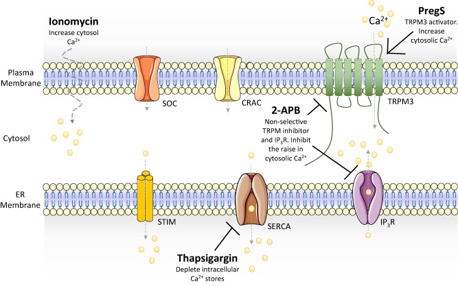
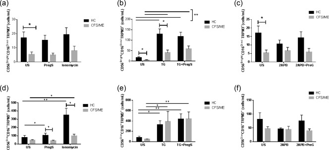
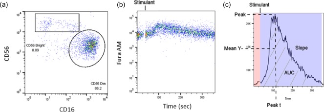
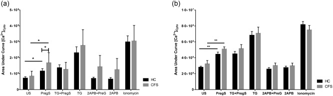
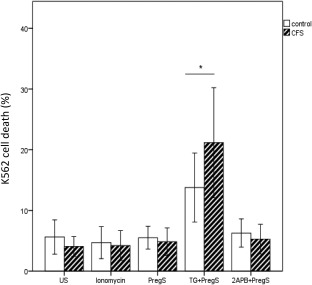
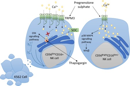
Transient receptor potential melastatin subfamily 3 (TRPM3) ion channels play a role in calcium (Ca2+ ) cell signalling. Reduced TRPM3 protein expression has been identified in chronic fatigue syndrome/myalgic encephalomyelitis (CFS/ME) patients. However, the significance of TRPM3 and association with intracellular Ca2+ mobilization has yet to be determined. Fifteen CFS/ME patients (mean age 48·82 ± 9·83 years) and 25 healthy controls (mean age 39·2 ± 12·12 years) were examined. Isolated natural killer (NK) cells were labelled with fluorescent antibodies to determine TRPM3, CD107a and CD69 receptors on CD56dim CD16+ NK cells and CD56bright CD16dim/- NK cells. Ca2+ flux and NK cytotoxicity activity was measured under various stimulants, including pregnenolone sulphate (PregS), thapsigargin (TG), 2-aminoethoxydiphenyl borate (2APB) and ionomycin. Unstimulated CD56bright CD16dim/- NK cells showed significantly reduced TRPM3 receptors in CFS/ME compared with healthy controls (HC). Ca2+ flux showed no significant difference between groups. Moreover, PregS-stimulated CD56bright CD16dim/- NK cells showed a significant increase in Ca2+ flux in CFS/ME patients compared with HC. By comparison, unstimulated CD56dim CD16+ NK cells showed no significant difference in both Ca2+ flux and TRPM3 expression. PregS-stimulated CD56dim CD16+ NK cells increased TRPM3 expression significantly in CFS/ME, but this was not associated with a significant increase in Ca2+ flux. Furthermore, TG-stimulated CD56dim CD16+ NK cells increased K562 cell lysis prior to PregS stimulation in CFS/ME patients compared with HC. Differential expression of TRPM3 and Ca2+ flux between NK cell subtypes may provide evidence for their role in the pathomechanism involving NK cell cytotoxicity activity in CFS/ME.
Keywords: cell surface molecules; inhibitory/activating receptors; natural killer cells.
© 2016 The Authors. Clinical & Experimental Immunology published by John Wiley & Sons Ltd on behalf of British Society for Immunology.
Figures
Similar articles
-
Potential pathophysiological role of the ion channel TRPM3 in myalgic encephalomyelitis/chronic fatigue syndrome (ME/CFS) and the therapeutic effect of low-dose naltrexone.J Transl Med. 2024 Jul 5;22(1):630. doi: 10.1186/s12967-024-05412-3. J Transl Med. 2024. PMID: 38970055 Free PMC article. Review.
-
Novel identification and characterisation of Transient receptor potential melastatin 3 ion channels on Natural Killer cells and B lymphocytes: effects on cell signalling in Chronic fatigue syndrome/Myalgic encephalomyelitis patients.Biol Res. 2016 May 31;49(1):27. doi: 10.1186/s40659-016-0087-2. Biol Res. 2016. PMID: 27245705 Free PMC article.
-
Loss of Transient Receptor Potential Melastatin 3 ion channel function in natural killer cells from Chronic Fatigue Syndrome/Myalgic Encephalomyelitis patients.Mol Med. 2018 Aug 14;24(1):44. doi: 10.1186/s10020-018-0046-1. Mol Med. 2018. PMID: 30134818 Free PMC article.
-
ERK1/2, MEK1/2 and p38 downstream signalling molecules impaired in CD56 dim CD16+ and CD56 bright CD16 dim/- natural killer cells in Chronic Fatigue Syndrome/Myalgic Encephalomyelitis patients.J Transl Med. 2016 Apr 21;14:97. doi: 10.1186/s12967-016-0859-z. J Transl Med. 2016. PMID: 27098723 Free PMC article.
-
Potential Implications of Mammalian Transient Receptor Potential Melastatin 7 in the Pathophysiology of Myalgic Encephalomyelitis/Chronic Fatigue Syndrome: A Review.Int J Environ Res Public Health. 2021 Oct 12;18(20):10708. doi: 10.3390/ijerph182010708. Int J Environ Res Public Health. 2021. PMID: 34682454 Free PMC article. Review.
Cited by
-
Potential pathophysiological role of the ion channel TRPM3 in myalgic encephalomyelitis/chronic fatigue syndrome (ME/CFS) and the therapeutic effect of low-dose naltrexone.J Transl Med. 2024 Jul 5;22(1):630. doi: 10.1186/s12967-024-05412-3. J Transl Med. 2024. PMID: 38970055 Free PMC article. Review.
-
Intra brainstem connectivity is impaired in chronic fatigue syndrome.Neuroimage Clin. 2019;24:102045. doi: 10.1016/j.nicl.2019.102045. Epub 2019 Oct 19. Neuroimage Clin. 2019. PMID: 31671321 Free PMC article.
-
Network Analysis of Symptoms Co-Occurrence in Chronic Fatigue Syndrome.Int J Environ Res Public Health. 2021 Oct 13;18(20):10736. doi: 10.3390/ijerph182010736. Int J Environ Res Public Health. 2021. PMID: 34682478 Free PMC article.
-
Current Research Provides Insight into the Biological Basis and Diagnostic Potential for Myalgic Encephalomyelitis/Chronic Fatigue Syndrome (ME/CFS).Diagnostics (Basel). 2019 Jul 10;9(3):73. doi: 10.3390/diagnostics9030073. Diagnostics (Basel). 2019. PMID: 31295930 Free PMC article.
-
Pathological Mechanisms Underlying Myalgic Encephalomyelitis/Chronic Fatigue Syndrome.Diagnostics (Basel). 2019 Jul 20;9(3):80. doi: 10.3390/diagnostics9030080. Diagnostics (Basel). 2019. PMID: 31330791 Free PMC article. Review.
References
-
- Fukuda K, Straus SE, Hickie I, Sharpe MC, Dobbins JG, Komaroff A. The chronic fatigue syndrome: a comprehensive approach to its definition and study. International Chronic Fatigue Syndrome Study Group. Ann Intern Med 1994; 121:953–9. - PubMed
-
- Stayer D, Scott V, Carter W. Low NK cell activity in chronic fatigue syndrome (CFS) and relationship to symptom severity. J Clin Cell Immunol 2015; 6:348.
-
- Draghi M, Yawata N, Gleimer M, Yawata M, Valiante NM, Parham P. Single‐cell analysis of the human NK cell response to missing self and its inhibition by HLA class I. Blood 2005; 105:2028–35. - PubMed
-
- Borrego F, Pena J, Solana R. Regulation of CD69 expression on human natural killer cells: differential involvement of protein kinase C and protein tyrosine kinases. Eur J Immunol 1993; 23:1039–43. - PubMed
MeSH terms
Substances
LinkOut - more resources
Full Text Sources
Other Literature Sources
Medical
Research Materials
Miscellaneous
