Testis-Specific GTPase (TSG): An oligomeric protein
- PMID: 27724860
- PMCID: PMC5057473
- DOI: 10.1186/s12864-016-3145-9
Testis-Specific GTPase (TSG): An oligomeric protein
Abstract
Background: Ras-related proteins in brain (Rab)-family proteins are key members of the membrane trafficking pathway in cells. In addition, these proteins have been identified to have diverse functions such as cross-talking with different kinases and playing a role in cellular signaling. However, only a few Rab proteins have been found to have a role in male germ cell development. The most notable functions of this process are performed by numerous testis-specific and/or germ cell-specific genes. Here, we describe a new Rab protein that is specifically expressed in male germ cells, having GTPase activity.
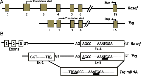
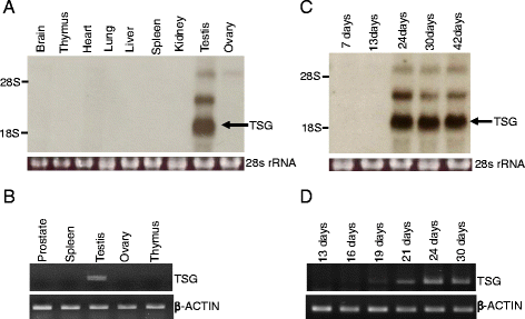
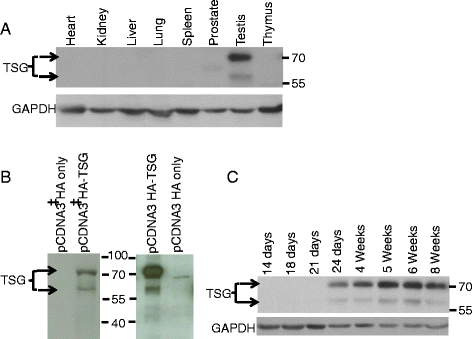
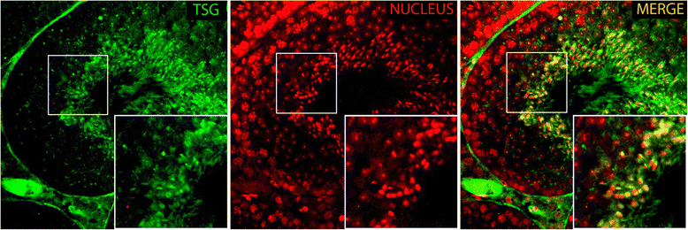
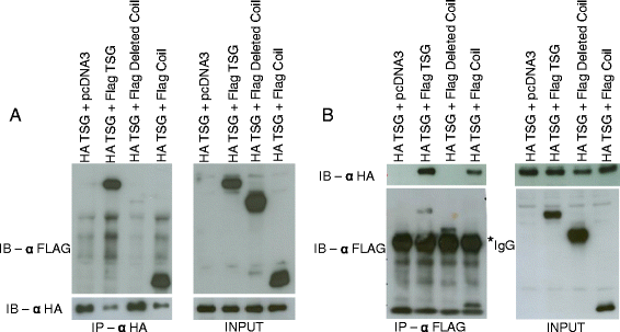
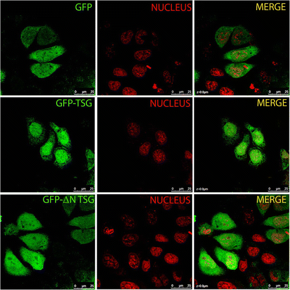
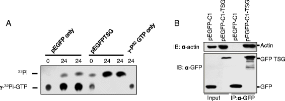
Results: Testis-specific GTPase (TSG) is a male-specific protein that is highly expressed in the testis. It has an ORF of 1593 base pairs encoding a protein of 530 amino acids. This protein appears in testicular cells approximately 24 days postpartum and is maintained thereafter. Immunohistochemistry of testicular sections indicates localized expression in germ cells, particularly elongating spermatids. TSG has a bipartite nuclear localization signal that targets the protein to the nucleus. The C-terminal region of TSG contains the characteristic domain of small Rab GTPases, which imparts GTPase activity. At the N-terminal region, it has a coiled-coil motif that confers self-interaction properties to the protein and allows it to appear as an oligomer in the testis.
Conclusion: TSG, being expressed in the male gonad in a developmental stage-specific manner, may have a role in male germ cell development. Further investigation of TSG function in vivo may provide new clues for uncovering the secrets of spermatogenesis.
Keywords: GTPase; RASEF; Testis.
Figures

Similar articles
-
Characterization of Rab45/RASEF containing EF-hand domain and a coiled-coil motif as a self-associating GTPase.Biochem Biophys Res Commun. 2007 Jun 8;357(3):661-7. doi: 10.1016/j.bbrc.2007.03.206. Epub 2007 Apr 13. Biochem Biophys Res Commun. 2007. PMID: 17448446
-
Sertoli-germ cell adherens junction dynamics in the testis are regulated by RhoB GTPase via the ROCK/LIMK signaling pathway.Biol Reprod. 2003 Jun;68(6):2189-206. doi: 10.1095/biolreprod.102.011379. Epub 2003 Jan 22. Biol Reprod. 2003. PMID: 12606349
-
Cellular functions of TC10, a Rho family GTPase: regulation of morphology, signal transduction and cell growth.Oncogene. 1999 Jul 1;18(26):3831-45. doi: 10.1038/sj.onc.1202758. Oncogene. 1999. PMID: 10445846
-
Fer kinase/FerT and adherens junction dynamics in the testis: an in vitro and in vivo study.Biol Reprod. 2003 Aug;69(2):656-72. doi: 10.1095/biolreprod.103.016881. Epub 2003 Apr 16. Biol Reprod. 2003. PMID: 12700184
-
The Emerging Role of MORC Family Proteins in Cancer Development and Bone Homeostasis.J Cell Physiol. 2017 May;232(5):928-934. doi: 10.1002/jcp.25665. Epub 2016 Nov 30. J Cell Physiol. 2017. PMID: 27791268 Review.
Cited by
-
Large Rab GTPases: Novel Membrane Trafficking Regulators with a Calcium Sensor and Functional Domains.Int J Mol Sci. 2021 Jul 19;22(14):7691. doi: 10.3390/ijms22147691. Int J Mol Sci. 2021. PMID: 34299309 Free PMC article. Review.
-
Transcriptome sequencing reveals the effects of circRNA on testicular development and spermatogenesis in Qianbei Ma goats.Front Vet Sci. 2023 Apr 27;10:1167758. doi: 10.3389/fvets.2023.1167758. eCollection 2023. Front Vet Sci. 2023. PMID: 37180060 Free PMC article.
-
Enhanced bovine genome annotation through integration of transcriptomics and epi-transcriptomics datasets facilitates genomic biology.Gigascience. 2024 Jan 2;13:giae019. doi: 10.1093/gigascience/giae019. Gigascience. 2024. PMID: 38626724 Free PMC article.
-
CRISPR/Cas9-mediated genome editing reveals 12 testis-enriched genes dispensable for male fertility in mice.Asian J Androl. 2022 May-Jun;24(3):266-272. doi: 10.4103/aja.aja_63_21. Asian J Androl. 2022. PMID: 34290169 Free PMC article.
-
Correlation between Rab3A Expression and Sperm Kinematic Characteristics.Dev Reprod. 2024 Mar;28(1):13-19. doi: 10.12717/DR.2024.28.1.13. Epub 2024 Mar 31. Dev Reprod. 2024. PMID: 38654977 Free PMC article. Korean.
References
Publication types
MeSH terms
Substances
LinkOut - more resources
Full Text Sources
Other Literature Sources
Molecular Biology Databases
Miscellaneous

