. 2016 Nov 21;39(4):466-479.
doi: 10.1016/j.devcel.2016.09.005. Epub 2016 Oct 6.

Acetylation of VGLL4 Regulates Hippo-YAP Signaling and Postnatal Cardiac Growth

Affiliations

Acetylation of VGLL4 Regulates Hippo-YAP Signaling and Postnatal Cardiac Growth

Zhiqiang Lin et al. Dev Cell. .

Abstract

Binding of the transcriptional co-activator YAP with the transcription factor TEAD stimulates growth of the heart and other organs. YAP overexpression potently stimulates fetal cardiomyocyte (CM) proliferation, but YAP's mitogenic potency declines postnatally. While investigating factors that limit YAP's postnatal mitogenic activity, we found that the CM-enriched TEAD1 binding protein VGLL4 inhibits CM proliferation by inhibiting TEAD1-YAP interaction and by targeting TEAD1 for degradation. Importantly, VGLL4 acetylation at lysine 225 negatively regulated its binding to TEAD1. This developmentally regulated acetylation event critically governs postnatal heart growth, since overexpression of an acetylation-refractory VGLL4 mutant enhanced TEAD1 degradation, limited neonatal CM proliferation, and caused CM necrosis. Our study defines an acetylation-mediated, VGLL4-dependent switch that regulates TEAD stability and YAP-TEAD activity. These insights may improve targeted modulation of TEAD-YAP activity in applications from cardiac regeneration to cancer.

Keywords: Hippo-YAP pathway; TEAD1; VGLL4; acetylation; cardiac; cardiomyocyte; degradation; necrosis; proliferation.


Figures

Figure 1
Figure 1. Developmental Changes in VGLL4-TEAD1 and YAP-TEAD1 Interaction in the Mouse Heart
(A) Immunoblot (IB) of protein extracts from adult mouse brain (B), heart (H), kidney (K), liver (Li), and lung (Lu). (B) Immunoblot (IB) of heart protein extracts from mice with the indicated postnatal (P) age in days. GAPDH internal control belonging to respective immunoblots is shown. (C) qRT-PCR measurement of Vgll4, Tead1, and Yap mRNA level in 3-day-old (P3) and 90-day-old (P90) hearts. *p < 0.05, n = 3. (D) VGLL4, TEAD1, and YAP expression in CMs and non-CMs. Adult hearts were dissociated by collagenase perfusion and then separated into CM and non-CM fractions. Protein extracts were immunoblotted with the indicated antibodies. (E) Age-dependent association of VGLL4 and TEAD1 in mouse heart. Tead1fb/+;R26BirA/+ heart extract was incubated with immobilized streptavidin (SA). Co-precipitated VGLL4 and TEAD1 were measured by immunoblotting. Tead1+/+;R26BirA/+ heart extract was used as a negative control. (F) Age-dependent association of YAP and TEAD1 in mouse heart. TEAD1 was precipitated from protein from P1, P8, or P50 mouse heart as in (D). Co-precipitated proteins were detected by immunoblotting. (G) Relative YAP or VGLL4 coIP with TEAD1, determined by quantification of (E). Precipitated proteins were normalized to TEAD1fb. *p < 0.05, n = 3. All error bars represent the SEM.
Figure 2
Figure 2. VGLL4 Overexpression Did Not Suppress Neonatal Cardiac Growth
P1 pups were injected subcutaneously with AAV9.GFP or AAV9.VGLL4-GFP. Control (Ctrl) mice were untreated. Hearts were analyzed at P8. (A) AAV9 expression constructs. AAV9.GFP and AAV9.VGLL4-GFP incorporate the cardiac troponin T (cTNT) promoter to drive selective CM expression. Heart protein immunoblots probed with GFP antibody demonstrated VGLL4-GFP fusion protein expression (asterisk). (B) Echocardiographic assessment of neonatal heart function. FS%, fractional shortening. n = 4. (C) Whole-mount images of hearts showing lack of substantial differences between groups. Scale bar, 2 mm. (D) Heart to body weight ratio was not significantly different (NS) between groups. n = 3. (E) Representative pH3 staining results. Scale bar, 50 μm. (F) Quantitation of pH3+ CMs. n = 3. NS, not significant. (G) Cell-cycle gene expression from P8 ventricular myocardium after treatment with AAV9.GFP or AAV9.VGLL4-GFP. Gene expression was measured by qRT-PCR and normalized to the AAV9.GFP group. n = 4. NS, not significant. All error bars represent the SEM.
Figure 3
Figure 3. VGLL4 TDU Domain Acetylation Decreased VGLL4-TEAD1 Interaction
(A) p300 bound and acetylated VGLL4. HEK293T cells were transfected with the indicated GFP and histone acetyltransferase (HAT; HA-tagged) expression plasmids. Proteins that co-precipitated with GFP were detected by immunoblotting (IB). K-Ac Ab, acetylated lysine-specific antibody. (B) VGLL4-K225 is the major VGLL4 acetylation site. VGLL4-GFP was overexpressed in HEK293T cells in the presence of p300, immunoprecipitated with GFP antibody, and analyzed by mass spectrometry. The area of the red circles is proportional to the fraction of peptides detected that contain the acetyl lysine residue indicated by the corresponding number. T1 and T2 represent the two TDU domains of VGLL4. (C) VGLL4 K225R mutation decreased VGLL4 acetylation. Wild-type (WT) or K255R mutated (R) VGLL4-GFP were co-expressed in HEK293T with p300, as indicated. VGLL4-GFP acetylation was detected by immunoprecipitation and western blot. Acetylation of VGLL4 K225R was normalized to wild-type VGLL4. *p < 0.05, n = 4. (D) Alignment of TDU domains from different proteins (top group) or from the first TDU domain of VGLL4 from different species (bottom group). The residue aligned with K225 of human VGLL4 is highlighted in red and indicated by an arrow. This residue is conserved in vertebrate VGLL4 but is not conserved across TDU domains. (E and F) VGLL4 K225 acetylation decreased VGLL4-TEAD1 interaction in vitro. Interaction between recombinant His-TEAD1[211–427] and synthetic, un-acetylated, or K225-acetylated VGLL4 TDU domain peptides was detected by immunoprecipitation and western blot (E) or by a nanoscale photonic interaction assay (F). (G) VGLL4[R] increased VGLL4-TEAD1 and decreased YAP-TEAD1 interaction in cultured cells. TEAD1fb and VGLL4-GFP expression plasmids were co-transfected into 293T cells. TEAD1 CoIP was carried out using FLAG antibody. The ratio between VGLL4 and TEAD1 in the immunoprecipitate was quantified by densitometry. *p < 0.05, n = 3. (H) p300 effect on YAP-TEAD1 transcriptional activity. 293T cells were co-transfected with the indicated plasmids plus pRL-TK. Left: 24 hr after transfection, cells were collected for luciferase activity measurement. Firefly luciferase activity was normalized to Renilla luciferase. *p < 0.05, n = 4. Right: western blot showing the expression of p300, Vgll4, and TEAD1fb in transduced cells. (I and J) Effect of VGLL4 acetylation on VGLL4-TEAD1 interaction in NRVM. The PLA was used to detect endogenous VGLL4-TEAD1 interaction in cultured NRVMs. Representative image (I) and quantification of TEAD1-VGLL4 interaction events in the nucleus (J) are shown. Each red dot was counted as an interaction event. Scale bar, 20 μm. ***p < 0.0001, Wilcoxon. (K) Endogenous levels of mVGLL4 (murine VGLL4) and mVGLL4-K216Ac (which corresponds to human K225Ac) in P6 and P60 heart. Hearts were lysed with denaturing buffer containing 2% SDS, and 100 μg of total protein was immunoblotted for total VGLL4 or VGLL4-K216Ac. Fold change of protein levels between P60 and P6 was determined by densitometry. *p < 0.05. (L) Endogenous levels of VGLL4 and VGLL4-K225Ac in human left ventricular myocardium, obtained from unused transplant donor hearts of the indicated ages. All error bars represent the SEM.
Figure 4
Figure 4. VGLL4 Overexpression Decreased TEAD1 Stability
(A) VGLL4 overexpression decreased TEAD1 protein level. Different doses of TEAD1 plasmids (indicated in μg) were co-transfected with 1.6 μg of VGLL4-GFP plasmid. Cells were collected for western blot 24 hr after transfection. (B and C) Generation and validation of TEAD1-Dendra2 construct. Tead1-Dendra2 plasmid was transfected into 293T cells. Western blot confirmed expression of TEAD1-Dendra2 fusion protein (B). TEAD1-Dendra2 merge fusion protein was green before illumination with 405 nm light. After 30 s of illumination, a fraction of TEAD1-Dendra2 exhibited red fluorescence (C). (D and E) Doxycycline (Dox)-inducible expression of VGLL4 caused TEAD1-Dendra2 degradation. pTEAD1-Dendra2 and pEF1a-rtTA were co-transfected into 293T cells along with pTetO empty vector (upper panel) or pTetO:HA-VGLL4 (lower panel). Twenty-four hours after transfection, Dox was added. Cells were analyzed at the indicated time points (D). Quantification of TEAD1-Dendra2 protein level is shown in (E). *p < 0.05, n = 3. (F) Time-lapse imaging of TEAD1-Dendra2 or Dendra2 proteins. Indicated plasmids were transfected into 293T cells. Twenty-four hours later, cells were treated with Dox. Four hours later, Dendra2 was photoconverted with 405-nm light. Relative red fluorescence intensity (RFI) was monitored for 3 hr by taking one image per minute. RFI was normalized to the value immediately after photoconversion. Plot shows average RFI signal over 10 min. n = 10. Experiment is representative of three independent repeats. (G) Dual luciferase assay of YAP-TEAD1 transcriptional activity. 293T cells were co-transfected with YAP[S127A], EF1a:rtTA, 8xGIITC-luciferase, pRL-TK internal control, and either TetO empty vector or TetO-VGLL4 as indicated. E64 was added as indicated. Twenty-four hours after transfection, cells were treated with Dox for the indicated number of hours, when cell extracts were analyzed for Firefly and Renilla luciferase activity. Relative luciferase activity was the ratio of Firefly to Renilla luciferase, normalized to empty vector at time 0. *p < 0.05, n = 4. (H) Model of VGLL4 regulation of YAP-TEAD1 activity. In the absence of VGLL4, YAP binds to TEAD1 to activate target gene expression. VGLL4 overexpression suppressed YAP-TEAD1 activity by both inhibiting TEAD1 transcriptional activity (i) and promoting TEAD1 degradation (ii). All error bars represent the SEM.
Figure 5
Figure 5. Abrogation of VGLL4-K225 Acetylation Unmasked Disruptive Effects of VGLL4 on YAP-TEAD Interaction and Neonatal Heart Maturation
P1 pups were treated with AAV9.VGLL4, AAV9.VGLL4[R] (containing the K225R mutation), or AAV.GFP. Hearts were examined at P8 or P12, as indicated. (A) Assay of cardiac TEAD1-interacting proteins. TEAD1 and its associated proteins were immunoprecipitated, and indicated proteins were detected by western blotting. Asterisk indicates the VGLL4-GFP band. (B) Endogenous p300 interacts with and acetylates VGLL4 in the neonatal heart. AAV9.GFP, AAV9.VGLL4-GFP, or AAV9.VGLL4[R]-GFP were administered to P1 mouse pups. p300 was immunoprecipitated from P8 heart extracts and probed with indicated antibodies. K-Ac Ab, acetyl lysine-specific antibody. Asterisk indicates acetylated VGLL4-GFP. Arrowhead indicates the VGLL4-GFP band, which runs just above the immunoglobulin heavy chain. (C and D) Echocardiographic measurement of left ventricular (LV) systolic function (fractional shortening, FS) (C) and diastolic LV wall thickness (D) at P8. *p < 0.05 compared with GFP control, n = 4. (E) Whole-mount (upper panels) and H&E-stained short-axis sections of AAV-transduced hearts at P12. Scale bars, 2 mm. (F) Heart to body weight ratio of AAV-transduced hearts at P8 or P12. *p < 0.05, n = 4. (G and H) Cardiac fibrosis was visualized by pirosirius red/fast green staining. Representative images (G) and quantification (H). Scale bar, 1 mm. *p < 0.05, n = 3. (I and J) qRT-PCR measurement of heart failure marker gene transcripts Myh6 and Nppa. Levels were normalized to GAPDH and expressed relative to the AAV9.GFP control group. *p < 0.05, n = 4. All error bars represent the SEM.
Figure 6
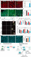
Figure 6. Acetylation-Deficient VGLL4[R] Decreased Cardiomyocyte Proliferation and Survival
AAV9.GFP, AAV9.VGLL4, or AAV9.VGLL4[R] were delivered to P1 pups, and hearts were examined at P8. (A and B) Measurement of CM necrosis. Rosa26mTmG (membrane localized RFP) P1 pups were treated with AAV9. Anti-myosin antibody MF20 was injected into mice at P7. At P8, mice were collected and intracellular MF20 antibody was detected by immunofluorescent staining. Representative images (A) and quantification (B). Scale bar, 50 μm. (C and D) Measurement of CM proliferation using pH3 immunofluorescence staining. (C) Representative images with boxed regions magnified in insets. Scale bar, 50 μm. (D) Quantification of pH3+ CMs. *p < 0.05, n = 3. (E and F) Clonal assay for CM proliferation. Confetti P1 pups were treated with mosaic dose of AAV9.Cre to label individual CMs, in addition to treatment with VGLL4 or control AAVs at the usual dose, which transduces nearly all CMs. Hearts were examined at P8. (E) Representative images with boxed regions magnified in insets. Scale bar, 50 μm. (F) quantification of clusters of adjacent, labeled CMs containing one color (monochromatic, potentially arising from proliferation) or two colors (bichromatic, arising from adjacent labeling events). Scale bar, 50 μm. *p < 0.05, n = 4. (G) qRT-PCR measurement of relative levels of YAP-TEAD canonical targets gene transcripts Aurka, Cdc20, and Ctgf in P12 heart. *p < 0.05, n = 3. (H and I) Measurement of cardiomyocyte cross-sectional area. CMs were outlined by wheat germ agglutinin staining. Representative images (H) and quantification of cross-sectional area (I). *p < 0.05, n = 3. (J) Model of VGLL4 regulation of heart growth. In normal newborn heart, predominant YAP-TEAD1 stimulates CM proliferation. The interaction between VGLL4 and TEAD1 is blunted by p300-mediated VGLL4 acetylation. Inhibition of VGLL4 acetylation, as in the K225R mutant, suppresses cardiac growth by both inhibiting YAP-TEAD1 interaction and decreasing TEAD1 stability. All error bars represent the SEM.

Similar articles

Cited by

References

    1. Bai H, Zhang N, Xu Y, Chen Q, Khan M, Potter JJ, Nayar SK, Cornish T, Alpini G, et al. Yes-associated protein regulates the hepatic response after bile duct ligation. Hepatology. 2012;56:1097–1107. - PMC - PubMed
    1. Barrett AJ, Kembhavi AA, Brown MA, Kirschke H, Knight CG, Tamai M, Hanada K. L-trans-Epoxysuccinyl-leucylamido(4-guanidino) butane (E-64) and its analogues as inhibitors of cysteine proteinases including cathepsins B, H and L. Biochem. J. 1982;201:189–198. - PMC - PubMed
    1. Camargo FD, Gokhale S, Johnnidis JB, Fu D, Bell GW, Jaenisch R, Brummelkamp TR. YAP1 increases organ size and expands un-differentiated progenitor cells. Curr. Biol. 2007;17:2054–2060. - PubMed
    1. Chan HM, La Thangue NB. p300/CBP proteins: HATs for transcriptional bridges and scaffolds. J. Cell Sci. 2001;114:2363–2373. - PubMed
    1. Chen HH, Mullett SJ, Stewart AF. Vgl-4, a novel member of the vestigial-like family of transcription cofactors, regulates alpha1-adrenergic activation of gene expression in cardiac myocytes. J. Biol. Chem. 2004;279:30800–30806. - PubMed

MeSH terms