. 2016 Nov 28;90(24):11106-11121.
doi: 10.1128/JVI.01310-16. Print 2016 Dec 15.

Foot-and-Mouth Disease Virus Viroporin 2B Antagonizes RIG-I-Mediated Antiviral Effects by Inhibition of Its Protein Expression

Affiliations

Foot-and-Mouth Disease Virus Viroporin 2B Antagonizes RIG-I-Mediated Antiviral Effects by Inhibition of Its Protein Expression

Zixiang Zhu et al. J Virol. .

Erratum in

Abstract

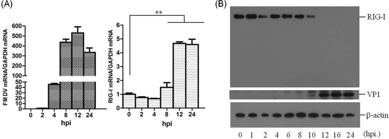
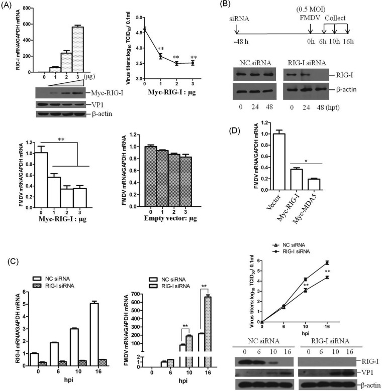
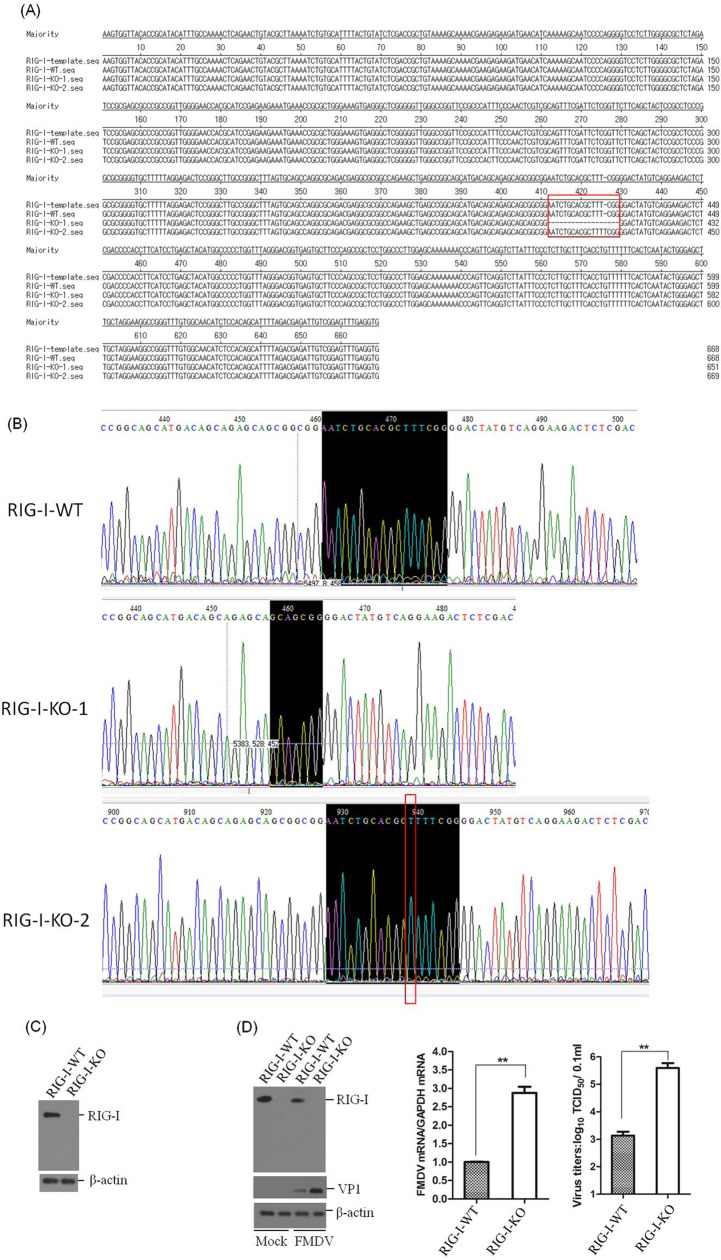
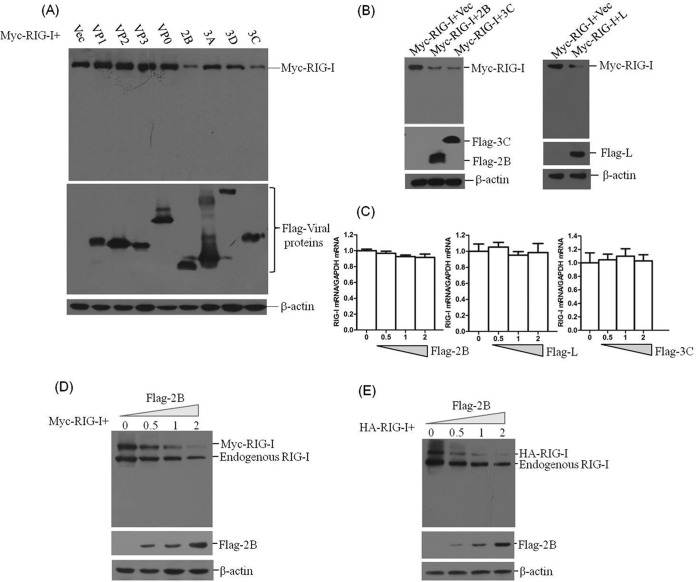
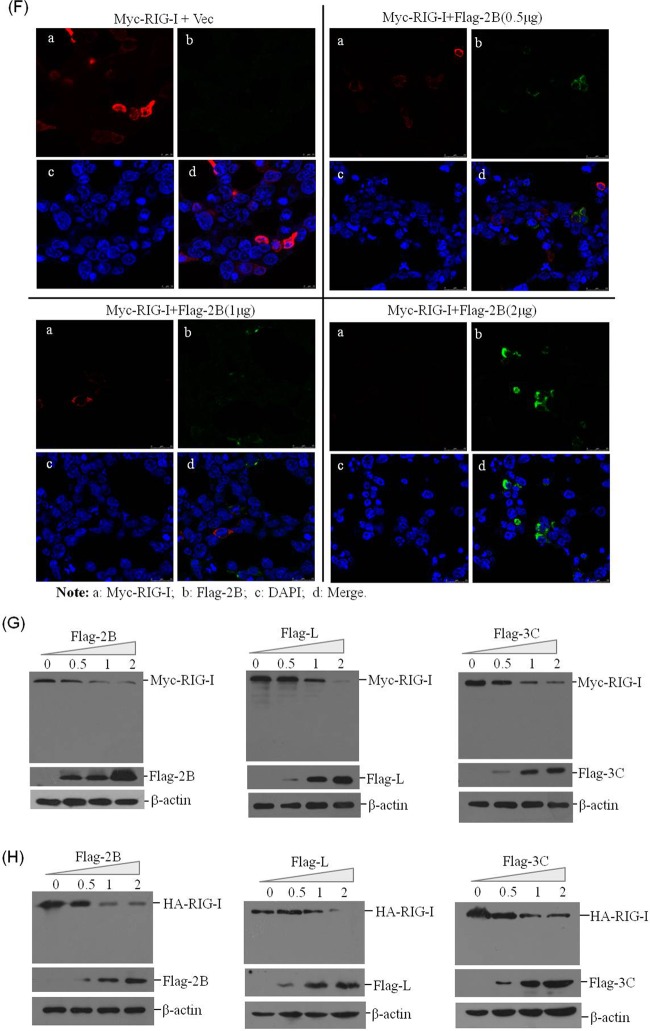
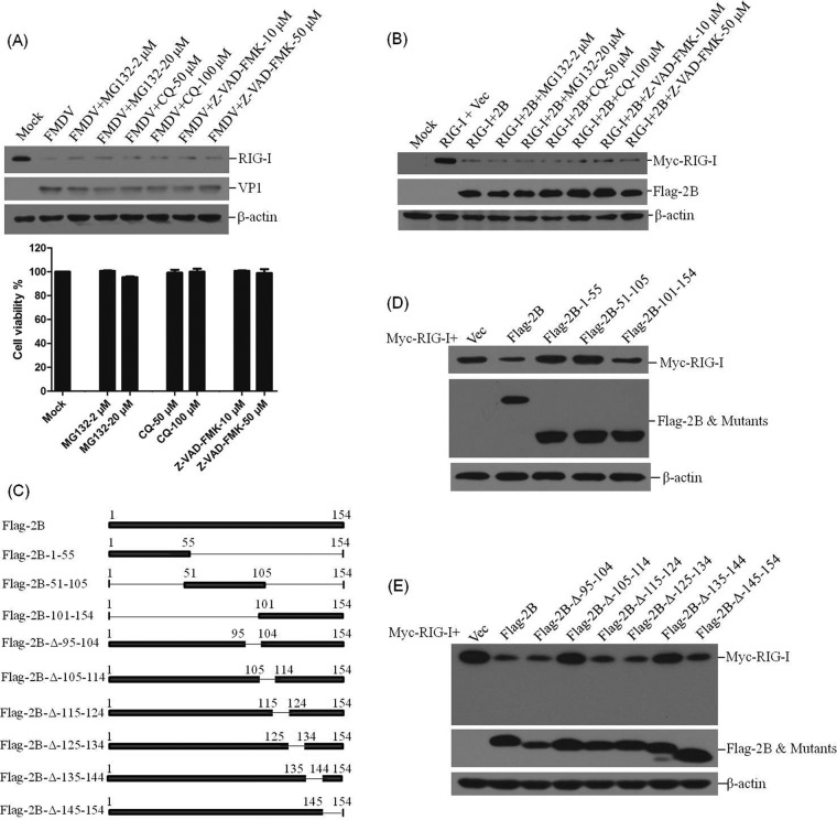
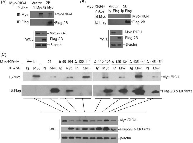
The role of retinoic acid-inducible gene I (RIG-I) in foot-and-mouth disease virus (FMDV)-infected cells remains unknown. Here, we showed that RIG-I inhibits FMDV replication in host cells. FMDV infection increased the transcription of RIG-I, while it decreased RIG-I protein expression. A detailed analysis revealed that FMDV leader proteinase (Lpro), as well as 3C proteinase (3Cpro) and 2B protein, decreased RIG-I protein expression. Lpro and 3Cpro are viral proteinases that can cleave various host proteins and are responsible for several of the viral polyprotein cleavages. However, for the first time, we observed 2B-induced reduction of host protein. Further studies showed that 2B-mediated reduction of RIG-I is specific to FMDV, but not other picornaviruses, including encephalomyocarditis virus, enterovirus 71, and coxsackievirus A16. Moreover, we found the decreased protein level of RIG-I is independent of the cleavage of eukaryotic translation initiation factor 4 gamma, the induction of cellular apoptosis, or the association of proteasome, lysosome, and caspase pathways. A direct interaction was observed between RIG-I and 2B. The carboxyl-terminal amino acids 105 to 114 and amino acids 135 to 144 of 2B were essential for the reduction of RIG-I, while residues 105 to 114 were required for the interaction. These data suggest the antiviral role of RIG-I against FMDV and a novel antagonistic mechanism of FMDV that is mediated by 2B protein.

Importance: This study demonstrated that RIG-I could suppress FMDV replication during virus infection. FMDV infection increased the transcriptional expression of RIG-I, while it decreased RIG-I protein expression. FMDV 2B protein interacted with RIG-I and induced reduction of RIG-I. 2B-induced reduction of RIG-I was independent of the induction of the cleavage of eukaryotic translation initiation factor 4 gamma or cellular apoptosis. In addition, proteasome, lysosome, and caspase pathways were not involved in this process. This study provides new insight into the immune evasion mediated by FMDV and identifies 2B as an antagonistic factor for FMDV to evade the antiviral response.


Figures

FIG 1
FIG 1
FMDV triggers RIG-I mRNA expression and decreases RIG-I protein abundance during viral infection. (A) PK-15 cells (5 × 10⁵ cells in each well) were grown in 35-mm culture dishes, the monolayers were infected with FMDV at an MOI of 0.5, and viral RNA (left panel) and RIG-I mRNA (right panel) were determined by qPCR at 0, 2, 4, 8, 12, and 24 hpi. **, P < 0.01, considered highly significant. The data represent results from one of the triplicate experiments. (B) Expression of endogenous RIG-I and FMDV VP1 proteins at the indicated time points postinfection (0, 1, 2, 4, 6, 8, 10, 12, 16, and 24 hpi) in PK-15 cells was determined by Western blotting. RIG-I was detected by rabbit anti-RIG-I polyclonal antibody. A mouse anti-VP1 polyclonal antibody prepared by our laboratory (unpublished data) was used to confirm the expression of viral protein, and mouse anti-β-actin was used to detect cellular β-actin.
FIG 2
FIG 2
RIG-I inhibits FMDV replication during virus infection. (A) PK-15 cells (5 × 10⁵ cells in each well) were transfected with increasing amounts of Myc-tagged RIG-I-expressing plasmids (0, 1, 2, or 3 μg), and the empty vector was used in the transfection process to ensure that the cells received the same amounts of total DNA plasmids. At 12 hpt, the cells were infected with equal amounts of FMDV (MOI of 0.5) for 16 h. The expression of RIG-I mRNA and viral RNA was examined by qPCR, and expression of Myc-RIG-I and FMDV VP1 protein was examined by Western blotting. The viral titers were determined by TCID50 assay. The effect of empty vector DNA on the viral replication was also evaluated by the transfection of 0, 1, 2, or 3 μg of empty vector in PK-15 cells. At 12 hpt, the cells were infected with equal amounts of FMDV (MOI of 0.5) for 16 h. The expression of viral RNA was determined by qPCR. (B) Schematics of the strategy used in the RNAi assay (upper panel) and confirmation of the efficiencies of a nontargeting siRNA (NC siRNA) and RIG-I siRNA in silencing RIG-I expression (lower panel). PK-15 cells (5 × 10⁵ cells in each well) were transfected with 150 nM NC siRNA or RIG-I siRNA, and the expression of RIG-I was determined by Western blotting at 0, 24, or 48 hpt. (C) PK-15 cells (5 × 10⁵ cells in each well) were transfected with 150 nM NC siRNA or RIG-I siRNA for 48 h, followed by infection with equal amounts of FMDV (MOI of 0.5) for 0, 6, 10, and 16 h. The expression of RIG-I mRNA and viral RNA was determined by qPCR (left panel). The expression of RIG-I and viral VP1 proteins was detected by Western blotting, and viral titers were determined by TCID50 assay (right panel). (D) PK-15 cells were transfected with 2 μg of empty vector, Myc-RIG-I, or Myc-MDA5 plasmids. At 12 hpt, the cells were infected with equal amounts of FMDV (MOI of 0.5) for 16 h. The viral RNA levels were determined by qPCR. All of the experiments were repeated three times, with similar results. The data represent results from one of the triplicate experiments. *, P < 0.05, considered significant; **, P < 0.01, considered highly significant.
FIG 3
FIG 3
Confirmation of the establishment of RIG-I knockout PK-15 cells. (A) Alignment of the RIG-I genomic nucleotide sequence of the published RIG-I reference sequence and of the RIG-I-WT, RIG-I-KO-1, and RIG-I-KO-2 sequences using LaserGene software. The red box indicates the regions that were mutated. (B) Confirmation of the genome editing by Sanger sequencing of the PCR amplicon from the RIG-I genome of the cell lines. (C) The established RIG-I-WT and RIG-I-KO cells were seeded in six-well culture plates (5 × 10⁵ cells in each well) and grown for 24 h, and the expression of RIG-I was detected by Western blotting. (D) RIG-I-WT and RIG-I-KO PK-15 cells were seeded in six-well culture plates (5 × 10⁵ cells in each well), and the monolayer cells were mock infected or infected with 0.5 MOI of FMDV for 10 h. The expression of RIG-I and viral VP1 proteins was detected by Western blotting. The expression of viral RNA was determined by qPCR, and viral titers were determined by TCID50 assay. All the experiments were repeated three times with similar results. The data represent results from one of the triplicate experiments. **, P < 0.01 considered highly significant.
FIG 4
FIG 4
2B protein induces the reduction of exogenous RIG-I expression. (A) HEK293T cells (5 × 10⁵ cells in each well) were transfected with Myc-tagged RIG-I-expressing plasmid (2 μg), along with various plasmids expressing Flag-tagged viral proteins (VP1, VP2, VP3, VP0, 2B, 3A, 3Dpol, or 3Cpro) or empty Flag vector plasmid (2 μg). The expression of Myc-RIG-I and Flag-tagged viral protein was detected by Western blotting at 36 hpt. (B) HEK293T cells (5 × 10⁵ cells in each well) were transfected with Myc-tagged RIG-I-expressing plasmid (2 μg), along with various plasmids expressing Flag-tagged viral proteins (2B, 3Cpro, or Lpro) or empty Flag vector (2 μg). The expression of Myc-RIG-I and Flag-tagged viral protein was detected by Western blotting at 36 hpt. (C) HEK293T cells (5 × 10⁵ cells in each well) were transfected with Myc-RIG-I-expressing plasmid (2 μg), along with increasing quantities of Flag-2B-, Flag-3C-, or Flag-L-expressing plasmids (0, 0.5, 1, or 2 μg). The empty vector was used in the transfection process to ensure that the cells received the same amounts of total plasmids. The expression of RIG-I mRNA was determined by qPCR analysis at 36 hpt. (D) HEK293T cells (5 × 10⁵ cells in each well) were transfected with Myc-RIG-I-expressing plasmid (2 μg), along with increasing quantities of Flag-2B-expressing plasmid (0, 0.5, 1, or 2 μg). The expression of Myc-RIG-I and Flag-2B was detected by Western blotting with anti-RIG-I and anti-Flag antibodies at 36 hpt. (E) HEK293T cells (5 × 10⁵ cells in each well) were transfected with HA-RIG-I-expressing plasmid (2 μg), along with increasing quantities of Flag-2B-expressing plasmid (0, 0.5, 1, or 2 μg). The expression of HA-RIG-I and Flag-2B was detected by Western blotting with anti-RIG-I and anti-Flag antibodies at 36 hpt. (F) HEK293T cells (3 × 10⁵ cells in each well) were transfected with Myc-RIG-I-expressing plasmid (2 μg), along with increasing quantities of Flag-2B-expressing plasmids (0, 0.5, 1, or 2 μg). The expression of Myc-RIG-I and Flag-2B was detected by IFA analysis at 36 hpt. Cells were double immunostained for Myc-RIG-I (red) and Flag-2B (green); cellular nuclei were counterstained with DAPI (blue). (G) HEK293T cells (5 × 10⁵ cells in each well) were transfected with Myc-RIG-I-expressing plasmid (2 μg), along with increasing quantities of Flag-2B-, Flag-L-, or Flag-3C-expressing plasmid (0, 0.5, 1, or 2 μg). The expression of Myc-RIG-I and Flag-tagged viral proteins was detected by Western blotting with anti-Myc and anti-Flag antibodies at 36 hpt. (H) HEK293T cells (5 × 10⁵ cells in each well) were transfected with HA-RIG-I-expressing plasmid (2 μg), along with increasing quantities of Flag-2B-, Flag-L-, or Flag-3C-expressing plasmids (0, 0.5, 1, or 2 μg). The expression of HA-RIG-I and Flag-tagged viral proteins was detected by Western blotting with anti-HA and anti-Flag antibodies at 36 hpt.
FIG 4
FIG 4
2B protein induces the reduction of exogenous RIG-I expression. (A) HEK293T cells (5 × 10⁵ cells in each well) were transfected with Myc-tagged RIG-I-expressing plasmid (2 μg), along with various plasmids expressing Flag-tagged viral proteins (VP1, VP2, VP3, VP0, 2B, 3A, 3Dpol, or 3Cpro) or empty Flag vector plasmid (2 μg). The expression of Myc-RIG-I and Flag-tagged viral protein was detected by Western blotting at 36 hpt. (B) HEK293T cells (5 × 10⁵ cells in each well) were transfected with Myc-tagged RIG-I-expressing plasmid (2 μg), along with various plasmids expressing Flag-tagged viral proteins (2B, 3Cpro, or Lpro) or empty Flag vector (2 μg). The expression of Myc-RIG-I and Flag-tagged viral protein was detected by Western blotting at 36 hpt. (C) HEK293T cells (5 × 10⁵ cells in each well) were transfected with Myc-RIG-I-expressing plasmid (2 μg), along with increasing quantities of Flag-2B-, Flag-3C-, or Flag-L-expressing plasmids (0, 0.5, 1, or 2 μg). The empty vector was used in the transfection process to ensure that the cells received the same amounts of total plasmids. The expression of RIG-I mRNA was determined by qPCR analysis at 36 hpt. (D) HEK293T cells (5 × 10⁵ cells in each well) were transfected with Myc-RIG-I-expressing plasmid (2 μg), along with increasing quantities of Flag-2B-expressing plasmid (0, 0.5, 1, or 2 μg). The expression of Myc-RIG-I and Flag-2B was detected by Western blotting with anti-RIG-I and anti-Flag antibodies at 36 hpt. (E) HEK293T cells (5 × 10⁵ cells in each well) were transfected with HA-RIG-I-expressing plasmid (2 μg), along with increasing quantities of Flag-2B-expressing plasmid (0, 0.5, 1, or 2 μg). The expression of HA-RIG-I and Flag-2B was detected by Western blotting with anti-RIG-I and anti-Flag antibodies at 36 hpt. (F) HEK293T cells (3 × 10⁵ cells in each well) were transfected with Myc-RIG-I-expressing plasmid (2 μg), along with increasing quantities of Flag-2B-expressing plasmids (0, 0.5, 1, or 2 μg). The expression of Myc-RIG-I and Flag-2B was detected by IFA analysis at 36 hpt. Cells were double immunostained for Myc-RIG-I (red) and Flag-2B (green); cellular nuclei were counterstained with DAPI (blue). (G) HEK293T cells (5 × 10⁵ cells in each well) were transfected with Myc-RIG-I-expressing plasmid (2 μg), along with increasing quantities of Flag-2B-, Flag-L-, or Flag-3C-expressing plasmid (0, 0.5, 1, or 2 μg). The expression of Myc-RIG-I and Flag-tagged viral proteins was detected by Western blotting with anti-Myc and anti-Flag antibodies at 36 hpt. (H) HEK293T cells (5 × 10⁵ cells in each well) were transfected with HA-RIG-I-expressing plasmid (2 μg), along with increasing quantities of Flag-2B-, Flag-L-, or Flag-3C-expressing plasmids (0, 0.5, 1, or 2 μg). The expression of HA-RIG-I and Flag-tagged viral proteins was detected by Western blotting with anti-HA and anti-Flag antibodies at 36 hpt.
FIG 5
FIG 5
2B protein induces the reduction of RIG-I and suppresses RIG-I-mediated signal transduction. (A) PK-15 cells (5 × 10⁵ cells in each well) were grown in six-well plates, and the monolayer cells were transfected with different doses of Flag-2B-expressing plasmids (0, 0.5, 1, or 2 μg). The expression of endogenous RIG-I and Flag-2B proteins was detected by Western blotting at 48 hpt. RIG-I was detected by using rabbit anti-RIG-I polyclonal antibody. (B) PK-15 cells (5 × 10⁵ cells in each well) were transfected with Flag-2B-expressing plasmid (2 μg). The cells were collected at 0, 4, 8, 12, 24, 36, and 48 hpt, and the cell lysates were analyzed by Western blotting to detect the expression levels of RIG-I and any possible cleaved bands. (C) PK-15 cells (5 × 10⁵ cells in each well) were transfected with Flag-2B-expressing plasmid (2 μg) or empty vector (2 μg) in the presence or absence of 5′ppp-dsRNA control or 5′ppp-dsRNA (1 μg/ml; InvivoGen). The expression of two ISGs (ISG15 and GBP1) was determined by qPCR assay at 24 hpt. The data represent results from one of the triplicate experiments. (D) HEK293T cells (5 × 10⁵ cells in each well) were transfected with HA-RIG-I-, HA-VISA-, HA-TBK1-, HA-MITA-, or HA-IRF3-expressing plasmids (2 μg), along with Flag-2B-expressing plasmid or empty vector (2 μg). The expression of HA-RIG-I, HA-VISA, HA-TBK1, HA-MITA, HA-IRF3, and Flag-2B was detected by Western blotting at 48 hpt. Mouse anti-HA antibody was used to detect HA-tagged proteins. (E) HEK293T cells (105 cells in each well) were seeded in 24-well plates, and the monolayer cells were transfected with Flag-2B-expressing plasmid (0.1 μg) or empty vector (0.1 μg), along with IFN-β luciferase reporter plasmid (0.05 μg). pRL-TK Renilla luciferase reporter plasmid (0.01 μg) was used in the reporter assay to normalize the transfection efficiency. At 24 h after transfection, the cells were left infected or uninfected with SeV (100 HAU/ml) for 16 h. A dual-specific luciferase assay kit was used to analyze the luciferase activities of firefly and Renilla. Empty vector plasmid was used in the transfection process to ensure that the cells received the same amounts of total plasmids. The data represent the means and standard deviations from three independent experiments. (F) HEK293T cells (105 cells in each well) were cotransfected with HA-RIG-I-expressing plasmid (0.1 μg) or empty vector (0.1 μg) and Flag-2B-expressing plasmid (0.1 μg) or empty Flag vector (0.1 μg), along with IFN-β luciferase reporter plasmid (0.1 μg). The pRL-TK Renilla luciferase reporter plasmid (0.01 μg) was used in the reporter assay to normalize the transfection efficiency. The dual-specific luciferase assay kit was used to analyze the luciferase activities of firefly and Renilla at 24 hpt as described for panel E. (G) FMDV 2B enhances virus replication in infected cells. PK-15 cells (5 × 10⁵ cells in each well) were transfected with Flag-2B-expressing plasmid (2 μg) or empty vector (2 μg). At 24 h after transfection, the cells were infected or uninfected with FMDV (MOI of 0.5) for 12 h. The expression of viral RNA was determined by qPCR assay. The viral VP1 proteins were detected by Western blotting. (H) FMDV 2B, Lpro, and 3Cpro enhance virus replication in infected cells. PK-15 cells (5 × 10⁵ cells in each well) were transfected with empty vector, Flag-2B-, Flag-3C-, or Flag-L-expressing plasmids (2 μg). At 24 h after transfection, the cells were infected with FMDV (MOI of 0.5) for 12 h. The viral titers were determined by using a TCID50 assay. All of the above-described experiments were repeated three times, with similar results. **, P < 0.01, considered highly significant.
FIG 6
FIG 6
2B does not induce cleavage of eIF4GI and cellular apoptosis. (A) PK-15 cells (5 × 10⁵ cells in each well) were transfected with 2 μg of Flag-L-expressing plasmid, and the cells were collected at 0, 16, 24, 36, and 48 hpt. The protein levels of eIF4GI were determined by Western blotting with anti-eIF4GI polyclonal antibody produced in rabbits, and Flag-L protein was detected by using anti-Flag antibody. (B) PK-15 cells were transfected with Flag-2B-expressing plasmid, as described for panel A, and the protein levels of eIF4GI were determined by Western blotting. (C) PK-15 cells (5 × 10⁵ cells in each well) were grown in each well of six-well plates. The monolayer cells were transfected with 2 μg of empty vector or 2 μg of Flag-2B-expressing plasmid using Lipofectamine 2000. Another well of PK-15 cells was infected with FMDV at an MOI of 0.05 as a positive control of apoptosis. The apoptosis status of the transfected and infected cells was analyzed by AnnV-PI staining and flow cytometric analysis at 24 hpt or hpi. The relative fold change in apoptosis of vector-transfected, Flag-2B-transfected, and virus-infected cells was determined. The experiments were repeated three times with similar results. (D) HEK293T cells (5 × 10⁵ cells in each well) were transfected with Myc-tagged RIG-I-expressing plasmid (2 μg), along with FMDV, EMCV, EV71, or CA16 Flag-2B-expressing plasmids (2 μg). The expression of Myc-RIG-I and Flag-tagged 2B proteins was detected by Western blotting at 36 hpt (left panel). The change in the abundance of Myc-RIG-I in the transfectants was determined by densitometric analysis using ImageJ software and normalized to β-actin (right panel). (E) Amino acid alignment of the 2B coding sequences of sequenced FMDV, EMCV, EV71, and CA16 genomes using LaserGene software (DNASTAR, Inc.). The GenBank accession numbers of the viral 2B sequences are AET43040.1 (FMDV), AFU64561.1 (EMCV), NP_740531 (EV71), and ALB74859.1 (CA16).
FIG 7
FIG 7
Effect of MG132, CQ, and Z-VAD-FMK on FMDV- and 2B-induced reduction of RIG-I, and the regions of 2B responsible for the activity. (A) PK-15 cells (5 × 10⁵ cells in each well) were mock infected or infected with FMDV (MOI of 0.5) and maintained in the presence or absence of the proteasome inhibitor MG132 (2 or 20 μM), the lysosome inhibitor CQ (50 or 100 μM), or the caspase inhibitor Z-VAD-FMK (10 or 50 μM) for 12 h. The expression of endogenous RIG-I and viral VP1 proteins was detected by Western blotting (upper panel). The cytotoxicity of MG132, CQ, and Z-VAD-FMK on PK-15 cells were also determined by MTS assay (lower panel). (B) HEK293T cells (5 × 10⁵ cells in each well) were cotransfected with Myc-RIG-I-expressing plasmid (2 μg) and empty vector (2 μg) or Flag-2B-expressing plasmid (2 μg) and maintained in the presence or absence of MG132 (2 or 20 μM), CQ (50 or 100 μM), or Z-VAD-FMK (10 or 50 μM) for 36 h. The expression of Myc-RIG-I and Flag-2B proteins was detected by Western blotting. (C) Schematics of a series of Flag-tagged truncated 2B constructs. (D) HEK293T cells (5 × 10⁵ cells in each well) were cotransfected with Myc-RIG-I-expressing plasmid (2 μg) and Flag-2B-expressing plasmid (2 μg), empty vector, or indicated 2B mutant-expressing plasmids. The expression of Myc-RIG-I and Flag-tagged proteins was detected at 36 hpt by Western blotting. (E) HEK293T cells (5 × 10⁵ cells in each well) were cotransfected with Myc-RIG-I-expressing plasmid (2 μg) and Flag-2B-expressing plasmid (2 μg), empty vector, or indicated 2B mutant-expressing plasmids. The expression of Myc-RIG-I and Flag-tagged proteins was detected at 48 hpt by Western blotting.
FIG 8
FIG 8
2B interacts with RIG-I. (A) 2 × 10⁶ HEK-293T cells were seeded in a 10-cm dish. The monolayer cells were cotransfected with 8 μg of Myc-RIG-I-expressing plasmid and 5 μg of empty Flag vector or 5 μg of Flag-2B-expressing plasmid. The cells were lysed at 24 hpt, and the lysates were immunoprecipitated with mouse anti-Myc or mouse normal IgG antibody and subjected to Western blotting. Whole-cell lysates (WCL) and immunoprecipitation (IP) antibody-antigen complexes were analyzed by immunoblotting (IB) using anti-Flag, anti-Myc, or anti-β-actin antibodies. (B) Similar transfection and immunoprecipitation experiments were performed as described above for panel A. However, the lysates were immunoprecipitated with rabbit anti-Flag antibody or rabbit normal IgG antibody and then subjected to Western blotting with anti-Myc or anti-Flag antibodies. (C) 2 × 10⁶ HEK293T cells were seeded in a 10-cm dish. The monolayer cells were cotransfected with Myc-RIG-I-expressing plasmid (8 μg) and empty Flag vector (5 μg), Flag-2B-expressing plasmid (5 μg), or a series of constructs expressing Flag-tagged truncated 2B (5 μg). The cells were lysed at 24 hpt, and the lysates were immunoprecipitated with mouse anti-Myc antibody or mouse normal IgG antibody and subjected to Western blotting. WCL and IP antibody-antigen complexes were analyzed by immunoblotting (IB) with anti-Flag, anti-Myc, or anti-β-actin antibodies.

Similar articles

Cited by

References

    1. Alexandersen S, Mowat N. 2005. Foot-and-mouth disease: host range and pathogenesis. Foot Mouth Dis Virus 288:9–42. doi:10.1007/3-540-27109-0_2. - DOI - PubMed
    1. Grubman MJ, Baxt B. 2004. Foot-and-mouth disease. Clin Microbiol Rev 17:465–493. doi:10.1128/CMR.17.2.465-493.2004. - DOI - PMC - PubMed
    1. Nayak A, Goodfellow IG, Belsham GJ. 2005. Factors required for the uridylylation of the foot-and-mouth disease virus 3B1, 3B2, and 3B3 peptides by the RNA-dependent RNA polymerase (3Dpol) in vitro. J Virol 79:7698–7706. doi:10.1128/JVI.79.12.7698-7706.2005. - DOI - PMC - PubMed
    1. Moffat K, Howell G, Knox C, Belsham GJ, Monaghan P, Ryan MD, Wileman T. 2005. Effects of foot-and-mouth disease virus nonstructural proteins on the structure and function of the early secretory pathway: 2BC but not 3A blocks endoplasmic reticulum-to-Golgi transport. J Virol 79:4382–4395. doi:10.1128/JVI.79.7.4382-4395.2005. - DOI - PMC - PubMed
    1. Moffat K, Knox C, Howell G, Clark SJ, Yang H, Belsham GJ, Ryan M, Wileman T. 2007. Inhibition of the secretory pathway by foot-and-mouth disease virus 2BC protein is reproduced by coexpression of 2B with 2C, and the site of inhibition is determined by the subcellular location of 2C. J Virol 81:1129–1139. doi:10.1128/JVI.00393-06. - DOI - PMC - PubMed

MeSH terms

Substances

LinkOut - more resources