. 2016 Dec;204(4):1407-1422.
doi: 10.1534/genetics.116.195099. Epub 2016 Oct 5.

The Identification of a Novel Mutant Allele of topoisomerase II in Caenorhabditis elegans Reveals a Unique Role in Chromosome Segregation During Spermatogenesis

Affiliations

The Identification of a Novel Mutant Allele of topoisomerase II in Caenorhabditis elegans Reveals a Unique Role in Chromosome Segregation During Spermatogenesis

Aimee Jaramillo-Lambert et al. Genetics. 2016 Dec.

Abstract

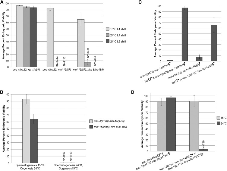
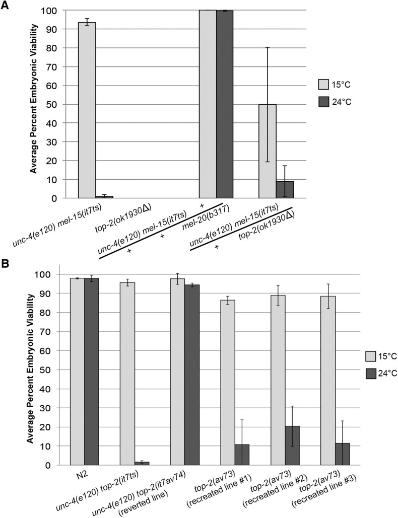
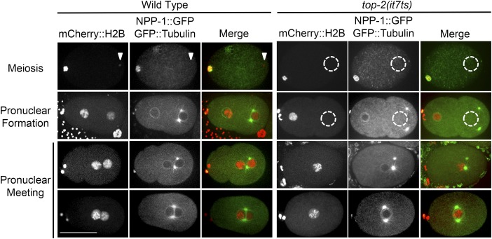
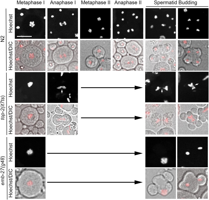
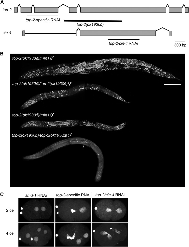
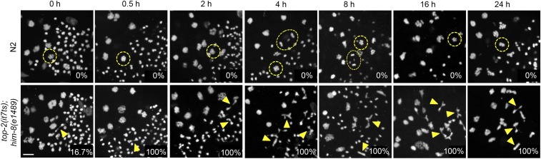
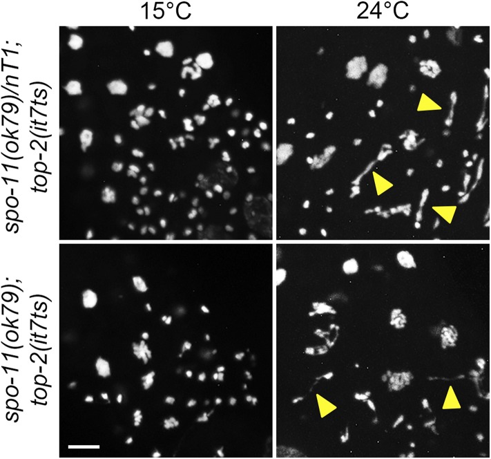
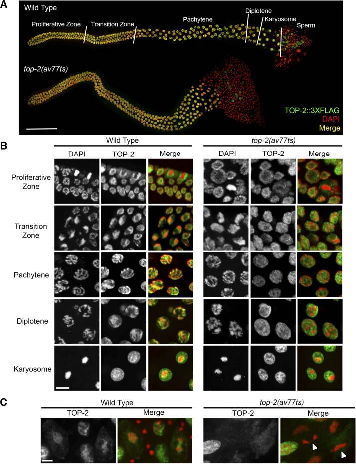
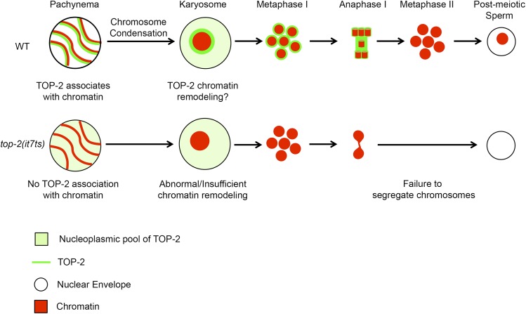
Topoisomerase II alleviates DNA entanglements that are generated during mitotic DNA replication, transcription, and sister chromatid separation. In contrast to mitosis, meiosis has two rounds of chromosome segregation following one round of DNA replication. In meiosis II, sister chromatids segregate from each other, similar to mitosis. Meiosis I, on the other hand, segregates homologs, which requires pairing, synapsis, and recombination. The exact role that topoisomerase II plays during meiosis is unknown. In a screen reexamining Caenorhabditis elegans legacy mutants isolated 30 years ago, we identified a novel allele of the gene encoding topoisomerase II, top-2(it7). In this study, we demonstrate that top-2(it7) males produce dead embryos, even when fertilizing wild-type oocytes. Characterization of early embryonic events indicates that fertilization is successful and sperm components are transmitted to the embryo. However, sperm chromatin is not detected in these fertilized embryos. Examination of top-2(it7) spermatogenic germ lines reveals that the sperm DNA fails to segregate properly during anaphase I of meiosis, resulting in anucleate sperm. top-2(it7) chromosome-segregation defects observed during anaphase I are not due to residual entanglements incurred during meiotic DNA replication and are not dependent on SPO-11-induced double-strand DNA breaks. Finally, we show that TOP-2 associates with chromosomes in meiotic prophase and that chromosome association is disrupted in the germ lines of top-2(it7) mutants.

Keywords: Caenorhabditis elegans; meiosis; spermatogenesis; top-2; topoisomerase II.


Figures

Figure 1
Figure 1
mel-15(it7) is a temperature-sensitive, paternal-effect embryonic lethal allele. (A) Average percent embryonic viability from unc-4(e120) rol-1(e91), unc-4(e120) mel-15(it7), and mel-15(it7); him-8(e1489) at 15 and 24°. The progeny of at least 22 hermaphrodites were scored for each strain at 15 and 24°. Error bars represent SD of the average percentage of three individual replicate experiments. N, total number of embryos and hatched larvae scored. For bars in which N is not stated, N > 2400. (B) Average percent embryonic viability from unc-4(e120) mel-15(it7ts) and mel-15(it7ts); him-8(e1489) hermaphrodites shifted to 24° during either spermatogenesis (L2/L3 to adult) or oogenesis (young adult). The progeny of at least 18 hermaphrodites were scored for each strain at each temperature shift. Error bars represent SD of the average percentage of three individual replicate experiments. (C) Embryonic lethality is rescued by mating with wild-type males. unc-4(e120) mel-15(it7ts) and mel-15(it7ts); him-8(e1489) hermaphrodites were mated with N2 males at 24°. Percent embryonic viability was calculated from the cross-progeny. (D) mel-15(it7ts) is a paternal-effect embryonic lethal allele. L4 males of each mutant were crossed with a single L4 fem-1(hc17ts) dpy-20(e1282) female at 15 or 24°. Percent embryonic viability was calculated from the cross-progeny.
Figure 2
Figure 2
mel-15(it7ts) paternal-effect embryonic lethality is caused by a point mutation in top-2. (A) Complementation tests were performed by crossing unc-4(e120) mel-15(it7ts) hermaphrodites with either top-2(ok1930Δ)/mIn1-gfp; him-8(e1489) or mel-20(b317)/mnC1-gfp; him-8(e1489) males. The average percent embryonic viability at 15 or 24° was calculated by scoring the progeny of F1 trans-heterozygous hermaphrodites. The number of hermaphrodites scored for each cross at 15° were: unc-4(e120) mel-15(it7ts)/mel-20(b317), n = 30; and unc-4(e120) mel-15(it7ts)/top-2(ok1930Δ), n = 42. The number of hermaphrodites scored for each cross at 24° were unc-4(e120) mel-15(it7ts)/mel-20(b317), n = 30; and unc-4(e120) mel-15(it7ts)/top-2(ok1930Δ), n = 40. The average percent embryonic viability for self-mating unc-4(e120) mel-15(it7ts) and top-2(ok1930Δ) hermaphrodites is also indicated (15°, n = 36 and 16, respectively; 24°, n = 21 and 20). Note: top-2(ok1930Δ) homozygotes are sterile Uncs and do not produce any progeny. Error bars indicate SD. (B) Average percent embryonic viability from N2 (wild type), unc-4(e120) top-2(it7ts), and the CRISPR/Cas9-mediated reverted line of unc-4(e120) top-2(it7ts) to wild type [unc-4(e120) top-2(av74)], and the three it7ts recreated lines in N2 [top-2(av73)] at 15 or 24°. The progeny of at least 28 hermaphrodites were scored for each strain at 15 and 24°. Error bars represent SD of the average percentage of three individual replicate experiments.
Figure 3
Figure 3
top-2(it7ts) embryos lack paternal DNA. Wild type (OCF8) and top-2(it7ts) hermaphrodites were shifted to 24° as L4s. After 24 hr, hermaphrodites were dissected and the embryos imaged. Red, chromatin (mCherry::H2B); Green, tubulin (GFP::TBA-2) and nuclear envelope (NPP-1::GFP). White triangle indicates paternal DNA. Dashed-line circle demarks area of missing paternal DNA. Two example images of pronuclear meeting are given. The number of wild-type embryos observed was 48. The number of top-2(it7ts) embryos observed was 47. Bar, 30 μm.
Figure 4
Figure 4
top-2(it7ts) male germ lines have chromosome-segregation defects. Images from live analysis of N2, top-2(it7ts); him-8(e1489), and emb-27(g48) males. Males were shifted to 24° as L3s. After 16 hr, male gonads were dissected and stained with Hoechst dye (red in merged panels) and visualized by DIC optics. White arrows indicate examples of chromatin bridges and atypical chromosome segregation. The number of germ lines scored for each genotype were: N2, n = 32; top-2(it7ts), n = 25; and emb-27(g48), n=21. Bar, 5 μm.
Figure 5
Figure 5
top-2 is required for multiple cell cycles and developmental stages. (A) Schematic comparing the gene structure of top-2 and its paralog cin-4. Thick black line below the top-2 gene structure indicates the extent of the ok1930Δ deletion. Thin black lines below the gene structures indicate the sequences targeted by either top-2-specific RNAi or the RNAi construct that targets sequences present in both genes. (B) Whole-mount top-2(ok1930Δ) and control top-2(ok1930Δ)/mIn1 hermaphrodites and males were prepared for imaging by methanol fixation and DAPI staining. A minimum of three animals of each genotype were imaged. Germ lines in the heterozygous control animals are outlined in white. Arrow points to the few mitotic germ cells generated in the top-2(ok1930Δ) homozygous mutant male. Bar, 100 μm. (C) top-2 RNAi causes segregation defects in the early embryo. L3 hermaphrodites expressing mCherry::H2B were plated on RNAi plates at 24° for 24 hr. Hermaphrodites were dissected and embryos imaged with a confocal microscope. 0% of two-cell (n = 16) and four-cell (n = 16) smd-1 RNAi embryos scored had the cross-eyed phenotype. Of the top-2-specific RNAi embryos scored, 75% of two cell (n = 32) and 81.6% of four cell (n = 38) had the cross-eyed phenotype. 62.7% of two-cell (n = 51) and 81.8% (n = 33) of four-cell top-2/cin-4 RNAi embryos had the cross-eyed phenotype. Bar, 30 μm.
Figure 6
Figure 6
top-2(it7ts) meiotic chromosome-segregation defects are not incurred during mitotic or premeiotic DNA replication. Young adult N2 or top-2(it7ts) males (16 hr post-L3) were shifted to 24° for the indicated time intervals, the gonads dissected, fixed, and stained with DAPI. Chromosome segregation was monitored by fluorescence microscopy. Images are from the spermatogenesis condensation and meiotic division zones. Examples of germ cells in metaphase (circles) and anaphase (ovals) are encircled in yellow. Yellow triangles indicate examples of chromatin bridges and incompletely separated chromosomes. The percentage of germ lines with at least one germ cell with chromosome-segregation defects is indicated in the bottom right corner for each time point. The number of N2 germ lines examined were: 0 hr, 15°, n = 9; 0.5 hr, n = 9; 2 hr, n = 8; 4 hr, n = 10; 8 hr, n = 9; 16 hr, n = 13; 24 hr, n = 15. The number of top-2(it7ts); him-8(e1489) germ lines examined were: 0 hr, 15°, n = 15; 0.5 hr, n = 15; 2 hr, n = 22; 4 hr, n = 14; 8 hr, n = 10; 16 hr, n = 12; 24 hr, n = 18. Bar, 5 μm.
Figure 7
Figure 7
top-2(it7ts) chromosome-segregation defects are not dependent on SPO-11-induced DSBs. Young adult spo-11(ok79)/nT1; top-2(it7ts) or spo-11(ok79); top-2(it7ts) males (16 hr post-L3) were incubated at either 15 or 24° for 4 hr, the germ lines dissected, fixed, and stained with DAPI. Chromosome segregation was monitored by fluorescence microscopy. Images are from the spermatogenesis condensation and meiotic division zones. Yellow triangles indicate examples of chromatin bridges and incompletely separated chromosomes. The number of germ lines with chromosome-segregation defects at 15°: spo-11(ok79)/nT1; top-2(it7ts), 0 of 7; spo-11(ok79); top-2(it7ts), 5 of 10. The number of germ lines with chromosome-segregation defects at 24°: spo-11(ok79)/nT1; top-2(it7ts), 16 of 17; spo-11(ok79); top-2(it7ts), 14 of 14. Bar, 5 μm.
Figure 8
Figure 8
TOP-2 localization in the male germ line. (A) Immunolocalization of TOP-2::3xFLAG (green) in wild type [top-2(av64)] and top-2(av77ts) (it7ts recreate) in entire dissected male germ lines counterstained with DAPI (red; colocalization, yellow). Young adult animals were shifted to 24° for 4 hr prior to dissection and staining. Stages of meiotic prophase are delineated by white lines on the wild-type germ line. Bar, 50 μm. (B) Representative images of Z-projected confocal sections through the proliferative zone, transition zone, pachytene nuclei, diplotene nuclei, and karyosome nuclei (24° for 4 hr) stained with anti-FLAG antibody (green) and counterstained with DAPI (red; colocalization, yellow). Bar, 5 μm. (C) Images of TOP-2::3xFLAG (green) immunolocalization in the meiotic division zone in wild-type and top-2(av77ts) male germ lines (24° for 4 hr) counterstained with DAPI (red). White triangles point to examples of chromatin bridges and incompletely separated chromosomes in the top-2(av77ts) mutant. Experiments were repeated a minimum of three times and a minimum of eight germ lines were examined for each condition. Bar, 5 μm.
Figure 9
Figure 9
Model for TOP-2 function in male meiosis. In wild-type germ lines TOP-2 first localizes along chromosome axes of germ-line nuclei during pachytene and then envelops the highly compact chromosome structure of karyosome nuclei, positioning TOP-2 to interact with DNA during chromosome condensation. TOP-2-mediated chromatin remodeling prepares the chromosomes for segregation during the meiotic divisions. In the top-2(it7ts) mutant, TOP-2 fails to localize along the chromosome tracks of pachytene nuclei and around the karyosome DNA. This results in insufficient chromatin remodeling leading to chromosome-segregation defects. WT, wild type.

Similar articles

Cited by

References

    1. Albertson D. G., Thomson J. N., 1993. Segregation of holocentric chromosomes at meiosis in the nematode, Caenorhabditis elegans. Chromosome Res. 1: 15–26. - PubMed
    1. Arribere J. A., Bell R. T., Fu B. X. H., Artiles K. L., Hartman P. S., et al. , 2014. Efficient marker-free recovery of custom genetic modifications with CRISPR/Cas9 in Caenorhabditis elegans. Genetics 198: 837–846. - PMC - PubMed
    1. Bembenek J. N., Verbrugghe K. J. C., Khanikar J., Csankovszki G., Chan R. C., 2013. Condensin and the spindle midzone prevent cytokinesis failure induced by chromatin bridges in C. elegans embryos. Curr. Biol. 23: 937–946. - PMC - PubMed
    1. Bhat M. A., Philp A. V., Glover D. M., Bellen H. J., 1996. Chromatid segregation at anaphase requires the barren product, a novel chromosome-associated protein that interacts with Topoisomerase II. Cell 87: 1103–1114. - PubMed
    1. Braun R. E., 2001. Packaging paternal chromosomes with protamine. Nat. Genet. 28: 10–12. - PubMed

Substances