. 2016 Oct 4:6:34557.
doi: 10.1038/srep34557.

ALB3 Insertase Mediates Cytochrome b6 Co-translational Import into the Thylakoid Membrane

Affiliations

ALB3 Insertase Mediates Cytochrome b6 Co-translational Import into the Thylakoid Membrane

Jarosław Króliczewski et al. Sci Rep. .

Abstract

The cytochrome b6 f complex occupies an electrochemically central position in the electron-transport chain bridging the photosynthetic reaction center of PS I and PS II. In plants, the subunits of these thylakoid membrane protein complexes are both chloroplast and nuclear encoded. How the chloroplast-encoded subunits of multi-spanning cytochrome b6 are targeted and inserted into the thylakoid membrane is not fully understood. Experimental approaches to evaluate the cytochrome b6 import mechanism in vivo have been limited to bacterial membranes and were not a part of the chloroplast environment. To evaluate the mechanism governing cytochrome b6 integration in vivo, we performed a comparative analysis of both native and synthetic cytochrome b6 insertion into purified thylakoids. Using biophysical and biochemical methods, we show that cytochrome b6 insertion into the thylakoid membrane is a non-spontaneous co-translational process that involves ALB3 insertase. Furthermore, we provided evidence that CSP41 (chloroplast stem-loop-binding protein of 41 kDa) interacts with RNC-cytochrome b6 complexes, and may be involved in cytochrome b6 (petB) transcript stabilization or processing.


Conflict of interest statement

Małgorzata Piskozub has paid employment at Amplicon Sp. z o. o. The following authors have no competing interests: Rafał Bartoszewski, Bożena Króliczewska, Jarosław Króliczewski.

Figures

Figure 1
Figure 1. Circular dichroism spectroscopy of native cytochrome b6.
(A) Native cytochrome b6 (solid line), denatured native cytochrome (dot line), Spectra are shown for the protein in buffer containing DDM. (B) The description is the same as in panel A.
Figure 2
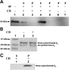
Figure 2. In vitro import of cytochrome b6 into thylakoid membrane.
(A) The integration of the cytochrome b6 into the thylakoid membrane in the presence or absence of stromal fraction (SF) was analysed with Western blot. Urea was used as chaotropic agents (CH). Lane 1, purified native cytochrome b6 as a control; lanes 2 and 3, supernatant (S) and membrane pellet (P) after insertion of native cytochrome b6; lanes 4–7, supernatant and membrane pellet after insertion of denatured cytochrome b6. Cytochrome b6 was isolated from Synechocystis sp. PCC 6803, biotin labelled and anti-biotin antibodies was used for detection. (B) Lane 1, molecular weight standard; Lanes 2–3, membrane fraction after ss-cytochrome b6 insertion with and without chaotropic extraction, respectively. Antibodies against N-terminal residues of cytochrome b6 were used (10 μg of total protein per each lane was applied). (C) Lanes 1 and 2, supernatant (S) and membrane pellet (P) after insertion of ss-cytochrome b6. Urea was used as a chaotropic agents (CH) and anti-biotin antibodies were used for protein detection. All the experiments were repeated twice and 10 μg of total protein per each lane was applied.
Figure 3
Figure 3. Thylakoid membrane fractions after insertion of spinach apocytochrome b6 and treatment with chaotropic agents (CH).
(A) The integration of the cytochrome b6 into the thylakoid membrane in the presence or absence of stromal fraction (SF) was analysed with Western blot. Lane 1, thylakoid membrane; lane 2, purified apocytochrome b6; lane 3 pelleted membrane fraction with inserted denatured (unfolded) protein; lane 4, pelleted membrane fraction with inserted denatured protein after chaotropic treatment with urea; lane 5, control, membrane fraction with inserted into membrane ss-apocytochrome b6; lanes 6 and 7, supernatant and membrane pellet, respectively after centrifugation of refolded apocytochrome b6 and inserted into membrane. The experiments were repeated twice and 10 μg of total protein per lane was applied. (B) Thylakoid membrane fractions after insertion of spinach ss-apocytochrome b6 and treatment with carboxypeptidase B. Lane 1, purified ss-apocytochrome b6; lane 2, thylakoid membrane with inserted ss-apocytochrome b6; lane 3, membrane treated with carboxypeptidase B (depicted with (C) after protein insertion; lanes 4 and 5, membrane and supernatant fraction with inserted denatured protein after carboxypeptidase B treatment; lane 6, supernatant fraction similar to lane 5, but membrane with inserted denatured protein was treated with urea and carboxypeptidase B, and an antibody against N-terminus of cytochrome b6 was used. Cytochrome b6 was biotin labelled and anti-biotin antibodies were used for detection with the exception of (B) line 6.
Figure 4
Figure 4. Circular dichroism spectroscopy of PsbW in aqueous buffer (unfolded) and DDM micelles (refolded).
Spectra are shown for the protein in aqueous buffer (dashed line) and after incorporation into DDM micelles (dotted line).
Figure 5
Figure 5. Thylakoid membrane fractions after insertion of PsbW.
The integration of the PsbW into the thylakoid membrane the presence or absence of stromal fraction was analysed by Western blot. Lane 1, thylakoid membrane before insertion; lanes 2–4 and 6–8, thylakoid membrane after insertion of PsbW; and lane 5, molecular weight standard. Antibodies against biotin were used for immunodetection. C - membrane treated with carboxypeptidase B after protein insertion, PK - membrane treated before protein insertion with proteinase K. On each lane, 10 μg of protein was applied. Identification of psbW protein in Western blot was also confirmed using MS.
Figure 6
Figure 6. Autoradiograph of cytochrome b6 expressed in cell-free assay in the presence of thylakoid membrane and stroma.
(A) Lane 1, thylakoid membrane as a control; lanes 2 and 3, translation of cytochrome b6 in the presence of thylakoid membrane and stromal fraction, supernatant (S) and membrane pellet (P) after fractionation; (B) Lanes 1 and 2 translation of cytochrome b6 in the presence of thylakoid membrane and stromal fraction, supernatant (S) and membrane pellet (P) after fractionation; lane 3 and 4, translation of cytochrome b6 in the presence of thylakoid membrane, stroma and cpSecY antibody, membrane pellet (P) and supernatant after fractionation; (C) Lane 1, thylakoid membrane as a control; lane 2 and 3, translation of cytochrome b6 in the presence of thylakoid membrane and stromal fraction, supernatant (S) and membrane pellet (P) after fractionation, lane 4 and 5, translation of cytochrome b6 in the presence of thylakoid membrane, stroma and cpSecY antibody, membrane pellet (P) and supernatant (S) after fractionation; lane 6 and 7 same as in lane 2 and 3 but endogenous RNA in stroma was removed by enzymatic digestion before use in translation reaction (reaction were performed in the presence of RNasin ribonuclease inhibitor).
Figure 7
Figure 7. Autoradiograph of cytochrome b6 expressed in cell free assay in the presence of thylakoid membrane and stroma.
(A) Lane 1, translation of cytochrome b6 - control; lane 2 and 3, translation of cytochrome b6 in the presence of thylakoid membrane, stromal fraction and ALB3 antibody, supernatant (S) and membrane pellet (P) after fractionation; lane 4, translation of cytochrome b6 in the presence of thylakoids membrane and stroma; lane 5 and 6, translation of cytochrome b6 in the presence of thylakoid membrane and stromal fraction, supernatant (S) and membrane pellet (P) after fractionation. (B) Lane 1, translation of cytochrome b6, membrane pellet fraction after fractionation - control; lane 2, translation of cytochrome b6 in the presence of 0.5 mM non-hydrolysable ATP analogue (AMP-PNP, 5′ adenylylimidodiphosphate), membrane pellet; lane 3, translation of cytochrome b6 in the presence of 0.1 mM GMP-PNP, membrane pellet; lane 4, translation of cytochrome b6 in the presence of 0.5 mM GMP-PNP.
Figure 8
Figure 8. A Model of targeting and insertion of cytochrome b6 into a thylakoid membrane by using the ALB3 insertase.
ALB3 insertase independently of cpSecY imports cytochrome b6 into the thylakoid membrane. The import process is co-translational and involves cpFtsY and cpSrp54.

Similar articles

Cited by

References

    1. Cramer W. A., Yamashita E. & Hasan S. S. In Encyclopedia of Biological Chemistry Vol. 4 (eds Lane M. D. & Lennarz W. J.) 167–171 (Academic Press, Waltham MA, 2013).
    1. Tikhonov A. N. The cytochrome b6f complex at the crossroad of photosynthetic electron transport pathways. Plant Physiol. Biochem. 81, 163–183, doi: 10.1016/j.plaphy.2013.12.011 (2014). - DOI - PubMed
    1. Joliot P. & Johnson G. N. Regulation of cyclic and linear electron flow in higher plants. Proc. Natl. Acad. Sci. USA 108, 13317–13322, doi: 10.1073/pnas.1110189108 (2011). - DOI - PMC - PubMed
    1. Fey V., Wagner R., Bräutigam K. & Pfannschmidt T. Photosynthetic redox control of nuclear gene expression. J. Exp. Bot. 56, 1491–1498, doi: 10.1093/jxb/eri180 (2005). - DOI - PubMed
    1. Chi W., Ma J. & Zhang L. Regulatory factors for the assembly of thylakoid membrane protein complexes. Philos. Trans. R. Soc. Lond. B Biol. Sci. 367, 3420–3429, doi: 10.1098/rstb.2012.0065 (2012). - DOI - PMC - PubMed

Publication types

MeSH terms

LinkOut - more resources