

A neurodegenerative perspective on mitochondrial optic neuropathies
- PMID: 27696015
- PMCID: PMC5106504
- DOI: 10.1007/s00401-016-1625-2
A neurodegenerative perspective on mitochondrial optic neuropathies
Abstract
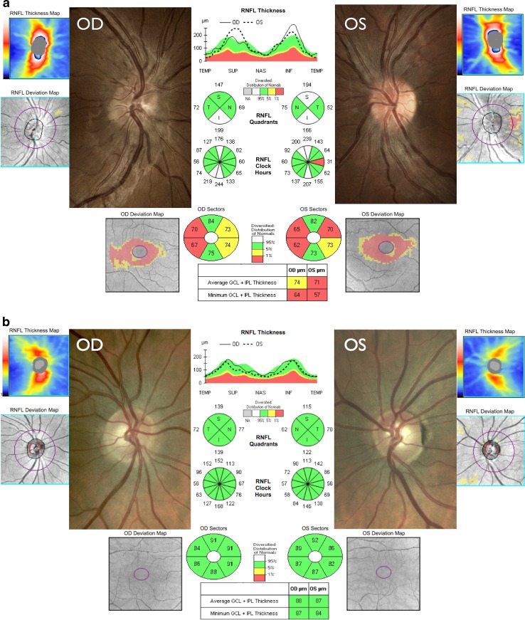
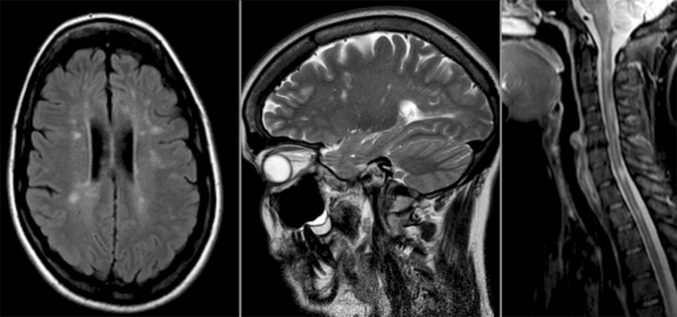
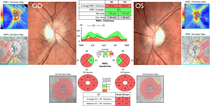
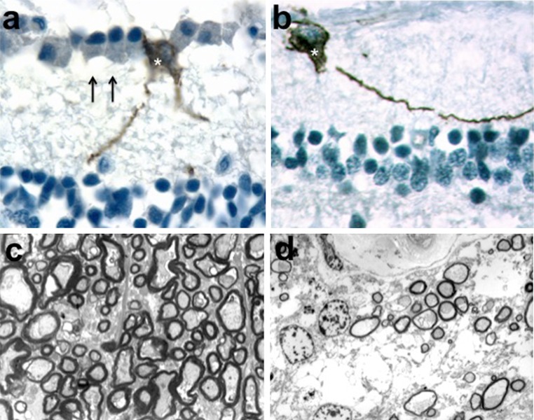
Mitochondrial optic neuropathies constitute an important cause of chronic visual morbidity and registrable blindness in both the paediatric and adult population. It is a genetically heterogeneous group of disorders caused by both mitochondrial DNA (mtDNA) mutations and a growing list of nuclear genetic defects that invariably affect a critical component of the mitochondrial machinery. The two classical paradigms are Leber hereditary optic neuropathy (LHON), which is a primary mtDNA disorder, and autosomal dominant optic atrophy (DOA) secondary to pathogenic mutations within the nuclear gene OPA1 that encodes for a mitochondrial inner membrane protein. The defining neuropathological feature is the preferential loss of retinal ganglion cells (RGCs) within the inner retina but, rather strikingly, the smaller calibre RGCs that constitute the papillomacular bundle are particularly vulnerable, whereas melanopsin-containing RGCs are relatively spared. Although the majority of patients with LHON and DOA will present with isolated optic nerve involvement, some individuals will also develop additional neurological complications pointing towards a greater vulnerability of the central nervous system (CNS) in susceptible mutation carriers. These so-called "plus" phenotypes are mechanistically important as they put the loss of RGCs within the broader perspective of neuronal loss and mitochondrial dysfunction, highlighting common pathways that could be modulated to halt progressive neurodegeneration in other related CNS disorders. The management of patients with mitochondrial optic neuropathies still remains largely supportive, but the development of effective disease-modifying treatments is now within tantalising reach helped by major advances in drug discovery and delivery, and targeted genetic manipulation.
Keywords: Dominant optic atrophy; Leber hereditary optic neuropathy; Mitochondrial diseases; Neurodegenerative diseases; OPA1; Retinal ganglion cell.
Conflict of interest statement
Compliance with ethical standards Financial disclosures PYWM holds a consultancy agreement with GenSight Biologics. VC holds consultancy agreements with GenSight Biologics, Santhera Pharmaceuticals, Stealth BioTherapeutics and Edison Pharmaceuticals.
Figures
Similar articles
-
Treatment strategies for inherited optic neuropathies: past, present and future.Eye (Lond). 2014 May;28(5):521-37. doi: 10.1038/eye.2014.37. Epub 2014 Mar 7. Eye (Lond). 2014. PMID: 24603424 Free PMC article. Review.
-
Mitochondrial optic neuropathies - disease mechanisms and therapeutic strategies.Prog Retin Eye Res. 2011 Mar;30(2):81-114. doi: 10.1016/j.preteyeres.2010.11.002. Epub 2010 Nov 26. Prog Retin Eye Res. 2011. PMID: 21112411 Free PMC article. Review.
-
Mitochondrial optic neuropathies.Handb Clin Neurol. 2023;194:23-42. doi: 10.1016/B978-0-12-821751-1.00010-5. Handb Clin Neurol. 2023. PMID: 36813316 Review.
-
Inherited mitochondrial optic neuropathies.J Med Genet. 2009 Mar;46(3):145-58. doi: 10.1136/jmg.2007.054270. Epub 2008 Nov 10. J Med Genet. 2009. PMID: 19001017 Free PMC article. Review.
-
Mitochondrial optic neuropathies: how two genomes may kill the same cell type?Biosci Rep. 2007 Jun;27(1-3):173-84. doi: 10.1007/s10540-007-9045-0. Biosci Rep. 2007. PMID: 17479363 Review.
Cited by
-
Intravitreal Gene Therapy vs. Natural History in Patients With Leber Hereditary Optic Neuropathy Carrying the m.11778G>A ND4 Mutation: Systematic Review and Indirect Comparison.Front Neurol. 2021 May 24;12:662838. doi: 10.3389/fneur.2021.662838. eCollection 2021. Front Neurol. 2021. PMID: 34108929 Free PMC article.
-
Gene therapy for primary mitochondrial diseases: experimental advances and clinical challenges.Nat Rev Neurol. 2022 Nov;18(11):689-698. doi: 10.1038/s41582-022-00715-9. Epub 2022 Oct 18. Nat Rev Neurol. 2022. PMID: 36257993 Review.
-
Loss of the mitochondrial i-AAA protease YME1L leads to ocular dysfunction and spinal axonopathy.EMBO Mol Med. 2019 Jan;11(1):e9288. doi: 10.15252/emmm.201809288. EMBO Mol Med. 2019. PMID: 30389680 Free PMC article.
-
Modelling autosomal dominant optic atrophy associated with OPA1 variants in iPSC-derived retinal ganglion cells.Hum Mol Genet. 2022 Oct 10;31(20):3478-3493. doi: 10.1093/hmg/ddac128. Hum Mol Genet. 2022. PMID: 35652445 Free PMC article.
-
Transcriptional profiling of retinal astrocytes identifies a specific marker and points to functional specialization.Glia. 2024 Sep;72(9):1604-1628. doi: 10.1002/glia.24571. Epub 2024 May 24. Glia. 2024. PMID: 38785355
References
-
- Adams JH, Blackwood W, Wilson J. Further clinical and pathological observations on Leber’s optic atrophy. Brain. 1966;89:15–26. - PubMed
-
- Alavi MV, Bette S, Schimpf S, Schuettauf F, Schraermeyer U, Wehrl HF, Ruttiger L, Beck SC, Tonagel F, Pichler BJ, et al. A splice site mutation in the murine Opa1 gene features pathology of autosomal dominant optic atrophy. Brain. 2007;130:1029–1042. - PubMed
-
- Alavi MV, Fuhrmann N, Nguyen HP, Yu-Wai-Man P, Heiduschka P, Chinnery PF, Wissinger B. Subtle neurological and metabolic abnormalities in an Opa1 mouse model of autosomal dominant optic atrophy. Exp Neurol. 2009;220:404–409. - PubMed
-
- Alexander C, Votruba M, Pesch UE, Thiselton DL, Mayer S, Moore A, Rodriguez M, Kellner U, Leo-Kottler B, Auburger G, et al. OPA1, encoding a dynamin-related GTPase, is mutated in autosomal dominant optic atrophy linked to chromosome 3q28. Nat Genet. 2000;26:211–215. - PubMed
Publication types
MeSH terms
Substances
Grants and funding
LinkOut - more resources
Full Text Sources
Other Literature Sources
