. 2016 Oct 3:6:34564.
doi: 10.1038/srep34564.

Phycocyanin Inhibits Tumorigenic Potential of Pancreatic Cancer Cells: Role of Apoptosis and Autophagy

Affiliations

Phycocyanin Inhibits Tumorigenic Potential of Pancreatic Cancer Cells: Role of Apoptosis and Autophagy

Gaoyong Liao et al. Sci Rep. .

Abstract

Pancreatic adenocarcinoma (PDA) is one of the most lethal human malignancies, and unresponsive to current chemotherapies. Here we investigate the therapeutic potential of phycocyanin as an anti-PDA agent in vivo and in vitro. Phycocyanin, a natural product purified from Spirulina, effectively inhibits the pancreatic cancer cell proliferation in vitro and xenograft tumor growth in vivo. Phycocyanin induces G2/M cell cycle arrest, apoptotic and autophagic cell death in PANC-1 cells. Inhibition of autophagy by targeting Beclin 1 using siRNA significantly suppresses cell growth inhibition and death induced by phycocyanin, whereas inhibition of both autophagy and apoptosis rescues phycocyanin-mediated cell death. Mechanistically, cell death induced by phycocyanin is the result of cross-talk among the MAPK, Akt/mTOR/p70S6K and NF-κB pathways. Phycocyanin is able to induce apoptosis of PANC-1 cell by activating p38 and JNK signaling pathways while inhibiting Erk pathway. On the other hand, phycocyanin promotes autophagic cell death by inhibiting PI3/Akt/mTOR signaling pathways. Furthermore, phycocyanin promotes the activation and nuclear translocation of NF-κB, which plays an important role in balancing phycocyanin-mediated apoptosis and autosis. In conclusion, our studies demonstrate that phycocyanin exerts anti-pancreatic cancer activity by inducing apoptotic and autophagic cell death, thereby identifying phycocyanin as a promising anti-pancreatic cancer agent.


Figures

Figure 1
Figure 1. Phycocyanin inhibits cell viability in tumor cells and has little effect in normal cells.
(A) Growth curves of capan-1, PANC-1, BxPC3, BGC-823, HepG2, H460, DU145 and MCF-7 cancer cell line in the presence of various concentration phycocyanin. (B) IC50 values for growth inhibition of different cancer cell lines including capan-1, PANC-1, BxPC3, BGC-823, HepG2, H460, DU145 and MCF-7 by phycocyanin. Different cancer cells were treated for 72 h with increasing concentrations of phycocyanin. Results are presented as the mean ± SD of at least three independent experiments, each performed in triplicate. (C) Growth curves of LO2, QSG-7701, AC-16, HK-2, HUVEC and NK-92 cell line in the presence of various concentration phycocyanin. (D) The suppressive effect of phycocyanin on the colony formation of PANC-1 cells.
Figure 2
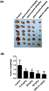
Figure 2. Phycocyanin inhibits pancreatic cancer tumor growth in vivo.
(A) Representative PANC-1 xenograft tumors from mice treated with vehicle, DDP (10 mg/kg) and phycocyanin (12.5-50 mg/kg). (B) Tumor weights of xenograft tumors from mice treated with vehicle, DDP (10 mg/kg) and phycocyanin (12.5–50 mg/kg). Data were presented as the mean ± SD, n = 6 for each group, *P < 0.05; **P < 0.01.
Figure 3
Figure 3. Phycocyanin blocks G2/M cell cycle progression and induces caspase 3 independent cell death in PANC-1 cells.
(A) The effect of phycocyanin on the cell cycle distribution of PANC-1 cells. PANC-1 cells treated with indicated concentrations of phycocyanin for 72 h were fixed, stained and analyzed for DNA content by FACS. (B) The distribution and percentage of cells in G0/G1, S and G2/M phase of the cell cycle were calculated and plotted. The statistically significant differences between the treated cells and the control group were indicated by *P < 0.05, **P < 0.01. (C) Nuclear shrinkage ratio was analyzed with cell scoring model of High Content Screening system software. The data shown were representative of three independent experiments. The statistically significant differences between the treated cells and the control group were indicated by *P < 0.05, **P < 0.01. (D) Flow cytometry analysis of PANC-1 cell apoptosis induced by phycocyanin for 72 h. (E) Following treatment with phycocyanin, early apoptotic cell population with Annexin V-positive and late apoptotic cell population with PI- positive PANC-1 cells increased in a dose-dependent manner. Mean ± SD of three assays. *P < 0.05, **P < 0.01, versus control. (F) Knockdown of caspase 3 by siRNA pool for 24 h followed by 10 μM phycocyanin treatment for 48 h decreases capase 3 expression in PANC-1 cells when compared to cells treated with lipofectamine alone (NS) or non-silencing siRNA (Control siRNA) and 10 μM phycocyanin. (G) Caspase 3 siRNA inhibited the activation of caspase 3 in PANC-1 cells induced by phycocyanin. Knockdown of caspase 3 by siRNA pool for 24 h followed by 10 μM phycocyanin treatment for 72 h decreases caspase 3 activation. The bars represent mean ± SD, n = 3, **P < 0.01, NA: No significance (t- test). (H) Dose-dependent growth cell inhibition by phycocyaninon in PANC-1 cells transfected with caspase 3 or control siRNA. PANC-1 cells transfected with caspase 3 or control siRNA were treated with indicated concentrations of phycocyanin for 48 h. Data were means ± SD of three independent experiments. *P < 0.05, **P < 0.01 (t-test).
Figure 4
Figure 4. Phycocyanin induces significant autosis in PANC-1 Cells.
(A) Phycocyanin induced dose-dependent Beclin 1 protein expression in PANC-1 cells. Western blot analysis of Beclin 1 expression after 0, 2.5, 5, 10, 20 and 40 μM phycocyanin treatment for 12 h and 24 h. (B) Phycocyanin induced time-dependent Beclin 1 protein expression in PANC-1 cells. Western blot analysis of Beclin 1 expression after 3, 6, 12, 24 and 48 h of treatment with 10 μM phycocyanin. (C) Fluorescence images of pancreatic cancer cells showing endogenous MAP-LC3 levels at different time points after phycocyanin treatment. Nuclei (blue) were labeled by DAPI. MAP-LC3 expression (green) was detected using an anti-MAP-LC3 polyclonal antibody. Goat anti-rabbit IgG/FITC were used as secondary antibody. Fluorescence images were obtained under a high content imaging system. Bar = 100 μm. (D) Quantification of the percentage of cells with focal MAP-LC3 at the indicated time after phycocyanin treatment. Error bars correspond to SD of three repeated wells, counting 200 cells each. *P < 0.05; **P < 0.01, 0 h versus phycocyanin-treated cells. (E) Effect of phycocyanin on expression of LC3 lipidation and Beclin 1 levels in pancreatic cancer cells were analyzed by Western blotting. Cells were treated with 10 μM phycocyanin for the indicated times. (F) Fluorescence visualization of lysosomal-dependent proteolysis upon cleavage and release of the fluorescent moiety of DQ-BSA (green) in phycocyanin-treated PANC-1 cells. Chloroquine is added to monitor DQ-BSA emission in cells with blocked lysosomal activity. Cells were simultaneously imaged in the presence of Lysotracker red (LTRred) to visualize the lysosomal compartment. Bar = 100 μm.
Figure 5
Figure 5. Inhibition of autophagy by Beclin 1 siRNA rescues phycocyanin-mediated cell death in PANC-1 cells.
(A) Knockdown of Beclin 1 by siRNA pool for 24 h followed by 10 μM phycocyanin treatment for 48 h decreases Beclin 1 expression in PANC-1 cells when compared to cells treated with non-silencing siRNA (Control siRNA) and 10 μM phycocyanin. (B) Treatment of PANC-1 with 10 μM phycocyanin for 24 h following a knockdown of Beclin 1 gene with siRNA pool for 48 h shows a significant decrease in MAP-LC3 punctate pattern as compared to cells treated with non-silencing siRNA (control siRNA) or lipofectamine alone followed by phycocyanin. (C) Quantification of the percentage of cells with focal MAP-LC3 at the indicated conditions. Error bars correspond to SD of three repeated wells, counting 200 cells each. *P < 0.05; **P < 0.01, 0 h versus phycocyanin-treated cells. (D) PANC-1 cells treated with 10 μM phycocyanin for 48 h following Beclin 1 only and Beclin1 and caspase 3 dual silencing showed a significant decrease of cell viability inhibition when compared to cells treated with control siRNA alone along with phycocyanin. The bars represent mean ± SD, n = 3, **control siRNA group vs Beclin 1 siRNA group, P < 0.01; ##“Beclin 1 siRNA” group vs “Beclin 1 siRNA + Caspase 3 siRNA” group, P < 0.01 (t test).
Figure 6
Figure 6. Involvement of the MAPK, PI3K/Akt/mTOR and NF-κB pathway in phycocyanin-induced cell death.
(A) Effect of phycocyanin on the MAPK signaling in PANC-1 cells. After treatments, the levels of Erk1/2, JNK and p38 MAPK and their phosphorylated forms were analyzed by Western blotting with indicated antibodies. Results shown were representative of at least three independent experiments. (B) Western blot analysis of pAkt, total Akt, p-mTOR, total mTOR, p-p70S6K and total p-p70S6K in PANC-1 cells. GAPDH was shown as a loading control. Data are representative from three independent experiments. (C) Phycocyanin time dependently activiated NF-κB signaling pathway and induced the nuclear localization of NF-κB. After treatments, the levels of IKKβ and IκB-α and their phosphorylated forms were analyzed by Western blotting with indicated antibodies. Cytosolic and nuclear proteins were subjected to 10% SDS-polyacrylamide gels followed by western blot analysis for NF-κB p65. The results were representative of three independent experiments.
Figure 7
Figure 7. Effect of phycocyanin on cell viability and apoptosis marker expression in PANC-1 cells in the case that the MAPK and the NF-κB pathway are separately and simultaneously suppressed.
(A) The nuclear localization of NF-κB after phycocyanin treatment was blocked by SN50. Cells were treated with 10 μM phycocyanin for different time with SN50 (10 μM). Cytosolic and nuclear proteins were used for western blot analysis using anti-NF-κB p65 antibody. (B) Effect of phycocyanin on PANC-1 cell viability treated with SN50. The data shown are representative of three independent experiments. The bars represented mean ± SD, n = 3. *P < 0.05, **P < 0.01, NA: No significance (t-test). (C) Western blot analysis of nucleus NF-κB, LC3 II/I, Beclin 1, procaspase, caspase and PARP expression in PANC-1 cells treated with 10 μM phycocyanin in absence or presence of SN50 (10 μM) for indicated time point. (D) Activation of p38 was effectively inhibited by the specific MEK inhibitor PD98059. (E) Activation of JNK was effectively inhibited by the specific MEK inhibitor PD98059. (F) Effect of phycocyanin on PANC-1 cell viability treated with PD98059. The data shown are representative of three independent experiments. The bars represented mean ± SD, n = 3, *P < 0.05, **P < 0.01, NA: No significance (t-test). (G) Western blot analysis of procaspase, caspase 3 and PARP expression in PANC-1 cells treated with 10 μM phycocyanin in absence or presence of PD98059 (5 μM) for 48 h. (H) Effect of phycocyanin on PANC-1 cell viability after inhibition of MAPK pathway alone and simultaneous inhibition of MAPK and NF-κB pathway. The bars represent mean ± SD, n = 3. Control group (NS) vs PD98059 group: *P < 0.05, **P < 0.01(t test); “PD98059” group vs “PD98059 + SN50” group: #P < 0.05, ##P < 0.01 (t-test).

Similar articles

Cited by

References

    1. Vincent A., Herman J., Schulick R., Hruban R. H. & Goggins M. Pancreatic cancer. The Lancet 378, 607–620 (2011). - PMC - PubMed
    1. Shaib Y. & Davila J. & EL‐SERAG, H. The epidemiology of pancreatic cancer in the United States: changes below the surface. Alimentary pharmacology & therapeutics 24, 87–94 (2006). - PubMed
    1. Zhou M., Fang Y., Xiang J. & Chen Z. Therapeutics Progression in Pancreatic Cancer and Cancer Stem Cells. Journal of Cancer Therapy 6, 237 (2015).
    1. Kuddus M., Singh P., Thomas G. & Al-Hazimi A. Recent developments in production and biotechnological applications of C-phycocyanin. BioMed research international 2013, 742859, 10.1155/2013/742859 (2013). - DOI - PMC - PubMed
    1. Romay C., Gonzalez R., Ledon N., Remirez D. & Rimbau V. C-phycocyanin: a biliprotein with antioxidant, anti-inflammatory and neuroprotective effects. Current protein & peptide science 4, 207–216 (2003). - PubMed

Publication types