. 2016 Oct 6;167(2):419-432.e16.
doi: 10.1016/j.cell.2016.09.011. Epub 2016 Sep 29.

Engineering T Cells with Customized Therapeutic Response Programs Using Synthetic Notch Receptors

Affiliations

Engineering T Cells with Customized Therapeutic Response Programs Using Synthetic Notch Receptors

Kole T Roybal et al. Cell. .

Abstract

Redirecting T cells to attack cancer using engineered chimeric receptors provides powerful new therapeutic capabilities. However, the effectiveness of therapeutic T cells is constrained by the endogenous T cell response: certain facets of natural response programs can be toxic, whereas other responses, such as the ability to overcome tumor immunosuppression, are absent. Thus, the efficacy and safety of therapeutic cells could be improved if we could custom sculpt immune cell responses. Synthetic Notch (synNotch) receptors induce transcriptional activation in response to recognition of user-specified antigens. We show that synNotch receptors can be used to sculpt custom response programs in primary T cells: they can drive a la carte cytokine secretion profiles, biased T cell differentiation, and local delivery of non-native therapeutic payloads, such as antibodies, in response to antigen. SynNotch T cells can thus be used as a general platform to recognize and remodel local microenvironments associated with diverse diseases.

Keywords: CARs; T cells; cancer; cellular engineering; immunotherapy; synthetic Notch; synthetic biology.


Figures

Figure 1
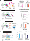
Figure 1. Engineering Antigen Triggered T cell Responses with Diverse SynNotch Receptors
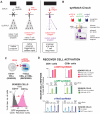
(A) TCRs and CARs activate kinase-based signaling cascades that drive the native T cell activation program providing little control over reshaping the T cell response. synNotch receptors recognize cell-surface antigens (e.g. disease related antigens) and directly regulate custom transcriptional programs with more precise control over the T cell response. Thus, in principle, synNotch receptors could be used to engineer a la carte responses. (B) synNotch receptors have a custom ligand binding domain that detects a cell- surface antigen of interest (e.g. scFvs targeted to CD19 or Her2 or nanobodies to GFP), the core regulatory region of Notch that controls proteolysis, and a cytoplasmic orthogonal transcription factor (e.g. Gal4 VP64). The corresponding response elements for the orthogonal transcription factor controlling custom transcriptional programs are also engineered into the T cell. (C) CD4+ AND CD8+ primary human T cells were engineered with the α-CD19 synNotch Gal4VP64 receptor and 5× Gal4 response elements controlling the expression of a BFP reporter. Histogram showing selective induction of the BFP reporter in α-CD19 synNotch receptor receiver CD4+ T cells in response to stimulation with sender cells with CD19- or CD19+ K562s. (D) CD4+ AND CD8+ primary human T cells were engineered with either the α- CD19, α-GFP (LaG17 or LaG16_2) nanobody, or α-Her2 (scFv affinity variants) synNotch Gal4VP64 receptors and 5× Gal4 response elements controlling the expression of a BFP reporter. The percentages of synNotch T cells that upregulate the BFP reporter after 24 hours of stimulation with the indicated sender cells is given (n ≥ 3 for all conditions, error bars = SEM).
Figure 2
Figure 2. synNotch Receptors can Drive Antigen-Induced Custom Cytokine Programs
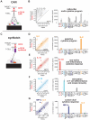
(A) CAR activation drives CD4+ T cells to produce a diverse set of cytokines. (B) A scatter plot showing the level (pg/mL) of 24 cytokines (see panel C for list of cytokines) produced by primary human α-CD19 CAR CD4+ T cells activated with target CD19+ K562 cells (y-axis) or negative control CD19- K562s (x-axis) after 24 hours of stimulation (n=3, error bars = SEM). The level of 24 cytokines produced by CD4+ α-CD19 CAR T cells stimulated by target CD19+ K562s (n=3, error bars = SEM). (C) CD4+ T cells were engineered with the α-CD19 synNotch Gal4VP64 receptor and the corresponding response elements controlling the expression of either IL- 2, IL-10, IL-12, or combined IL-2/MIP-1α. The cells were co-cultured with target CD19+ K562s or CD19- non-target K562s. (D) Scatterplots showing the production of synNotch regulated cytokines in response to CD19+ vs. CD19- K562 stimulation (n=3, error bars = SEM). The level of cytokines produced by α-CD19 synNotch T cells driving IL-2, IL-10, Flexi IL-12, or IL-2/MIP-1α production in response to CD19+ K562 cells is given. Only the synNotch regulated cytokines were produced above background levels. For synNotch CD4+ T cells driving IL-12 production, IFNγ was also produced as IL-12 can cause CD4+ T cells to differentiate into Th1 IFNγ-producing T cells (n=3, error bars = SEM).
Figure 3
Figure 3. synNotch Receptors Can Drive Antigen-Dependent Skewing of T Cell Differentiation to the Anti-Tumor Th1 Fate
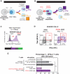
(A) When CD4+ T cells are activated through engagement of pathogen-derived peptides presented by MHC molecules on antigen-presenting cells they differentiate into particular T cell subtypes depending on the infection. Th1 and Th2 are canonical CD4+ T cell fates that drive different immune responses. Th1 cells express the transcription factor Tbet, produce IFNγ, and aid in cellular immunity and tumor clearance. Th2 cells produce IL-4, an important cytokine for stimulation of antibody production by B cells. (B) CD4+ α-CD19 synNotch T cells were engineered to regulate the expression Tbet and thus Th1 fate choice by T cells. The synNotch T cells were co-cultured with target CD19+ or non-target CD19- K562 cells for 11 days to determine if synNotch driven Tbet expression could skew CD4+ T cells to Th1 fate in a CD19- dependent manner. (C) Histograms showing the selective expression of Tbet T2A EGFP after 24 hours of CD4+ α-CD19 synNotch T cells with CD19+ K562s (representative of at least 3 experiments). (D) Two dimensional dot plots of intracellular stained CD4+ α-CD19 synNotch Gal4VP64 T cells for Tbet and IFNγ after 11 days of culture with either CD19+ or CD19- K562s. T cells were stimulated with PMA/Ionomycin for 4 hrs prior to staining to drive cytokine production (representative of at least 3 experiments). (E) The percentage of IFNγ+ (Th1) T cells after 11 days of the indicated treatment (n ≥ 3 for all treatments, error bars = SEM, significance determined by Student’s t-test, n.s. p≥0.05).
Figure 4
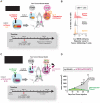
Figure 4. Customized Killer T Cells – synNotch Driven TRAIL Production
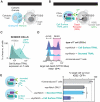
(A) CD8+ cytotoxic T cells recognize infected cells via their TCR and directly kill the infected cell by creating pores in the cell with perforin allowing for the delivery of granzymes that initiate programmed cell death. (B) CD4+ T cells were engineered with the α-GFP synNotch controlling the expression of the apoptotic regulator TRAIL in response to surface GFP. (C) Histograms showing the selective expression of surface TRAIL after 24 hours of CD4+ α-GFP synNotch T cell incubation with surface GFP+ K562s. (D) Histograms showing surface GFP+ K562 cell death via uptake of the dead stain SYTOX blue after 24hr. co-culture with the indicated T cell type (T cell:Target Cell Ratio = 1:1). (E) Percentage target cell survival calculated from replicate data shown in panel D (n=4, error bars = SEM, significance determined by Student’s t-test ** p≤0.01)
Figure 5
Figure 5. synNotch T Cell Delivery of Immunotherapeutics – Antibodies, Adjuvants, and Immunosuppressive Agents
(A) CD4+ T cells were engineered with the α-GFP synNotch controlling the expression of Pembrolizumab (α-PD-1 HA) and a myc-tagged α-CTLA4 scFv (both antibodies were expressed as a single transcript with a T2A sequence between). After 24 hours of stimulation of the T cells with surface GFP+ or GFP- K562s, the supernatant was collected and used to stain PD-1+ or CTLA4+ K562s. Secreted antibody binding to target cells was monitored via flow cytometry after secondary staining with α-HA A647 (α-PD-1) or α-myc A647 (CTLA-4 scFV) antibodies (representative of 3 replicates). (B) CD4+ T cells were engineered with the α-GFP synNotch receptor controlling the expression of Blinatumomab, an α-CD19/CD3 bi-specific antibody that retargets T cells to CD19+ tumors. Histograms showing CD69 (activation marker) expression on the synNotch T cells after co-culture with surface GFP+ only, CD19+ only, or surface GFP+CD19+ K562s. The T cells strongly activate in the presence of the surface GFP+CD19 K562s and a small percentage of the T cells activate when incubated with CD19+ only K562s due to low levels of basal leakage of Blinatumomab expression (representative of 3 independent experiments). (C) CD4+ T cells were engineered with the α-GFP synNotch receptor controlling the expression of Flagellin. Supernatant was harvested from the T cells after co- culture with surface GFP+ or GFP- K562s and added to hTLR5 HEK-blue secreted alkaline phosphatase (SEAP) reporter cells. After 24 hours SEAP activity was monitored and the level of Flagellin in the supernatant was measured a purified Flagellin standard (n=3, error bars = SD). (D) CD4+ T cells were engineered with the α-CD19 synNotch receptor controlling the expression of PD-L1 and IL-10. Quantification of the percentage of synNotch T cells that express PD-L1 and intracellular IL-10 after co-culture with CD19+ or CD19- K562s for 24 hrs is given. The amount of IL-10 in the supernatant was also determined by ELISA (n=3, error bars = SEM).
Figure 6
Figure 6. In vivo Local Expression of Cytokines and Bi-specifc Tumor- targeted Antibodies in Solid Tumors Via synNotch T cells
(A) NSG mice were subcutaneously injected with CD19- non-target K562s and target CD19+ in the left and right flank, respectively. α-CD19 synNotch T cells in control of IL-2 iRES mCherry expression were injected into the mice after tumors were established and tumors were harvested at the indicated time point to determine whether the synNotch T cells had infiltrated the tumor and expression of IL-2 and mCherry reporter was induced. (B) Histograms of IL-2 IRES mCherry reporter levels in tumor and spleen infiltrated CD4+ synNotch T cells injected i.v. showing selective expression of the mCherry reporter in target CD19+ tumors (data representative of 3 replicate mice). (C) NSG mice were subcutaneously injected with CD19- non-target K562s and target CD19+ in the left and right flank, respectively. α-GFP synNotch T cells in control of Blinatumomab (α-CD19/CD3 BiTE) expression were injected i.v. into the mice 4 days after tumor implantation. The tumors were measured by caliper over for 25 days. (D) Bilateral CD19+ and GFP/CD19+ K562 tumor growth curves in mice treated with CD4+ and CD8+ T cells engineered with the α-GFP synNotch receptor controlling Blinatumomab (α-CD19/CD3 BiTE) expression. The dual antigen GFP/CD19+ tumor is selectively cleared (n=5 mice, error = SEM, significance determined by Student’s t-test ** p≤0.01).
Figure 7
Figure 7. SynNotch Circuits Allow Versatile Reprogramming of T cells to Monitor and Selectively Modulate Their Microenvironment
(A) synNotch receptors can drive diverse behaviors in primary human T cells. We show that synNotch receptors can drive custom cytokine production profiles, effectively deliver non-native therapeutics, and control T cell differentiation, all in an antigen-dependent and T cell activation independent manner. (B) synNotch are sufficient to target T cells in vivo to locally produce a therapeutic payload. Future T cell therapies could utilize synNotch receptors to target T cells to disease-related or tissue-specific antigens for local delivery of therapeutics that are ineffective or toxic as systemically administered agents in humans.

Comment in

Similar articles

Cited by

References

    1. Akira S, Takeda K. Toll-like receptor signalling. Nat. Rev. Immunol. 2004;4:499–511. - PubMed
    1. Amarnath S, Mangus CW, Wang JCM, Wei F, He A, Kapoor V, Foley JE, Massey PR, Felizardo TC, Riley JL, et al. The PDL1-PD1 axis converts human TH1 cells into regulatory T cells. Sci Transl Med. 2011;3:111ra120–111ra120. - PMC - PubMed
    1. Anderson R, Macdonald I, Corbett T, Hacking G, Lowdell MW, Prentice HG. Construction and biological characterization of an interleukin-12 fusion protein (Flexi-12): delivery to acute myeloid leukemic blasts using adeno-associated virus. Hum. Gene Ther. 1997;8:1125–1135. - PubMed
    1. Barrett DM, Singh N, Porter DL, Grupp SA, June CH. Chimeric antigen receptor therapy for cancer. Annu. Rev. Med. 2014a;65:333–347. - PMC - PubMed
    1. Barrett DM, Teachey DT, Grupp SA. Toxicity management for patients receiving novel T-cell engaging therapies. Curr. Opin. Pediatr. 2014b;26:43–49. - PMC - PubMed

Publication types

MeSH terms