. 2016 Sep 29;4(1):105.
doi: 10.1186/s40478-016-0377-5.

Progression of motor neuron disease is accelerated and the ability to recover is compromised with advanced age in rNLS8 mice

Affiliations

Progression of motor neuron disease is accelerated and the ability to recover is compromised with advanced age in rNLS8 mice

Krista J Spiller et al. Acta Neuropathol Commun. .

Abstract

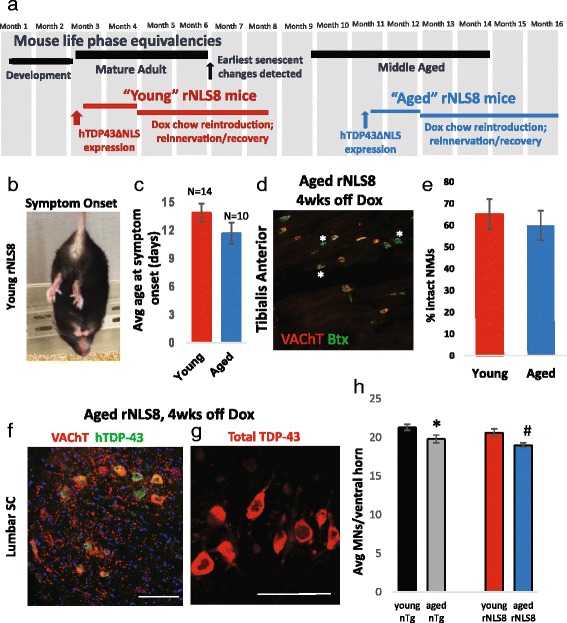
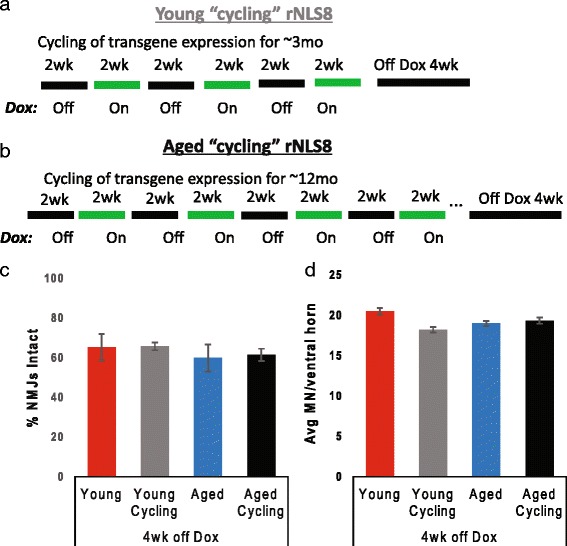
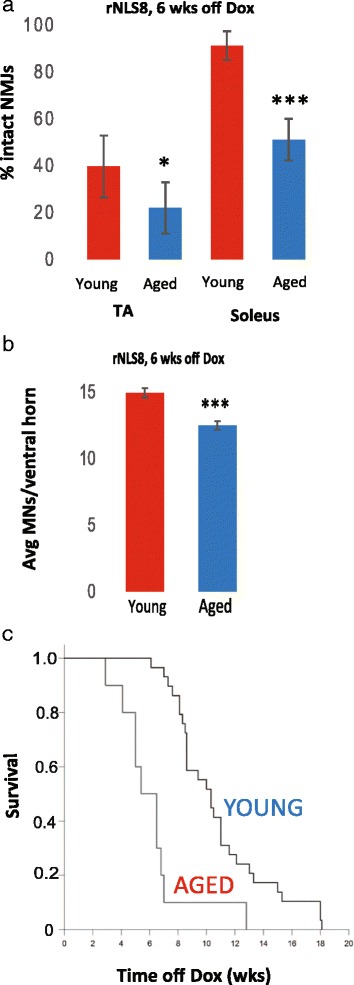
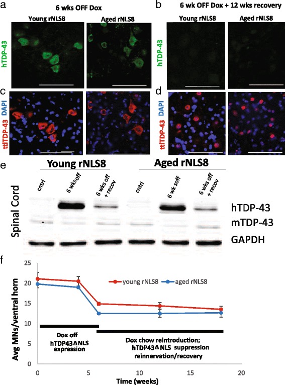
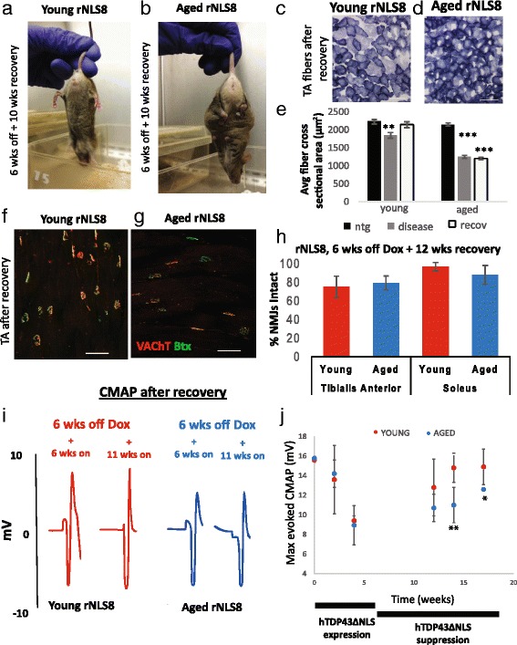
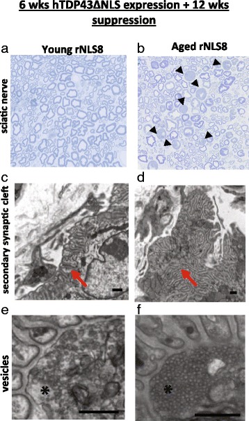
In order to treat progressive paralysis in ALS patients, it is critical to develop a mouse that closely models human ALS in both pathology and also in the timing of these events. We have recently generated new TDP-43 bigenic mice (called rNLS8) with doxycycline (Dox)-suppressible expression of human TDP-43 (hTDP-43) harboring a defective nuclear localization signal (hTDP-43∆NLS) under the control of the NEFH promoter. Our previous studies characterized the pathology and disease course in young rNLS8 mice following induction of neuronal hTDP-43ΔNLS. We now seek to examine if the order and timing of pathologic events are changed in aged mice. We found that the expression of hTDP-43∆NLS in 12+ month old mice did not accelerate the appearance of neuromuscular abnormalities or motor neuron (MN) death in the lumbar spinal cord (SC), though disease progression was accelerated. However, following suppression of the transgene, important differences between young and aged rNLS8 mice emerged in functional motor recovery. We found that recovery was incomplete in aged mice relative to their younger treatment matched counterparts based on gross behavioral measures and physiological recordings from the animals' gastrocnemius (GC) muscles, despite muscle reinnervation by surviving MNs. This is likely because the reinnervation most often only resulted in partial nerve and endplate connections and the muscle's junctional folds were much more disorganized in aged rNLS8 mice. We believe that these studies will be an important basis for the future design and evaluation of therapies designed to slow denervation and promote re-innervation in adult ALS patients.

Keywords: Amyotrophic lateral sclerosis; Motor neuron; Neuromuscular junction; Reinnervation; TDP-43; rNLS mice.


Figures

Fig. 1
Fig. 1
Aging does not affect ALS-like MN disease onset in rNLS8 mice. a Experimental timeline with life phase equivalencies indicated. b Representative image of a young rNLS8 mouse at symptom onset (notice hindlimb clasping). c Aged rNLS8 mice do not show a difference in onset of symptoms compared to young mice as assessed by appearance of one or more of the following phenotypes: clasping of hindlimbs, tremor, weight loss, and/or hunched posture. Mean ± SEM, p = 0.14. d Representative muscle cryosection from the TA of an aged rNLS8 mouse at 4 wks off Dox, with overlap of VAChT positive motor terminals (red) and acetylcholine receptors (α-bungarotoxin, BTX, green) as an indicator of innervated motor endplates. Denervated NMJs are marked with white asterisks. Scale bar = 100 μm. e After 4 wks of transgene expression, aged Tg mice show no difference in TA innervation when compared to young Tg mice. Data are mean ± SD (n = 3–4 for both young and aged Tg mice; p = 0.38). f Representative immunostaining for VAChT (red, to label MNs) and hTDP-43 (green) on a cryosection of lumbar SC from an aged rNLS8 mouse at 4 weeks off Dox shows that the majority of MNs express the transgene. g When staining with a TDP-43 antibody that labels both hTDP-43 and endogenous mTDP-43, widespread nuclear clearance is observed at this 4 wks off Dox time-point, similar to young rNLS8 SC [32]. Scale bar = 100 μm. h A slight decrease is observed in the number of lumbar SC MNs in young versus aged nTg mice (compare black vs grey bar, p = 0.05), but this MN loss is not exacerbated by 4 wks of transgene expression in rNLS8 animals (compare grey vs. blue bar, p = 0.1). p = 0.05, compared to young nTg; # p < 0.05, compared to young rNLS8 mice
Fig. 2
Fig. 2
Cell stress does not accelerate disease onset. a-b Experimental scheme in which rNLS8 animals were weaned and then exposed to repeated cycles of hTDP-43ΔNLS expression (for 2 wks) followed by suppression (for 2 wks) by feeding them chow with or without Dox. This lasted for 3 months in the “young cycling” group (a), or 12 months in the “aged cycling” group (b) prior to removing Dox continuously for 4 wks. c-d Repeated transgene expression and suppression, designated “cycling”, for either 3 months (grey bar) or for a whole year (black bar) does not exacerbate TA denervation (c) or lumbar MN loss (d) at 4 weeks off Dox compared, to young or aged mice that are off Dox without cycling (red and blue bars). Data are mean ± SD (n = 3–4 for all groups of mice)
Fig. 3
Fig. 3
Differences emerge between young and aged rNLS8 mice as the ALS-like disease progresses. a After 6 weeks of transgene expression, aged mice show differences in both TA and soleus innervation compared to young mice, p = 0.04 and p = 8.3E-4, n = 3–4. b Aged mice also have fewer lumbar MNs than young rNLS8 mice at 6 weeks off Dox. Data are mean ± SD (n = 3–4 for both young and aged mice; p = 1.2E-04), p < 0.05, ***p < 0.001. c Aging significantly accelerates time to death after transgene induction in rNLS8 mice. Median time to death is 6.5 weeks for aged mice vs. 10.3 weeks for young mice. (n = 10 for aged mice, n = 29 for young mice, Log rank test statistic = 18.7, p < 0.001)
Fig. 4
Fig. 4
Suppressing the hTDP-43ΔNLS transgene restores endogenous mTDP-43 and halts MN loss in both young and aged rNLS8 mice. a Representative pictures of lumbar SC of young (left) and aged (right) rNLS8 mice at 6 wks off Dox show high, cytoplasmic expression of hTDP-43 (green). b After 12 wks back on Dox, both young and aged rNLS8 mice show a clearance of this cytoplasmic hTDP-43. c When the same cryosections were stained with a TDP-43 antibody that labels both mouse and human TDP-43 or total (ttl) TDP-43 (red), nuclear clearance of TDP-43 is obvious in both young and aged rNLS8 mice at 6 wks off Dox. d After 12 wks of transgene suppression, there is a return of endogenous mTDP-43 to the nucleus in young and aged mice. Scale bars = 100 μm. e Representative immunoblots showing RIPA-soluble hTDP-43∆NLS, mTDP-43 and GAPDH in rNLS8 SC before and after recovery compared to age-matched nTg control (cntrl) mice. There is no difference in the clearance of hTDP-43 or restoration of endogenous mouse TDP-43 between young and old animals. f Timeline of MN loss at L3–L5 levels of the SC in young (red) and aged (blue) rNLS8 mice showing that there is a precipitous decline in MN numbers while the transgene is expressed, which is completely halted by transgene suppression. Mean ± SD, n = 3–4 per time-point
Fig. 5
Fig. 5
Suppressing the hTDP- 43ΔNLS transgene does not reverse muscle atrophy and dysfunction despite apparent muscle re-innervation in Tg aged mice. a-b Pictures of the phenotype of a representative young (a) and aged (b) rNLS8 mouse after 6 weeks of hTDP-43ΔNLS expression followed by 10 wk of suppression. Note that the young mouse splays its limbs normally, while the aged mouse is still clasping. c-e Representative pictures of muscle fibers from TAs of young (c) and aged (d) rNLS8 mice after 6 wks of transgene expression and 12 wks of subsequent suppression, stained with succinate dehydrogenase. Scale bars = 100 μm. e In both young and aged rNLS8 mice, average cross sectional fiber area in the TA muscle decreases during disease (6 wks off Dox, grey bar), reflecting muscle atrophy after denervation. However, in the young rNLS8 mice, after transgene suppression, the TA is re-innervated, and the average fiber size returns to baseline levels. Conversely, in the aged rNLS8 mice, despite apparent muscle re-innervation, the muscle remains significantly smaller than nTg controls. Mean ± SEM; **p < 0.01, ***p < 0.001. f-h After suppression of transgene expression for 12 wks, aged rNLS8 mice show the same level of re-innervation of TA and Sol muscles compared to young rNLS8 mice. f-g. Representative muscle cryosections from the TA of a young (f) and aged (g) rNLS8 mouse after long transgene suppression. Scale bars = 100 μm. h Quantification of intact NMJs in young (red) and aged (blue) TA and Sol after recovery, n = 3–4, mean ± SD. i-j Evoked CMAPs in the GC muscle after stimulation of the sciatic nerve in young (red) and aged (blue) rNLS8 mice that are back on Dox after 6 wks off show a more robust motor recovery in young animals. i Individual traces from the same animals at two different time-points showing the M-wave. j CMAP measurements significantly decrease with hTDP-43 expression, but can recover following transgene suppression and muscle re- innervation. Unlike young rNLS8 mice, the aged mice never return to their original, pre-disease baseline and have significantly lower maximum evoked CMAPs from the GC than young rNLS8 mice after 8 and 10 week of recovery. Data are mean ± SD, n = 3–7 animals per genotype, *p < 0.05, **p < 0.01
Fig. 6
Fig. 6
Functional motor recovery is likely diminished in rNLS8 mice because of impairments at the NMJ. a-b Representative pictures of sciatic nerves from a young (a) and aged (b) rNLS8 mice after 12 wks of transgene suppression, cut transversely at 1 μm and stained with toluidine blue and imaged by light microscopy at 40× to show axon morphology. The black arrowheads point to degenerating axons in the aged sciatic nerve section. c-f At the EM level, NMJ ultrastructural differences are apparent between the young and aged recovered rNLS8 mice. c-d While the TA of the young rNLS8 mouse still has junctional folds that are arranged in an organized way, mostly linearly, (see red arrow in c) the aged junctional folds are more often highly disorganized (see red arrow in d). Further, the NMJs of the young animal were more often completely re-innervated, whereas many of the junctional folds of the aged animal were only partially supplied by a nerve. e-f The synaptic vesicles were packed much more densely in the nerve terminal of the aged mouse (* in f) compared to the young mouse (* in e). Scale bars, 500 nm

Similar articles

Cited by

References

    1. Alfieri JA, Pino NS, Igaz LM. Reversible behavioral phenotypes in a conditional mouse model of TDP-43 proteinopathies. J Neurosci. 2014;34:15244–59. doi: 10.1523/JNEUROSCI.1918-14.2014. - DOI - PMC - PubMed
    1. Balice-Gordon RJ. Age-related changes in neuromuscular innervation. Muscle Nerve Suppl. 1997;5:S83–7. doi: 10.1002/(SICI)1097-4598(1997)5+<83::AID-MUS20>3.0.CO;2-Z. - DOI - PubMed
    1. Banker BQ, Kelly SS, Robbins N. Neuromuscular transmission and correlative morphology in young and old mice. J Physiol. 1983;339:355–77. doi: 10.1113/jphysiol.1983.sp014721. - DOI - PMC - PubMed
    1. Beghi E, Logroscino G, Chio A, Hardiman O, Mitchell D, Swingler R, Traynor BJ, Consortium E The epidemiology of ALS and the role of population-based registries. Biochim Biophys Acta. 2006;1762:1150–7. doi: 10.1016/j.bbadis.2006.09.008. - DOI - PubMed
    1. Black MM, Lasek RJ. Slowing of the rate of axonal regeneration during growth and maturation. Exp Neurol. 1979;63:108–19. doi: 10.1016/0014-4886(79)90188-2. - DOI - PubMed

LinkOut - more resources