. 2016 Oct 25;7(43):70699-70714.
doi: 10.18632/oncotarget.12208.

miR-135a promotes gastric cancer progression and resistance to oxaliplatin

Affiliations

miR-135a promotes gastric cancer progression and resistance to oxaliplatin

Lin-Hai Yan et al. Oncotarget. .

Abstract

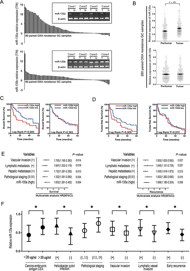
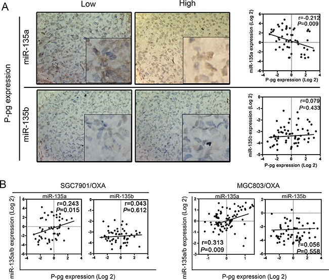
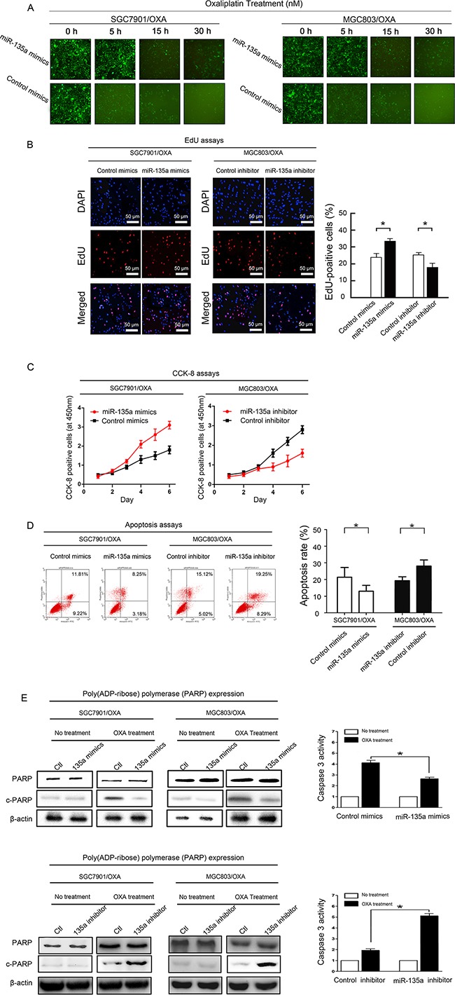
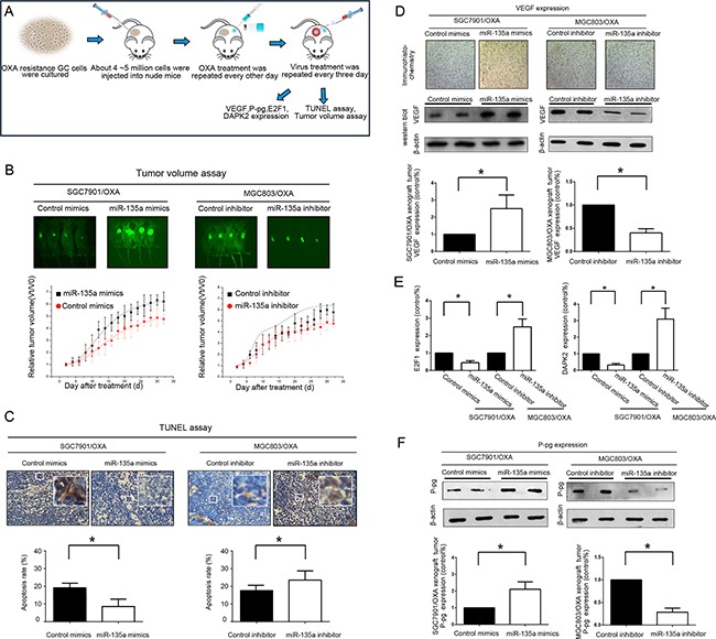
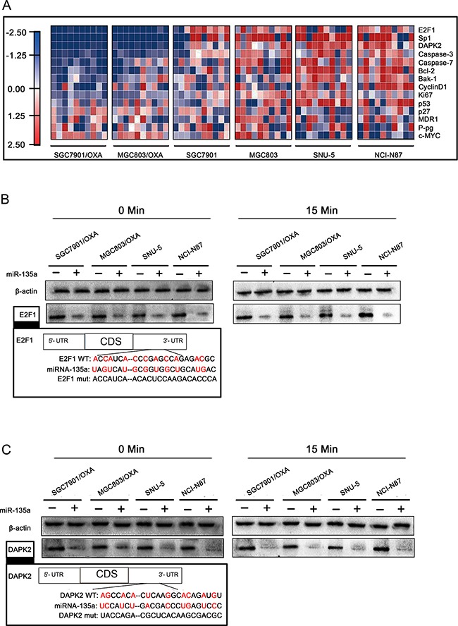
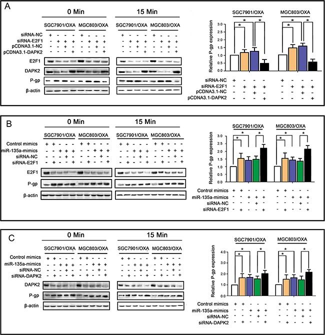
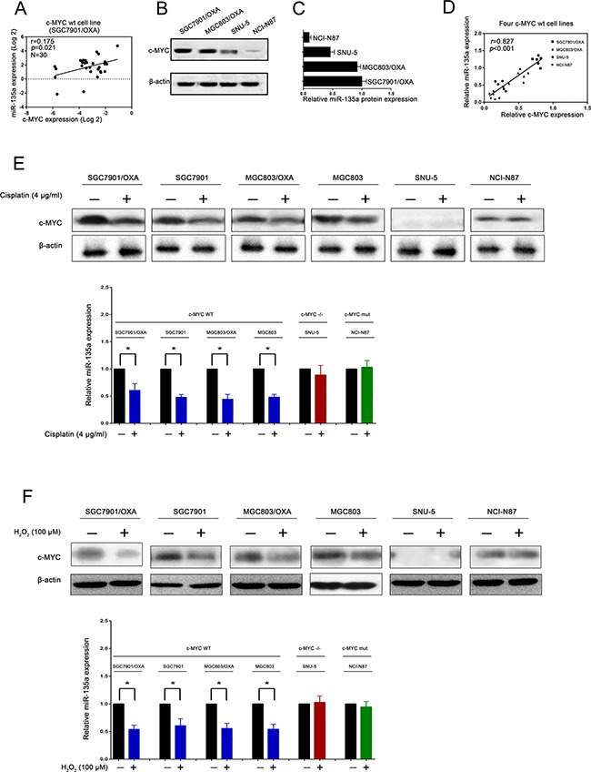
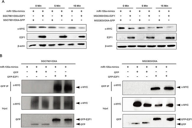
Resistance to oxaliplatin (OXA)-based chemotherapy regimens continues to be a major cause of gastric cancer (GC) recurrence and metastasis. We analyzed GC samples and matched non-tumorous control stomach tissues from 280 patients and found that miR-135a was overexpressed in GC samples relative to control tissues. Tumors with high miR-135a expression were more likely to have aggressive characteristics (high levels of carcino-embryonic antigen, vascular invasion, lymphatic metastasis, and poor differentiation) than those with low levels. Patients with greater tumoral expression of miR-135a had shorter overall survival times and times to disease recurrence. Furthermore, miR-135a, which promotes the proliferation and invasion of OXA-resistant GC cells, inhibited E2F transcription factor 1 (E2F1)-induced apoptosis by downregulating E2F1 and Death-associated protein kinase 2 (DAPK2) expression. Our results indicate that higher levels of miR-135a in GC are associated with shorter survival times and reduced times to disease recurrence. The mechanism whereby miR-135a promotes GC pathogenesis appears to be the suppression of E2F1 expression and Sp1/DAPK2 pathway signaling.

Keywords: gastric cancer; transcription factor E2F1.


Conflict of interest statement

COMPETING INTERESTS

The authors have declared that no competing interests exist.

Figures

Figure 1
Figure 1. The miR-135 family is frequently upregulated in GC and is associated with poor prognosis
A. miR-135a and miR-135b expression were significantly greater in GC tissues than in the corresponding adjacent tissues based on qRT-PCR. B. Relative expression of miR-135a and miR-135b in paired GC tissue samples (n=280). miR-135a expression was significantly upregulated in tumors compared with the corresponding adjacent non-tumorous stomach tissues. C, D. Elevated miR-135a levels negatively correlated with the overall survival and tumor-free survival of GC patients, whereas no substantial difference was observed for miR-135b. E. A multivariate analysis of the hazard ratios (HRs) revealed that the upregulation of miR-135a may be an independent prognostic factor for the overall survival (OS) and recurrence-free survival rates (based on the Cox multivariate proportional hazards regression model). The HRs are presented as the mean (95% confidence interval). The variables included in the multivariate analysis were selected based on the results of univariate analysis. F. Relative expression of miR-135a in 280 human GC samples with or without high levels of serum carcino-embryonic antigen (CEA), helicobacter pylori infection, pathological staging, vascular invasion, lymphatic vessel metastasis, and early recurrence. The data were from three independent experiments, *P < 0.05.
Figure 2
Figure 2. Relationship between miR-135 family levels and P-gp expression in GC patients
A. The relationship between miR-135 family levels and P-gp expression in 80 GC samples was determined with linear regression models. B. The relationship between miR-135 family levels and P-gp mRNA expression in OXA-resistant GC cell lines. The data were from three independent experiments.
Figure 3
Figure 3. miR-135a promotes OXA resistance in GC cells
A. Transfection of a miR-135a mimic (containing a segment of GFP) into OXA-resistant GC cell lines increased cellular viability, as revealed by fluorescence microscopy at 0, 5, 15, and 30 hours (×100). B. EdU staining indicating that SGC7901/OXA cell proliferation and the percentage of EdU-positive SGC7901/OXA cells increased when miR-135a was upregulated; likewise, MGC803/OXA cell proliferation and the percentage of EdU-positive MGC803/OXA cells decreased when miR-135a was downregulated. C. After 48 h of transfection with a miR-135a mimic or inhibitor, the growth rates of SGC7901/OXA and MGC803/OXA cells were measured with CCK8 assays. D. miR-135a inhibits apoptosis of GC cells: representative flow cytometry analysis of Annexin V- and Propidium iodide-stained SGC7901/OXA cells and MGC803/OXA cells treated with a miR-135a mimic or inhibitor (left panels). Quantification of apoptotic cells (Annexin V/PI) was performed on two independent cell populations (right panels). E. Overexpression of miR-135a inhibits OXA-induced apoptosis: SGC7901/OXA and MGC803/OXA cells were transfected with 100 nM pre-miR-negative (Control) or pre-miR-135a (miR-135a mimic) and then treated with 40 nM OXA for 48 h. Western blotting of cell lysates was performed with antibodies against cleaved PARP or total PARP, and β-actin was used as a control (left panels). SGC7901/OXA cells transfected with 100 nM pre-miR-negative (Ctl) or pre-miR-135a (miR-135a mimic) were seeded onto 96-well plates at 8×10³ cells per well. After 6 h, cells were treated with or without 40 nM OXA for 48 h. The activity of caspase 3 was measured with a Caspase-Glo 3/7 kit (right panels). The data were from three independent experiments, *P < 0.05.
Figure 4
Figure 4. Lentiviral administration of miR-135a suppresses OXA-resistant GC cell growth in vivo
A. A suspension of SGC7901/OXA or MGC803/OXA cells was injected i.p. into mice. Virus or OXA (10 mg/kg) was administered i.p. every two or three days. Tumors were treated until they reached approximately five times their original volume. B. Green fluorescence photographs illustrating representative features and growth curves of SGC7901/OXA or MGC803/OXA tumors in nude mice after injection with LV-GFP-miR-135a mimic, LV-GFP-control mimic, LV-GFP-miR-135a inhibitor, or LV-GFP-control inhibitor. The relative tumor volume was evaluated at two-day intervals in comparison with day 0, when the virus treatment was performed. C.The percentage of apoptotic cells in GC tissue was analyzed by TUNEL. D. Immunohistochemistry staining for VEGF in tumor tissues from mice with subcutaneous OXA-resistant GC cell implantation. Cells with positive staining were counted from 10 different visual fields. Western blotting was performed with an antibody against total VEGF, and β-Actin was used as a loading control. E. After administration of the miR-135a-expressing lentivirus, the levels of E2F1 and DAPK2 protein in the implanted tumor tissue were analyzed by Western blotting. F. After administration of the miR-135a-expressing lentivirus, the levels of P-gp protein in the implanted tumor tissue were analyzed by Western blotting. The data were from three independent experiments, *P < 0.05.
Figure 5
Figure 5. miR-135a downregulates E2F1 and DAPK2 in OXA-resistant GC cells
A. Heat maps of gene expression changes after transfection with the miR-135a mimic, as revealed by qRT-PCR. The red, white, and blue right-hand panel indicates the log2 of expression ratios after miR-135a mimic transfection. White represents a 1:1 ratio, red indicates upregulation, and blue indicates downregulation. B, C. After 48 h of miR-135a mimic transfection, E2F1 and DAPK2 protein levels with OXA treatment for 0 and 15 minutes were analyzed by Western blotting with anti-E2F1, anti-DAPK2, and anti-β-actin antibodies.
Figure 6
Figure 6. miR-135a promotes OXA resistance in GC cells by suppressing the E2F1/DAPK2 signaling pathway
A. E2F1 siRNA enhanced P-gp expression, while DAPK2 overexpression reversed this effect, as revealed by qRT-PCR (right panels) and Western blotting (left panels). B. Similar effects of the miR-135a mimic and siRNA-E2F1 on P-gp expression, with possible synergistic effects when transfected together, as revealed by qRT-PCR (right panels) and Western blotting (left panels). C. Similar effects of the miR-135a mimic and siRNA-DAPK2 on P-gp expression, with possible synergistic effects when transfected together, as revealed by qRT-PCR (right panels) and Western blotting (left panels). The data were expressed as the mean ± S.E.M. of three independent experiments, *P < 0.05.
Figure 7
Figure 7. miR-135a promoting oxaliplatin resistance in gastric cancer cells is regulated by c-MYC
A. Relationship between miR-135a and c-MYC expression levels in SGC7901/OXA cells. B, C, D. Relationship between miR-135a and c-MYC expression levels in oxaliplatin resistance and normal gastric cancer cells. E, F. Expression analysis of c-MYC and miR-135a level changes in different gastric cancer cells after treatment of cisplatin and H2O2, respectively. The data were expressed as the means ± S.E.M. of three independent experiments. *P < 0.05.
Figure 8
Figure 8. miR-135a promoting oxaliplatin resistance in gastric cancer cells is regulated by E2F1/DAPK2/P-pg axis
A. Expression analysis of c-MYC and miR-135a level changes in different gastric cancer cells after siRNA-c-MYC transfection, protein expression level for 0, and 5 minutes was analyzed by western blot with anti- MYC, and anti-β-actin antibodies. B. After treatment of siRNA-c-MYC, c-MYC downstream factor protein extracts were analyzed by western blot with anti-MYC, anti- E2F1, anti-DAPK2, anti-P-pg and anti-β-actin antibodies. C. The effect of miR-135a overexpression to rescue the siRNA-c-MYC-mediated suppression of P-pg, as revealed by qRT-PCR. D. SGC7901/OXA and MGC803/OXA cells were stained for P-pg expression, representative images. The data were expressed as the means ± S.E.M. of three independent experiments. *P < 0.05 and **P < 0.01.
Figure 9
Figure 9. Association of E2F1 with c-MYC in miR-135a-induced oxaliplatin resistance
A. The indicated GC cells were exposed to miR-135a mimics for 0, 5, or 15 minutes, the expressions of E2F1 and c-MYC were analyzed by Western blotting. B. The GC cells were immunoprecipitated using GFP, GFP-E2F1 and c-MYC, and cell lysates were analyzed by Western blotting.

Similar articles

Cited by

References

    1. Yu S, Yang CS, Li J, You W, Chen J, Cao Y, Dong Z, Qiao Y. Cancer Prevention Research in China. Cancer Prev Res (Phila) 2015;8:662–674. - PubMed
    1. Mickevicius A, Ignatavicius P, Markelis R, Parseliunas A, Butkute D, Kiudelis M, Endzinas Z, Maleckas A, Dambrauskas Z. Trends and results in treatment of gastric cancer over last two decades at single East European centre: a cohort study. BMC surg. 2014;14:98. - PMC - PubMed
    1. Shang Y, Feng B, Zhou L, Ren G, Zhang Z, Fan X, Sun Y, Luo G, Liang J, Wu K, Nie Y, Fan D. The miR27b-CCNG1-P53-miR-508-5p axis regulates multidrug resistance of gastric cancer. Oncotarget. 2016;7:538–549. doi: 10.18632/oncotarget.6374. - DOI - PMC - PubMed
    1. Riquelme I, Letelier P, Riffo-Campos AL, Brebi P, Roa JC. Emerging Role of miRNAs in the Drug Resistance of Gastric Cancer. Int J Mol Sci. 2016;17:E424. - PMC - PubMed
    1. Cheng Y, Jutooru I, Chadalapaka G, Corton JC, Safe S. The long non-coding RNA HOTTIP enhances pancreatic cancer cell proliferation, survival and migration. Oncotarget. 2015;6:10840–10852. doi: 10.18632/oncotarget.3450. - DOI - PMC - PubMed

MeSH terms