. 2016 Sep 28:6:34141.
doi: 10.1038/srep34141.

Persistence of fan-shaped keratocytes is a matrix-rigidity-dependent mechanism that requires α5β1 integrin engagement

Affiliations

Persistence of fan-shaped keratocytes is a matrix-rigidity-dependent mechanism that requires α5β1 integrin engagement

Maryam Riaz et al. Sci Rep. .

Abstract

Despite the importance of matrix rigidity on cell functions, many aspects of the mechanosensing process in highly migratory cells remain elusive. Here, we studied the migration of highly motile keratocytes on culture substrates with similar biochemical properties and rigidities spanning the range between soft tissues (~kPa) and stiff culture substrates (~GPa). We show that morphology, polarization and persistence of motile keratocytes are regulated by the matrix stiffness over seven orders of magnitude, without changing the cell spreading area. Increasing the matrix rigidity leads to more F-actin in the lamellipodia and to the formation of mature contractile actomyosin fibers that control the cell rear retraction. Keratocytes remain rounded and form nascent adhesions on compliant substrates, whereas large and uniformly distributed focal adhesions are formed on fan-shaped keratocytes migrating on rigid surfaces. By combining poly-L-lysine, fibronectin and vitronectin coatings with selective blocking of αvβ3 or α5β1 integrins, we show that αVβ3 integrins permit the spreading of keratocytes but are not sufficient for polarization and rigidity sensing that require the engagement of α5β1 integrins. Our study demonstrates a matrix rigidity-dependent regulation of the directional persistence in motile keratocytes and refines the role of αvβ3 and α5β1 integrins in the molecular clutch model.


Figures

Figure 1
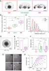
Figure 1. Matrix stiffness controls morphology and polarization of migrating cells.
(A) Differential Interference Contrast (DIC) images of representative fish epithelial keratocytes migrating on culture substrates of different stiffnesses (E = 1.5 kPa in red, 9 kPa in blue, 110 kPa in green, 3 MPa in orange and 70 GPa in purple). (B) The variation of cell morphology, as determined by the analysis of aligned cell outlines, is shown for populations of crawling cells plated on substrates of various stiffnesses (80 ≤ n ≤ 160 for each population). The mean cell shape is presented in bold line for each population of cells with one standard deviation away from the mean in each direction. (C) Evolution of the lamellipodial curvature as a function of the substrate stiffness. The inset shows a semi-log scale. (D) Distribution of the aspect ratios of large populations of keratocytes plated on substrates of varying stiffness. The inset shows a semi-log scale. (E) Histogram of the cell count (%) as a function of the substrate stiffness. Lines are gaussian fits. (F) Distribution of the cell area of live keratocytes plated on different matrix rigidities (85 ≤ n ≤ 160 for each population). N.S indicates that no statistical difference was observed. (G) Histogram of the percentage of polarized cells within large populations of individual keratocytes plated on various matrix stiffnesses. The number of cells is indicated at the bottom of the bars. (H) The typical migration of a keratocyte from a soft (9 kPa) to a stiff (230 kPa) region is superimposed on the fluorescent image of beads embedded in the stiffer region of the hydrogel (see Movie S1). Red lines represent the cell boundary. White arrow indicates the direction of the gradient of rigidity from the softer (9 kPa) to the stiffer region (230 kPa). Scale bar is 50 μm. Temporal evolutions of (I) the cell aspect ratio and (J) the instantaneous cell velocity during the crossing event presented in (H).
Figure 2
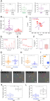
Figure 2. Migration is controlled by the matrix stiffness.
(A) Superimposed migration trajectories of individual keratocytes plated for 30 min on flat substrates with various rigidities (red: 1.5 kPa, blue: 9 kPa, green: 110 kPa, orange: 3 MPa and purple: 70 GPa) and similar FN coatings. Initial positions of the crawling cells were superimposed at the origin for comparison. All plots range from −350 μm to + 350 μm on both the x- and y- axis. (B) Evolution of migration curvature as a function of the matrix stiffness. 85 ≤ n ≤ 160 for each population. (C) Evolution of the lamellipodial curvature as a function of the migration curvature. (D) Variation of the motility coefficient, μ, as a function of the substrate stiffness. The green mark represents the range of Young’s moduli of the internal side of fish scales. Data are shown as mean ± s.d.
Figure 3
Figure 3. Matrix stiffness affects actin and myosin distribution patterns.
Immunostained images of individual keratocytes plated on soft (left column, in red), intermediate (center column, in green), and stiff (right column, in purple) culture substrates. (A) Cross-sectional confocal views (xz) and (B) normal views (xy) of keratocytes labeled for actin with fluorescent phalloidin. The color-coded average distribution of actin is shown for n = 11 (soft), n = 13 (intermediate) and n = 14 cells (stiff). (C) Normal views of keratocytes immunolabeled for myosin and color-coded average distribution of myosin for n = 9 (soft), n = 10 (intermediate) and n = 12 cells (stiff). (D) Merge images of (B) and (C). The front and rear division schematically presented in (E) was used to estimate the front to rear actin ratio (F) and myosin ration (G) for soft (in red), intermediate (in green) and stiff (in purple) substrates. (H) The distribution of actin was measured along a line from the cell rear (point 0) to the cell front (point 100) for keratocytes plated on soft (in red), intermediate (in green) and stiff (in purple) culture substrates. (I) The distribution of myosin was measured from the left side (point 0) to the right side (point 100) for crawling cells plated on soft (red), intermediate (green) and stiff (purple) culture substrates. Black arrows in (H) and (J) indicate the highest fluorescent signals. Scale bars are 5 μm.
Figure 4
Figure 4. Cell-substrate adhesions are modulated by matrix stiffness.
(A) Typical inverted images of keratocytes plated on soft (left, in red), intermediate (center, in green), and stiff (right, in purple) culture substrates and immunolabeled for vinculin. (B) Evolution of the ratio of vinculin area to cell area with the substrate rigidity. (C) Cumulative frequencies of the adhesion areas for soft (in red), intermediate (in green) and stiff (in purple) matrices. Solid lines are gaussian fits. (D) Inverted image of a keratocyte plated on a stiff substrate coated with PLL and immunolabeled for vinculin. (E) Ratio of the vinculin area to the cell area measured for keratocytes plated on stiff substrates coated with PLL (n = 16) and FN (n = 19). (F) Evolution of the cell aspect ratio on soft (in red), intermediate (in green) and stiff (in purple) substrates coated with FN (plain bars) and PLL (hashed bars). (G) Image sequence of crawling cells plated on a stiff substrate coated with PLL that polarized and migrated after the addition of fibronectin (t = 02 min 30 sec.) in the culture media. (H) Temporal evolution of the aspect ratio of crawling cells plated on a stiff substrate coated with PLL in response to the addition of fibronectin (red arrow) in the culture media. *p < 0.05, **p < 0.01, ***p < 0.001 and n.s. not significant.
Figure 5
Figure 5. α5β1 integrin engagement is required for keratocyte mechanosensing.
(A) Effect of a α5β1 antibody treatment on the persistence length of individual keratocytes plated on soft (in red), intermediate (in green) and stiff (in purple) FN-coated substrates. (B) Comparison of the cell aspect ratio of keratocytes plated on PLL-coated stiff substrates after addition of FN in the culture media (in purple) and with the addition of FN in the culture media of keratocytes treated with α5β1 antibody and plated on PLL-coated stiff substrates (in purple). (C) Typical trajectories described by keratocytes on stiff substrate after a α5β1 antibody treatment (in red). Temporal variation of (D) the migration curvature (E) the migrating velocity of keratocytes plated on FN-coated stiff substrates and treated with a α5β1 antibody. (F) Ratio of the vinculin area to the cell area measured for keratocytes migrating on stiff FN-coated substrates (in purple) and treated with α5β1 antibody (dashed red bar). (G) Projected area, (H) cell aspect ratio and (I) polarized fraction of individual keratocytes plated on stiff substrates coated with FN (in orange) and VN (in blue). (J) Sequence of the migration of 6 keratocytes plated on a stiff VN-coated surface in response to FN added in the culture media at t = 42 min 50 sec. Evolution of the total migration length of keratocytes migrating on (K) soft and (L) stiff substrates coated with VN after addition of FN in the culture media. *p < 0.05, **p < 0.01 and n.s. not significant.
Figure 6
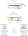
Figure 6. Schematic of the role of α5β1 and αVβ3 integrins in the focal adhesion clutch model.
Integrins are coupled to F-actin via linker proteins, such as vinculin, talin and paxilin, that move backward (red arrows) in response to pushing forces (in green) exerted by actin polymerization and pulling forces (in orange) exerted by actomyosin contractility. Cell spreading requires only αVβ3 integrin engagement. (A) On soft matrices, keratocytes have a low aspect ratio, are characterized by a low fraction of polarized cells, form nascent adhesions located at the cell periphery, have a low persistence and a high lamellipodial curvature. (B) On stiff matrices, keratocytes have a high aspect ratio with mature focal adhesions, have a high fraction of polarized cells, a high persistence and a low lamellipodial curvature.

Similar articles

Cited by

References

    1. Lauffenburger D. A. & Horwitz A. F. Cell migration: A physically integrated molecular process. Cell 84, 359–369 (1996). - PubMed
    1. Reffay M. et al. Interplay of RhoA and mechanical forces in collective cell migration driven by leader cells. Nat. Cell Biol. 16, 217–223 (2014). - PubMed
    1. Friedl P. & Glimour D. Collective cell migration in morphogenesis, regeneration and cancer. Nat. Rev. Mol. Cell Bio. 10, 445–457 (2009). - PubMed
    1. Roca-Cusachs P., Sunyer R. & Trepat X. Mechanical guidance of cell migration: lessons from chemotaxis. Curr. Opin. Cell Biol. 25, 543–549 (2013). - PubMed
    1. Plotnikov S. V., Pasapera A. M., Sabass B. & Waterman C. M. Force fluctuations within focal adhesions mediate ECM-rigidity sensing to guide directed cell migration. Cell 151, 1513–1527 (2012). - PMC - PubMed

LinkOut - more resources