

Naturally occurring compounds acting as potent anti-metastatic agents and their suppressing effects on Hedgehog and WNT/β-catenin signalling pathways
- PMID: 27669681
- PMCID: PMC6529111
- DOI: 10.1111/cpr.12299
Naturally occurring compounds acting as potent anti-metastatic agents and their suppressing effects on Hedgehog and WNT/β-catenin signalling pathways
Abstract
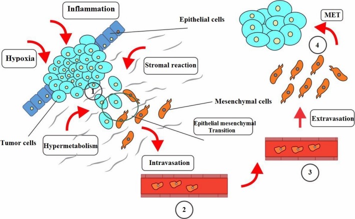
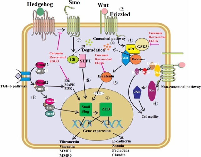
Despite numerous remarkable achievements in the field of anti-cancer therapy, tumour relapse and metastasis still remain major obstacles in improvement of overall cancer survival, which may be at least partially owing to epithelial-mesenchymal transition (EMT). Multiple signalling pathways have been identified in EMT; however, it appears that the role of the Hedgehog and WNT/β-catenin pathways are more prominent than others. These are well-known preserved intracellular regulatory pathways of different cellular functions including proliferation, survival, adhesion and differentiation. Over the last few decades, several naturally occurring compounds have been identified to significantly obstruct several intermediates in Hedgehog and WNT/β-catenin signalling, eventually resulting in suppression of signal transduction. This article highlights the current state of knowledge associated with Hedgehog and WNT/β-catenin, their involvement in metastasis through EMT processes and introduction of the most potent naturally occurring agents with capability of suppressing them, eventually overcoming tumour relapse, invasion and metastasis.
© 2016 John Wiley & Sons Ltd.
Figures
Similar articles
-
Crosstalk between Wnt/β-catenin and Hedgehog/Gli signaling pathways in colon cancer and implications for therapy.Cancer Biol Ther. 2015;16(1):1-7. doi: 10.4161/15384047.2014.972215. Cancer Biol Ther. 2015. PMID: 25692617 Free PMC article. Review.
-
Cinobufacini Inhibits Colon Cancer Invasion and Metastasis via Suppressing Wnt/β-Catenin Signaling Pathway and EMT.Am J Chin Med. 2020;48(3):703-718. doi: 10.1142/S0192415X20500354. Epub 2020 Apr 24. Am J Chin Med. 2020. PMID: 32329642
-
The role of nutraceuticals in the regulation of Wnt and Hedgehog signaling in cancer.Cancer Metastasis Rev. 2010 Sep;29(3):383-94. doi: 10.1007/s10555-010-9233-4. Cancer Metastasis Rev. 2010. PMID: 20711635 Free PMC article. Review.
-
IWR-1 inhibits epithelial-mesenchymal transition of colorectal cancer cells through suppressing Wnt/β-catenin signaling as well as survivin expression.Oncotarget. 2015 Sep 29;6(29):27146-59. doi: 10.18632/oncotarget.4354. Oncotarget. 2015. PMID: 26450645 Free PMC article.
-
MicroRNA-148a suppresses epithelial-mesenchymal transition and invasion of pancreatic cancer cells by targeting Wnt10b and inhibiting the Wnt/β-catenin signaling pathway.Oncol Rep. 2017 Jul;38(1):301-308. doi: 10.3892/or.2017.5705. Epub 2017 Jun 6. Oncol Rep. 2017. Retraction in: Oncol Rep. 2023 Mar;49(3):56. doi: 10.3892/or.2023.8493. PMID: 28586066 Retracted.
Cited by
-
Hydroxytyrosol inhibits cancer stem cells and the metastatic capacity of triple-negative breast cancer cell lines by the simultaneous targeting of epithelial-to-mesenchymal transition, Wnt/β-catenin and TGFβ signaling pathways.Eur J Nutr. 2019 Dec;58(8):3207-3219. doi: 10.1007/s00394-018-1864-1. Epub 2018 Nov 21. Eur J Nutr. 2019. PMID: 30460610
-
Triple-negative breast cancer: understanding Wnt signaling in drug resistance.Cancer Cell Int. 2021 Aug 10;21(1):419. doi: 10.1186/s12935-021-02107-3. Cancer Cell Int. 2021. PMID: 34376211 Free PMC article. Review.
-
Roles of Wnt Signaling Pathway and ROR2 Receptor in Embryonic Development: An Update Review Article.Epigenet Insights. 2022 Jan 31;15:25168657211064232. doi: 10.1177/25168657211064232. eCollection 2022. Epigenet Insights. 2022. PMID: 35128307 Free PMC article.
-
Wnt/β-catenin signaling in cancers and targeted therapies.Signal Transduct Target Ther. 2021 Aug 30;6(1):307. doi: 10.1038/s41392-021-00701-5. Signal Transduct Target Ther. 2021. PMID: 34456337 Free PMC article. Review.
-
Astragalus polysaccharide inhibits breast cancer cell migration and invasion by regulating epithelial‑mesenchymal transition via the Wnt/β‑catenin signaling pathway.Mol Med Rep. 2020 Apr;21(4):1819-1832. doi: 10.3892/mmr.2020.10983. Epub 2020 Feb 12. Mol Med Rep. 2020. PMID: 32319619 Free PMC article.
References
-
- Siegel R, Naishadham D, Jemal A. Cancer statistics, 2012. CA Cancer J Clin. 2012;62:10–29. - PubMed
Publication types
MeSH terms
Substances
LinkOut - more resources
Full Text Sources
Other Literature Sources
