. 2016 Nov 1;7(44):71255-71273.
doi: 10.18632/oncotarget.12104.

Gamma-interferon-inducible, lysosome/endosome-localized thiolreductase, GILT, has anti-retroviral activity and its expression is counteracted by HIV-1

Affiliations

Gamma-interferon-inducible, lysosome/endosome-localized thiolreductase, GILT, has anti-retroviral activity and its expression is counteracted by HIV-1

Yoshinao Kubo et al. Oncotarget. .

Abstract

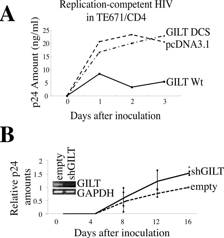
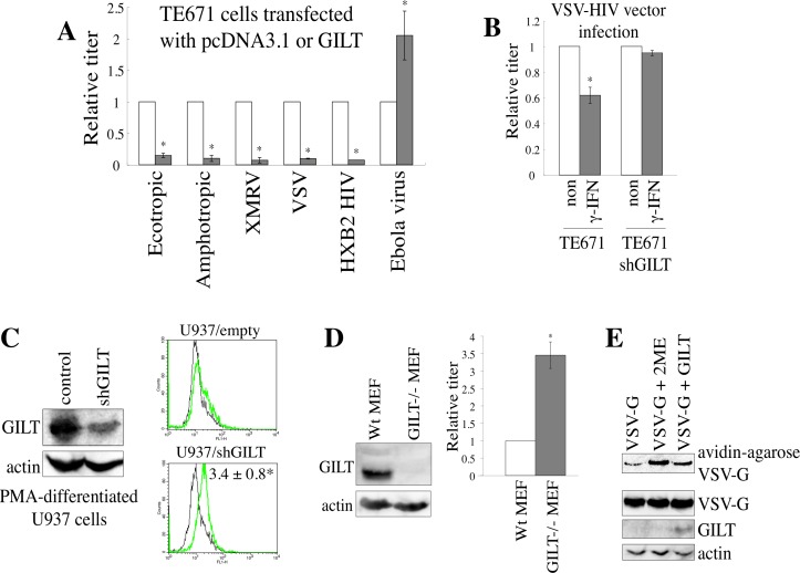
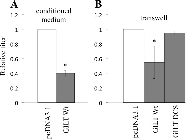
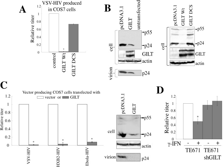
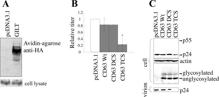
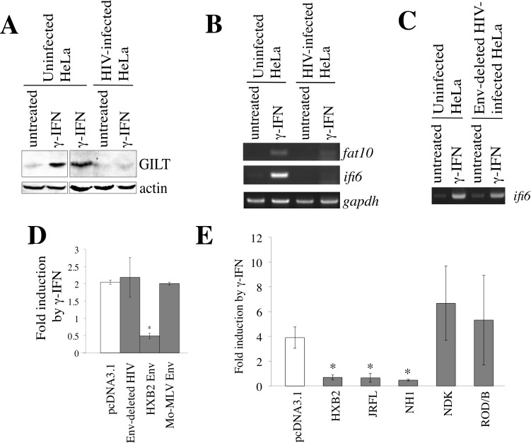
The mechanism by which type II interferon (IFN) inhibits virus replications remains to be identified. Murine leukemia virus (MLV) replication was significantly restricted by γ-IFN, but not human immunodeficiency virus type 1 (HIV-1) replication. Because MLV enters host cells via endosomes, we speculated that certain cellular factors among γ-IFN-induced, endosome-localized proteins inhibit MLV replication. We found that γ-IFN-inducible lysosomal thiolreductase (GILT) significantly restricts HIV-1 replication as well as MLV replication by its thiolreductase activity. GILT silencing enhanced replication-defective HIV-1 vector infection and virion production in γ-IFN-treated cells, although γ-IFN did not inhibit HIV-1 replication. This result showed that GILT is required for the anti-viral activity of γ-IFN. Interestingly, GILT protein level was increased by γ-IFN in uninfected cells and env-deleted HIV-1-infected cells, but not in full-length HIV-1-infected cells. γ-IFN-induced transcription from the γ-IFN-activation sequence was attenuated by the HIV-1 Env protein. These results suggested that the γ-IFN cannot restrict HIV-1 replication due to the inhibition of γ-IFN signaling by HIV-1 Env. Finally, we found that 4,4'-dithiodipyridine (4-PDS), which inhibits S-S bond formation at acidic pH, significantly suppresses HIV-1 vector infection and virion production, like GILT. In conclusion, this study showed that GILT functions as a host restriction factor against the retroviruses, and a GILT mimic, 4-PDS, is the leading compound for the development of novel concept of anti-viral agents.

Keywords: antiviral; endosome; gamma-interferon; retroviruses; thiolreductase.


Conflict of interest statement

CONFLICTS OF INTEREST

There is no conflict of interest.

Figures

Figure 1
Figure 1. γ-IFN restricts MLV replication but not HIV-1 replication
A. TE671/mCAT1 cells were treated with γ-IFN (0.2 μg/ml) for 1 day, and then inoculated with the Moloney MLV. The MLV titers were measured 3 days after the inoculation. B. TE671/CD4 cells were treated with γ-IFN (0.2 g/ml) for 1 day, and then inoculated with the HIV-1 LAI strain. The p24 amounts in culture supernatants were measured 3 days after the inoculation. These experiments were repeated three times. Means are indicated. Error bars show standard deviations. Asterisks indicate statistically significant differences.
Figure 2
Figure 2. GILT inhibits MLV replication
A. TE671/mCAT1 cells were transfected with pcDNA3.1, wild type GILT, or the DCS mutant, and inoculated with Mo-MLV. Lysates of the inoculated cells were analyzed by western blotting, using the indicated antibodies. B. Amino acid sequences near the thiolreductase active site are indicated. The GILT DCS mutant contains the substitutions of two cysteine residues to serine. C. TE671/mCAT1 cells stably transduced by the empty or shGILT-expressing lentivirus vector were treated with γ-IFN (0.2 μg/ml), and inoculated with Mo-MLV. Three days after the inoculation, Mo-MLV titers were measured by XC plaque assay (n = 3). Error bars show standard deviations. Asterisks indicate statistically significant differences. D. TE671 cells were transfected by pcDNA3.1 or GILT. TE671 cells transduced by the empty or shGILT-expressing vector were treated with γ-IFN (0.2 μg/ml). GILT and actin proteins were detected by western immunoblotting, and the intensities of the proteins were measured by a densitometer. The amounts of GILT were normalized by the actin levels. The amounts of GILT in pcDNA3.1-transfected cells are always set to 1, and relative values are indicated (n = 3).
Figure 3
Figure 3. GILT restricts HIV-1 replication
A. TE671/CD4 cells were transfected with pcDNA3.1, wild type GILT, or the GILT DCS mutant, and inoculated with the HIV-1 LAI strain. HIV-1 Gag p24 levels in the supernatants were measured. This experiment was repeated three times, and a representative result is shown. B. Primary MDMs transduced by the empty or shGILT-expressing lentivirus vector were inoculated with the HIV-1 AD8 strain. The amounts of Gag p24 in the supernatants were measured (n = 4). The amounts of p24 in the empty vector-transduced MDMs 16 days after the inoculation are always set to 1, and relative values are indicated. Asterisks indicate statistically significant differences.
Figure 4
Figure 4. GILT inhibits viral entry by digesting S-S bonds of viral Env protein
A. TE671 cells transfected with pcDNA3.1 (open bars) or GILT (closed bars) were inoculated with HIV-1 vector pseudotyped with indicated viral Env proteins. Relative values to titers in the pcDNA3.1-transfected cells are indicated (n = 3). Asterisks indicate statistically significant differences. B. Control and shGILT-expressing TE671 cells were treated with γ-IFN (0.2 μg/ml), and were inoculated with the VSV-pseudotyped vector. Relative values to titers in the untreated cells are indicated (n = 3). Asterisks indicate statistically significant differences. C. Control and shGILT-expressing U937 cells were treated with PMA. Lysates from the differentiated cells were analyzed by western blotting (left panel). The PMA-differentiated cells were inoculated with the VSV-pseudotyped HIV-1 vector encoding GFP, and the inoculated cells were analyzed by flow cytometry. Relative values to numbers of GFP-positive cells in control U937 macrophages are indicated (n = 3) (right panel). D. Cell lysates prepared from MEFs of GILT-knockout and wild type mice were subjected to western immunoblotting (left panel). The MEF2 were inoculated with the amphotropic MLV vector. Relative values to transduction titers in the wild type MEFs are indicated (n = 3). E. COS7 cells were transfected by VSV-G, together with pcDNA3.1 or GILT. COS7 cells transfected by VSV-G and pcDNA3.1 were treated with 2-mercaptoethanol (2ME) as a positive control. Proteins with free cysteine residues were isolated, and analyzed by western blotting (see Materials and Methods) (upper panel). Cell lysates from the transfected cells were analyzed by western blotting (three lower panels). Representative results are indicated.
Figure 5
Figure 5. Secreted GILT inhibits viral infection
A. VSV-pseudotyped HIV-1 vector was diluted with culture supernatant from the pcDNA3.1- or GILT-transfected COS7 cells, and inoculated to HeLa cells. B. Target HeLa cells were cultured with the pcDNA3.1- or GILT-transfected COS7 cells in transwells. The HeLa cells were inoculated with amphotropic MLV-pseudotyped HIV-1 vector. Relative values to titers in the pcDNA3.1-transfected cells are indicated (n = 3). Asterisks indicate statistically significant differences.
Figure 6
Figure 6. GILT inhibits HIV-1 virion production
A. COS7 cells were transfected with the VSV-pseudotyped HIV-1 vector construction plasmids, together with pcDNA3.1, wild type GILT, or the DCS mutant. Culture supernatants from the transfected cells were used to inoculate HeLa cells, and titers were measured. Relative values to titers in the pcDNA3.1-transfected cells are indicated (n = 3). Asterisks indicate statistically significant differences. B. Cell lysates and virion pellets from the transfected cells were analyzed by western blotting. C. COS7 cells were transfected with the VSV-, HXB2-, or Ebola virus-pseudotyped HIV-1 vector construction plasmids together with pcDNA3.1 or wild type GILT, and transduction titers of culture supernatant from the transfected cells were measured. Relative values to titers in pcDNA3.1-transfected cells are indicated (n = 3). Cell lysates and virion pellets from the Ebola virus-pseudotyped HIV-1-producing cells were analyzed by western blotting. D. Control and shGILT-expressing TE671 cells were transfected with the VSV-pseudotyped HIV-1 vector construction plasmids, and were cultured with or without γ-IFN (0.2 μg/ml). Culture supernatants from the cells were used to inoculate HeLa cells, and the titers were measured (n = 3).
Figure 7
Figure 7. GILT inhibits HIV-1 virion production by digesting S-S bonds in CD63
A. COS7 cells stably expressing CD63-HA were transfected with pcDNA3.1 or GILT. Proteins with free cysteine residues were isolated, and analyzed by western blotting (upper panel). CD63 protein levels in cell lysates were also analyzed by western blotting (lower panel) B. COS7 cells were transfected with VSV-pseudotyped HIV-1 vector construction plasmids, together with pcDNA3.1, HA-tagged wild type CD63, or the DCS or TCS mutant, and culture supernatants from the transfected cells were used to inoculate HeLa cells. Relative values to titers in the pcDNA3.1-transfected cells are indicated (n = 3). Asterisks indicate statistically significant differences. C. Cell lysates and virion pellets from the transfected cells were analyzed by western blotting.
Figure 8
Figure 8. CD63 is involved in inhibition of virion formation by GILT
A. HeLa cells were transduced by the shCD63-expressing lentiviral vector. Cell surface expression of CD63 in the shCD63- or empty vector-transduced cells was analyzed by flow cytometer. Relative values to mean of fluorescence intensities (MFIs) in the empty vector-transduced cells are indicated (n = 3). Asterisks indicate statistically significant differences. B. The empty or chCD63 vector-transduced cells were transfected with VSV-pseudotyped HIV-1 vector construction plasmids together with pcDNA3.1 or GILT. Transduction titers of the culture supernatants from the transfected cells were measured. Relative values to titers in the pcDNA3.1-transfected, empty vector-transduced cells are indicated (n = 3). C. Cell lysates and virion pellets prepared from the transfected cells were analyzed by western blotting.
Figure 9
Figure 9. GILT decreases the infectivity of released MLV particles
A. COS7 cells were transfected with the MLV vector construction plasmids, together with pcDNA3.1 or wild type GILT. The culture supernatants of the transfected cells were used to inoculate TE671 cells. Relative values to titers in pcDNA3.1-transfected cells are indicated (n = 3). Asterisks indicate statistically significant differences. B. COS7 cells were transfected with the MLV vector construction plasmids, together with pcDNA3.1, wild type GILT, or the DCS mutant. Cell lysates and virion pellets from the transfected cells were analyzed by western blotting, using anti-MLV p30 and anti-GILT antibodies. C. COS7 cells were transfected with the MLV vector construction plasmids, together with pcDNA3.1, the C-terminally HA-tagged wild type CD63, or the DCS or TCS mutant. Cell lysates and virion pellets from the transfected cells were analyzed by western blotting, using anti-MLV Gag, anti-HA, and anti-actin antibodies. D. COS7 cells were transfected with the MLV vector construction plasmids, together with pcDNA3.1 or wild type GILT. Cell lysates and virion pellets from the transfected cells were analyzed by western blotting, using anti-MLV p30 (CA), anti-SU, anti-TM, anti-GILT, and anti-actin antibodies. The C-terminal R peptide of the MLV TM protein is cleaved after virion budding. Therefore, the R peptide-containing TM (TM+R) and the R peptide-deficient TM (TM-R) proteins were detected in cell lysates and virion pellets, respectively.
Figure 10
Figure 10. HIV-1 Env inhibits γ-IFN signaling
A. HeLa cells were inoculated with VSV-pseudotyped replication-competent HIV-1 LAI, and then treated with γ-IFN (0.2 μg/ml). Cell lysates from the infected and uninfected HeLa cells were analyzed by western blotting. B. HeLa cells were inoculated with the VSV-pseudotyped HIV-1 LAI strain, and then treated with γ-IFN (0.2 μg/ml). Total RNA samples were prepared from the treated cells, and fat10, ifi6, and gapdh mRNA levels were measured by RT-PCR. C. HeLa cells were inoculated with the VSV-pseudotyped wild type or env-deleted HIV-1. The ifi6 and gapdh mRNAs were measured by RT-PCR. D. and E. HeLa cells were transfected with CMV-R-Luc and GAS-F-Luc, together with indicated plasmids. The transfected cells were treated with γ-IFN (0.2 μg/ml), and Renilla and firefly luciferase activities were measured. The fold induction of firefly luciferase per Renilla luciferase by γ-IFN is indicated (n = 3). Asterisks indicate statistically significant differences.
Figure 11
Figure 11. 4,4′-dithiodipyridine (4-PDS) restricts the entry and virion production of HIV-1 vector
A. Chemical structure of 4-PDS is indicated. B. 293T/CD4 target cells were pre-treated with 4-PDS, and inoculated with the HXB2 Env-containing HIV-1 vector. Relative values to transduction titers in solvent (ethanol)-treated cells are indicated (n = 3). Error bars show standard deviations. Asterisks indicate statistically significant differences. C. 293T cells pretreated with 4-PDS were inoculated with the Ebola virus-pseudotyped HIV-1 vector. D. 293T cells were transfected with the VSV-pseudotyped HIV-1 vector construction plasmids. The transfected cells were treated with 4-PDS or DTNB (30 μM) for 1 day, 24 hr after the transfection. The cells were washed with medium to remove the chemicals, and cultured for 5 hr. Transduction titers of the culture supernatants were measured. Relative values to titers in equal volume of ethanol-treated cells are indicated (n = 3). Cell lysates from the treated cells were analyzed by western blotting.
Figure 12
Figure 12. Mechanism by which GILT inhibits retrovirus replication
A. A viral particle is internalized to an endosome of a host cell. In the endosome, GILT digests S-S bonds of the viral Env proteins, and attenuates the infection. Secreted GILT also digests S-S bonds of the viral Env proteins. B. GILT does not inhibit MLV virion formation, but decreases infectivity of released MLV particles. GILT digests S-S bonds of viral Env protein in the released viral particle. C. In GILT-negative cells, HIV-1 Gag protein forms a complex with CD63, and is transported to cell surface. Finally, virions are formed and released. In GILT-expressing cells, CD63 S-S bonds are digested, and HIV-1 Gag protein cannot form a complex with disulfide bond-deficient CD63. The free HIV-1 Gag protein is degraded by an unknown mechanism.

Similar articles

Cited by

References

    1. Sheehy AM, Gaddis NC, Choi JD, Malim MH. Isolation of a human gene that inhibits HIV-1 infection and is suppressed by the viral Vif protein. Nature. 2002;418:646–650. - PubMed
    1. Neil SJ, Zang T, Bieniasz PD. Tetherin inhibits retrovirus release and is antagonized by HIV-1 Vpu. Nature. 2008;451:425–430. - PubMed
    1. Lu J, Pan Q, Rong L, He W, Liu SL, Liang C. The IFITM proteins inhibit HIV-1 infection. Journal of Virology. 2011;85:2126–2137. - PMC - PubMed
    1. Goujon C, Moncorge O, Bauby H, Doyle T, Ward CC, Schaller T, Hue S, Barclay WS, Schulz R, Malim MH. Human MX2 is an interferon-induced post-entry inhibitor of HIV-1 infection. Nature. 2013;502:559–562. - PMC - PubMed
    1. Krapp C, Hotter D, Gawanbacht A, McLaren PJ, Kluge SF, Stuzel CM, Mack K, Reith E, Engelhart S, Ciuffi A, Hornung V, Sauter D, Teleni A, Kirchhoff F. Guanylate binding protein (GBP) 5 is an interferon-inducible inhibitor of HIV-1 infectivity. Cell Host Microbe. 2016;19:504–514. - PubMed

LinkOut - more resources