. 2016 Oct 4;113(40):11196-11201.
doi: 10.1073/pnas.1613630113. Epub 2016 Sep 19.

Structure of the unliganded form of the proprotein convertase furin suggests activation by a substrate-induced mechanism

Affiliations

Structure of the unliganded form of the proprotein convertase furin suggests activation by a substrate-induced mechanism

Sven O Dahms et al. Proc Natl Acad Sci U S A. .

Abstract

Proprotein convertases (PCs) are highly specific proteases required for the proteolytic modification of many secreted proteins. An unbalanced activity of these enzymes is connected to pathologies like cancer, atherosclerosis, hypercholesterolaemia, and infectious diseases. Novel protein crystallographic structures of the prototypical PC family member furin in different functional states were determined to 1.8-2.0 Å. These, together with biochemical data and modeling by molecular dynamics calculations, suggest essential elements underlying its unusually high substrate specificity. Furin shows a complex activation mechanism and exists in at least four defined states: (i) the "off state," incompatible with substrate binding as seen in the unliganded enzyme; (ii) the active "on state" seen in inhibitor-bound furin; and the respective (iii) calcium-free and (iv) calcium-bound forms. The transition from the off to the on state is triggered by ligand binding at subsites S1 to S4 and appears to underlie the preferential recognition of the four-residue sequence motif of furin. The molecular dynamics simulations of the four structural states reflect the experimental observations in general and provide approximations of the respective stabilities. Ligation by calcium at the PC-specific binding site II influences the active-site geometry and determines the rotamer state of the oxyanion hole-forming Asn295, and thus adds a second level of the activity modulation of furin. The described crystal forms and the observations of different defined functional states may foster the development of new tools and strategies for pharmacological intervention targeting furin.

Keywords: activation; conformational transition; serine-protease; specificity.


Conflict of interest statement

The authors declare no conflict of interest.

Figures

Fig. 1.
Fig. 1.
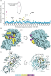
Analysis of substrate induced structural rearrangements of furin. (A) Overall structure of human unliganded furin (catalytic and the P-domains are in gold and blue, respectively). The catalytic residues (cyan stick model), the substrate binding pockets (labeled S1–S5), and bound ions (purple sphere, Na+; green spheres, Ca2+) are indicated. (B) The Cα-displacement values (intervals <0.3 Å, 0.3–0.8 Å, and >0.8 Å are highlighted in cyan, yellow, and magenta, respectively) were mapped to the molecular surface.
Fig. S1.
Fig. S1.
Structural analysis of furin in its unliganded vs. its MI-52–bound states. (A) Plot of the Cα-displacement values of the Cα atoms covering all amino acids of furin (intervals <0.3 Å, 0.3–0.8 Å, and >0.8 Å are highlighted in cyan, yellow, and magenta, respectively). (B) Mapping of the respective values to the molecular surface and colored according to A. (C) Cartoon representation colored according to A. Bound ions are shown as light gray (Na+) and dark gray (Ca2+) spheres.
Fig. S2.
Fig. S2.
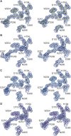
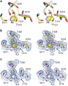
Electron-density maps observed for the active-site residues and the alignment template of human furin. Stereo panels show the structures in stick representation. The 2FoFc simulated annealing composite-omit electron density maps are given as blue-colored mesh, which is contoured at 1.0 σ. (A) Unliganded furin. (B) Furin complexed with MI-52. (C) Unliganded furin in presence of EDTA. (D) Furin in presence of EDTA and complexed with MI-52.
Fig. 2.
Fig. 2.
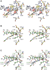
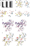
Structural comparison of substrates binding to the active sites of furin and to subtilisin Carlsberg. (A) Structural alignment of selected residues of unliganded furin (yellow carbons) and inhibitor-bound furin (gray carbons; inhibitor: ball-and-stick). Steric clashes between bound inhibitor/substrate and unliganded furin are highlighted as red line patterns. (B) Structural alignment of unliganded subtilisin (orange carbons) and inhibitor-bound subtilisin (green carbons). (C) Structural alignment of inhibitor-bound subtilisin (green carbons) and furin (gray-colored stick model). Important interactions are always highlighted by dashes.
Fig. S3.
Fig. S3.
Stereo representation of the structural comparison of substrate binding by furin and subtilisin Carlsberg. Detailed view of the alignment template and the active-site residues of inhibitor-bound furin (protein: gray-colored stick model; inhibitor: gray-colored ball-and-stick model), unliganded furin (yellow-colored stick model), unliganded subtilisin (light orange-colored stick model), and eglin-c (shown P5–P1 segment Ser-Pro-Val-Thr-Leu) bound subtilisin (protein: light green-colored stick model; inhibitor: dark green-colored ball-and-stick model). (A) Structural alignment of selected residues of unliganded furin and inhibitor-bound furin. Steric clashes between bound inhibitor/substrate and unliganded furin are highlighted as red line patterns around the respective atoms. (B) Structural alignment of unliganded subtilisin and inhibitor-bound subtilisin. Important interactions of the active-site residues and between the alignment template and the inhibitor are highlighted as gray and yellow dashes, respectively. (C) Structural alignment of inhibitor-bound subtilisin and furin. Important homologous amino acids of the proteases are labeled in green (subtilisin) and gray (furin).
Fig. 3.
Fig. 3.
PCA. (A) Projections of furin (squares) and subtilisin Carlsberg (triangles) on the first two principal components. Only human furin structures (green) were used to compute the covariance matrix. Murine furin structures (blue) are shown for comparison only. Open and closed markers represent unliganded and inhibitor-bound structures, respectively. (Inset) The proportion of the structural variation encoded within each principal component. (B) Histograms of MD-simulation frames projected on the first principal component for each simulated system: furin unliganded state (red), furin inhibitor-bound (blue), furin inhibitor-removed (green), subtilisin unliganded state (black), substilisin inhibitor-bound (cyan), and subsitilisn inhibitor-removed (magenta). Projections of the crystallographic structures are shown on the x axis as in A. (Inset) Examples of MD trajectories.
Fig. S4.
Fig. S4.
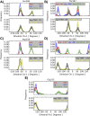
Rotameric states from MD simulations. Histograms are computed from five independent runs of each simulated system: unliganded furin (red), furin with inhibitor MI-52 (blue), unliganded furin in the presence of EDTA (green), unliganded furin with Thr367’s χ1 initially set at 60° (black), unliganded furin in the presence of EDTA and with Asp258 protonated (yellow). For residues Ser368 (A), Thr367 (B), Asp258 (C), and Asn295 (D) the measured quantity is the χ1 dihedral-angle, whereas for Gly255 (E) it is the ψ dihedral-angle. The information is split into two panels for clarity. The Upper panel contains plots from the simulations of unliganded furin, furin with inhibitor, and unliganded furin in the presence of EDTA structures; the Lower panel contains plots corresponding to simulations of unliganded furin with Thr367's χ1 initially set at 60° and unliganded furin in the presence of EDTA with Asp258 protonated. Each curve represents an average histogram (solid lines) with 1 SD as upper limit (error bar). The averages were computed from five independent runs. The values in each inset correspond to the values found in the crystallographic structures.
Fig. S5.
Fig. S5.
Substrate-induced conformational changes of the sodium site in furin. Stereo panels show a superposition of selected residues of unliganded furin (yellow, protein: stick model; nonbonded atoms: spheres) and inhibitor-bound furin (gray, protein: stick model; inhibitor: ball-and-stick; nonbonded atoms: spheres). (A) Superposition of the unliganded inhibitor-bound states. (B and C) Electron-density maps observed for the sodium binding site. Sodium ions and water molecules are given as big and small spheres, respectively. The 2FoFc simulated annealing composite-omit electron-density maps are given as blue-colored mesh and are contoured at 1.0 σ. (B) Unliganded furin. (C) Furin complexed with MI-52.
Fig. S6.
Fig. S6.
Binding of calcium to furin and its structural effects. (A) Bar diagram of the anomalous signal observed for the calcium binding sites at high (1 mM) and low (formally 1 nM, but see SI Materials and Methods for details) Ca2+ concentrations as well as in the presence of EDTA. (B–D) The coordinating amino acids are given for Ca binding sites Ca-I (B), Ca-II (C), and Ca-III (D) as stick model (yellow carbons correspond to high calcium concentration, cyan carbons correspond to the presence of EDTA) together with selected water molecules (red spheres). The Ca2+ ions (green spheres) are shown together with the anomalous electron-density map (countoured at 6.0 σ and given as orange mesh) at high calcium concentration or in presence of EDTA. At low Ca2+-concentration, Ca2+-(III) is displaced by Na+ (purple sphere). (E) Stereo representation of the superposition of EDTA and MI-52 soaked furin (purple carbons, purple inhibitor) with non-EDTA–treated furin (gray). Selected hydrogen bonds are highlighted as dashes according to Fig. 2. Coordinative bonds of Ca2+-(II) are highlighted as gray dashes. Calcium ions and water molecules are represented by large and small spheres, respectively (coloring according to the carbon atoms of the respective structures). (F) Superposition of unliganded furin (yellow) and additionally EDTA-treated unliganded furin (cyan). Basically, identical structural alterations as shown in Fig. 2A are observed. Steric clashes with the superposed inhibitor (according to Fig. 2A and Fig. S4) and with N295 in its inhibitor-bound conformation are highlighted as red line pattern. Coordinative bonds of Ca2+-(II) are highlighted as yellow dashes. Calcium ions and water molecules are represented by large and small spheres, respectively (coloring according to the carbon atoms of the respective structures).
Fig. S7.
Fig. S7.
Rmsd curves for the simulated systems. In the panels are shown the average rmsd curves (solid lines) with 1 SD as upper and lower limits (error bars). The averages were computed out of five and four independent runs for furin and substilisin systems, respectively. In all cases, the starting protein-fold is preserved.
Fig. 4.
Fig. 4.
Schematic energy landscapes according to different scenarios. Furin, in an inhibitor-free condition (red solid line), resides most of the time at the bottom of an energy well adopting an off-state conformation, whereas its catalytically active on state is not energetically stable. In the presence of an inhibitor (blue solid line), the energy landscape is modified and the furin–ligand interaction stabilizes the on-state conformation. Furin and the ligand are represented as continuous gray/red and cyan surfaces, respectively. Black dashed lines represent the corresponding energy states.

Similar articles

Cited by

References

    1. Thomas G. Furin at the cutting edge: From protein traffic to embryogenesis and disease. Nat Rev Mol Cell Biol. 2002;3(10):753–766. - PMC - PubMed
    1. Artenstein AW, Opal SM. Proprotein convertases in health and disease. N Engl J Med. 2011;365(26):2507–2518. - PubMed
    1. Seidah NG, Prat A. The biology and therapeutic targeting of the proprotein convertases. Nat Rev Drug Discov. 2012;11(5):367–383. - PubMed
    1. Becker GL, et al. Highly potent inhibitors of proprotein convertase furin as potential drugs for treatment of infectious diseases. J Biol Chem. 2012;287(26):21992–22003. - PMC - PubMed
    1. Garten W, et al. Processing of viral glycoproteins by the subtilisin-like endoprotease furin and its inhibition by specific peptidylchloroalkylketones. Biochimie. 1994;76(3-4):217–225. - PubMed