. 2016 Nov 18;84(12):3388-3398.
doi: 10.1128/IAI.00732-16. Print 2016 Dec.

Thermal Unfolding of the Pertussis Toxin S1 Subunit Facilitates Toxin Translocation to the Cytosol by the Mechanism of Endoplasmic Reticulum-Associated Degradation

Affiliations

Thermal Unfolding of the Pertussis Toxin S1 Subunit Facilitates Toxin Translocation to the Cytosol by the Mechanism of Endoplasmic Reticulum-Associated Degradation

Tuhina Banerjee et al. Infect Immun. .

Abstract

Pertussis toxin (PT) moves from the host cell surface to the endoplasmic reticulum (ER) by retrograde vesicular transport. The catalytic PTS1 subunit dissociates from the rest of the toxin in the ER and then shifts to a disordered conformation which may trigger its export to the cytosol through the quality control mechanism of ER-associated degradation (ERAD). Functional roles for toxin instability and ERAD in PTS1 translocation have not been established. We addressed these issues with the use of a surface plasmon resonance system to quantify the cytosolic pool of PTS1 from intoxicated cells. Only 3% of surface-associated PTS1 reached the host cytosol after 3 h of toxin exposure. This represented, on average, 38,000 molecules of cytosolic PTS1 per cell. Cells treated with a proteasome inhibitor contained larger quantities of cytosolic PTS1. Stabilization of the dissociated PTS1 subunit with chemical chaperones inhibited toxin export to the cytosol and blocked PT intoxication. ERAD-defective cell lines likewise exhibited reduced quantities of cytosolic PTS1 and PT resistance. These observations identify the unfolding of dissociated PTS1 as a trigger for its ERAD-mediated translocation to the cytosol.


Figures

FIG 1
FIG 1
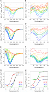
Chemical chaperones inhibit the thermal unfolding of PTS1. The temperature-induced unfolding of PTS1 was monitored by far-UV CD (A, C, E, and G) and near-UV CD (B, D, F, and H). Both measurements were conducted nearly simultaneously on the same sample. The change in color from blue to red denotes a change in temperature from 20°C to 50°C for untreated PTS1 (A and B), PTS1 treated with 10% glycerol (C and D), or PTS1 treated with 100 μM PBA (E and F). (G and H) Thermal unfolding profiles for untreated PTS1 (red), glycerol-treated PTS1 (blue), and PBA-treated PTS1 (green) derived from the data presented in panels A to F. Tm values for each condition are presented in the corresponding color. For far-UV CD analysis, the mean residue molar ellipticities at 220 nm ([θ]220) were plotted as a function of temperature. For near-UV CD, the mean residue molar ellipticities at 280 nm ([θ]280) were plotted as a function of temperature.
FIG 2
FIG 2
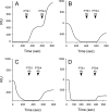
PBA and glycerol do not inhibit disassembly of the PT holotoxin. PT was appended to a GD1a-coated SPR sensor, and a baseline measurement corresponding to the mass of the sensor-bound holotoxin was set as 0 RIU. The experiment was then initiated by adding various reagents to the perfusion buffer at 0 s. The reagents were removed after ∼200 s and replaced with sequential perfusions of PTS1 and PTS4 antibodies (Ab), as indicated by the arrowheads. (A) Addition of ATP; (B) addition of ATP and CHAPS; (C) addition of ATP, CHAPS, and PBA; (D) addition of ATP, CHAPS, and 10% glycerol.
FIG 3
FIG 3
PTS1 but not PTS4 can be detected in the cytosol and extracellular medium of intoxicated cells. (A) CHO cells treated with digitonin to selectively permeabilize the plasma membrane were separated into intact membrane (pellet [P]) and cytosolic (supernatant [S]) fractions by centrifugation. To demonstrate the fidelity of the fractionation procedure, Western blot analysis was used to track the distributions of PDI, a soluble ER resident protein, and Hsp90, a cytosolic protein, in both fractions. (B and C) CHO cells pulse-loaded at 4°C with 1 μg/ml of PT were chased for 3 h at 37°C in toxin-free medium. Membrane pellet and cytosolic supernatant fractions from digitonin-permeabilized cells were collected at the end of the pulse and the end of the chase. Both fractions were perfused over SPR sensors coated with anti-PTS1 (B) or anti-PTS4 (C) antibodies. The pellet fraction was solubilized in 1% Triton X-100 before perfusion. Unintoxicated cells and cells intoxicated in the presence of BfA were used as negative controls. Ligand was removed from the perfusion buffer after 200 s. (D and E) CHO cells pulse-loaded at 4°C with 1 μg/ml of PT were chased for 3 h at 37°C in toxin-free medium. The extracellular medium collected at the end of the pulse and the end of the chase was perfused over SPR sensors coated with anti-PTS1 (D) or anti-PTS4 (E) antibody. Unintoxicated cells and cells intoxicated in the presence of BfA were used as negative controls. Ligand was removed from the perfusion buffer after 200 s. Each individual condition tested in panels B to E was derived from the same cell population. Data are presented in multiple panels due to the use of different antibodies and different signal strengths for intracellular versus extracellular PTS1.
FIG 4
FIG 4
Chemical chaperones inhibit PTS1 delivery to the cytosol and PT intoxication. (A and B) CHO cells pulse-loaded at 4°C with PT were chased for 3 h at 37°C in toxin-free medium containing 10% glycerol (A) or 100 μM PBA (B). Intoxicated cells chased in the absence of drug treatment were used as positive controls, while unintoxicated cells and cells intoxicated in the presence of BfA were used as negative controls. To detect the translocated toxin, cytosolic fractions from digitonin-permeabilized cells were perfused over an SPR slide coated with an anti-PTS1 antibody. PTS1 standards were perfused over the sensor as additional controls; only the 1-ng and 0.1-ng controls are shown (for scaling purposes). Ligand was removed from the perfusion buffer after 200 s. (C) CHO cells pulse-loaded at 4°C with PT were chased for 3 h at 37°C in toxin-free medium containing 10% glycerol or 100 μM PBA. Unintoxicated cells, intoxicated cells chased in the absence of drug treatment, and cells intoxicated in the presence of BfA were used as controls. Cell extracts generated by detergent lysis were incubated at 25°C for 1 h with purified PTS1 and biotin-NAD, the donor molecule for the ADP-ribosylation reaction. Since Giα can only be ADP-ribosylated by PTS1 at one site, its in vivo modification due to productive intoxication will prevent subsequent in vitro modification with biotin-labeled ADP-ribose. Western blot analysis was used to detect biotin-labeled Giα or the GAPDH loading control. Lane 1, intoxicated cells; lane 2, unintoxicated cells; lane 3, cells intoxicated in the presence of BfA; lane 4, cells intoxicated in the presence of glycerol; lane 5, cells intoxicated in the presence of PBA. (D) Signals for biotin-labeled Giα were normalized to those for the GAPDH loading control and then expressed as percentages of the values from the unintoxicated control cells. The averages ± standard deviations of results from 3 to 5 independent experiments per condition are shown.
FIG 5
FIG 5
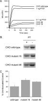
Attenuated ERAD activity inhibits PTS1 translocation and PT intoxication. (A) Wild-type CHO cells and two mutant CHO cell lines with attenuated ERAD-mediated translocation to the cytosol (CHO 16 and CHO 46) were incubated with PT at 4°C for 30 min. Unbound toxin was removed, and the cells were chased for 3 h at 37°C in toxin-free medium. Cytosolic fractions from digitonin-permeabilized cells and known quantities of PTS1 standards were then perfused over an SPR slide coated with an anti-PTS1 antibody. (B and C) Wild-type and mutant CHO cells incubated at 4°C for 30 min in the absence or presence of PT were chased for 3 h at 37°C in toxin-free medium. Cell extracts generated by detergent lysis were incubated at 25°C for 1 h with purified PTS1 and biotin-NAD, the donor molecule for the ADP-ribosylation reaction. (B) Western blot analysis was used to detect biotin-labeled Giα (lower band) or the actin loading control (upper band). (C) Signals for biotin-labeled Giα were normalized to the actin loading control and then expressed as percentages of the values from the corresponding unintoxicated control cells. The averages ± ranges for results from 2 to 4 independent experiments per condition are shown.

Similar articles

Cited by

References

    1. Spooner RA, Smith DC, Easton AJ, Roberts LM, Lord JM. 2006. Retrograde transport pathways utilised by viruses and protein toxins. Virol J 3:26. doi:10.1186/1743-422X-3-26. - DOI - PMC - PubMed
    1. Teter K. 2013. Toxin instability and its role in toxin translocation from the endoplasmic reticulum to the cytosol. Biomolecules 3:997–1029. doi:10.3390/biom3040997. - DOI - PMC - PubMed
    1. Brodsky JL, Skach WR. 2011. Protein folding and quality control in the endoplasmic reticulum: recent lessons from yeast and mammalian cell systems. Curr Opin Cell Biol 23:464–475. doi:10.1016/j.ceb.2011.05.004. - DOI - PMC - PubMed
    1. Hazes B, Read RJ. 1997. Accumulating evidence suggests that several AB-toxins subvert the endoplasmic reticulum-associated protein degradation pathway to enter target cells. Biochemistry 36:11051–11054. doi:10.1021/bi971383p. - DOI - PubMed
    1. Deeks ED, Cook JP, Day PJ, Smith DC, Roberts LM, Lord JM. 2002. The low lysine content of ricin A chain reduces the risk of proteolytic degradation after translocation from the endoplasmic reticulum to the cytosol. Biochemistry 41:3405–3413. doi:10.1021/bi011580v. - DOI - PubMed

LinkOut - more resources