. 2016 Aug 18;1(13):e87023.
doi: 10.1172/jci.insight.87023.

ADAM17 substrate release in proximal tubule drives kidney fibrosis

Affiliations

ADAM17 substrate release in proximal tubule drives kidney fibrosis

Eirini Kefaloyianni et al. JCI Insight. .

Abstract

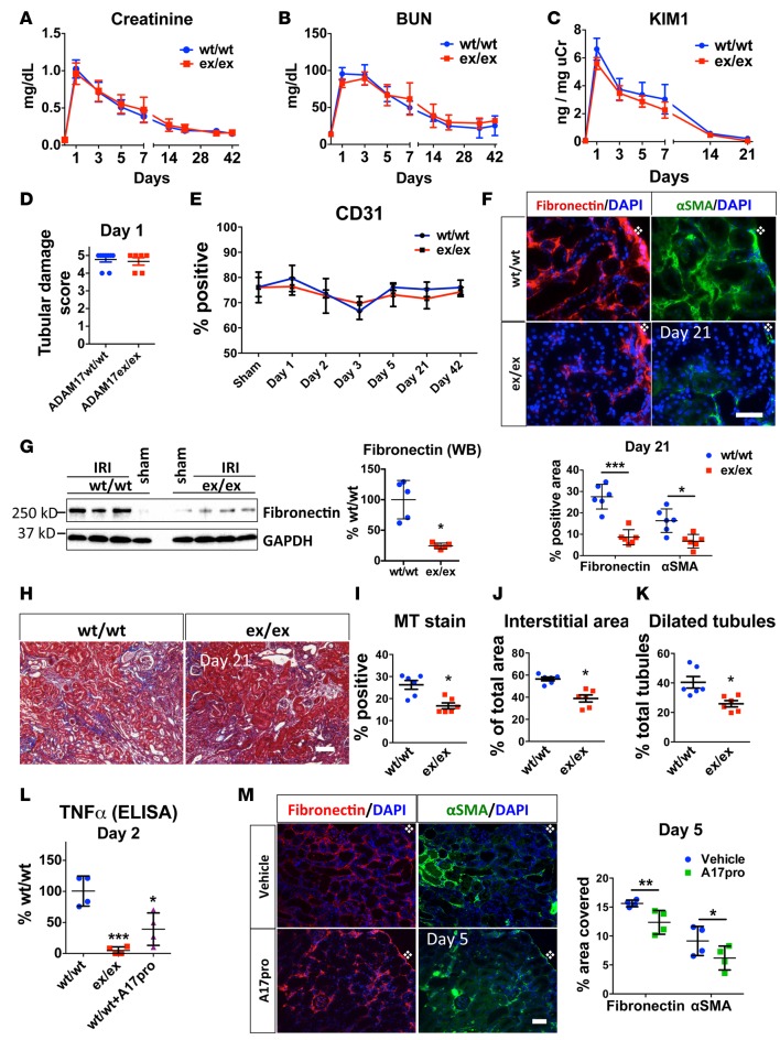
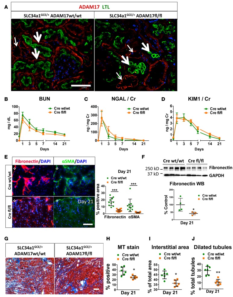
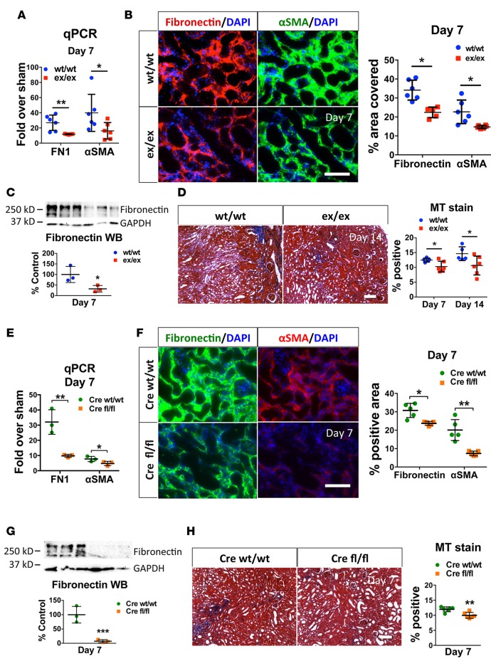
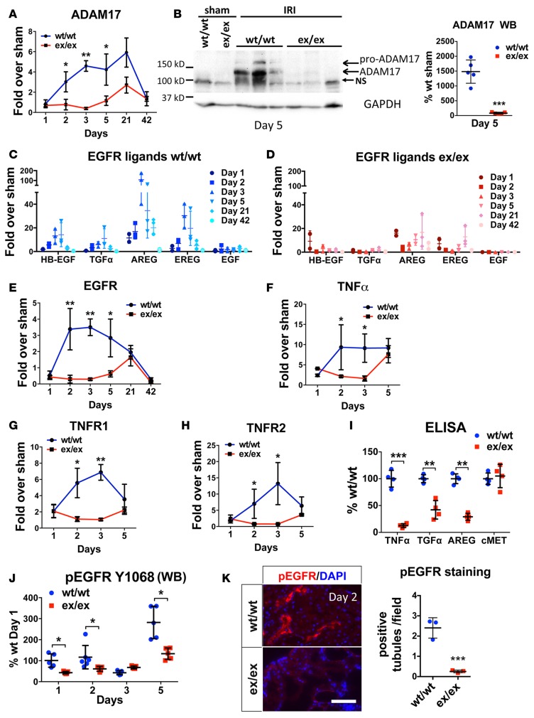
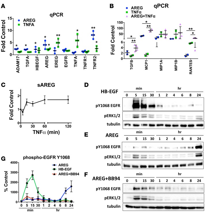
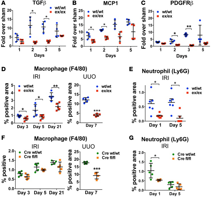
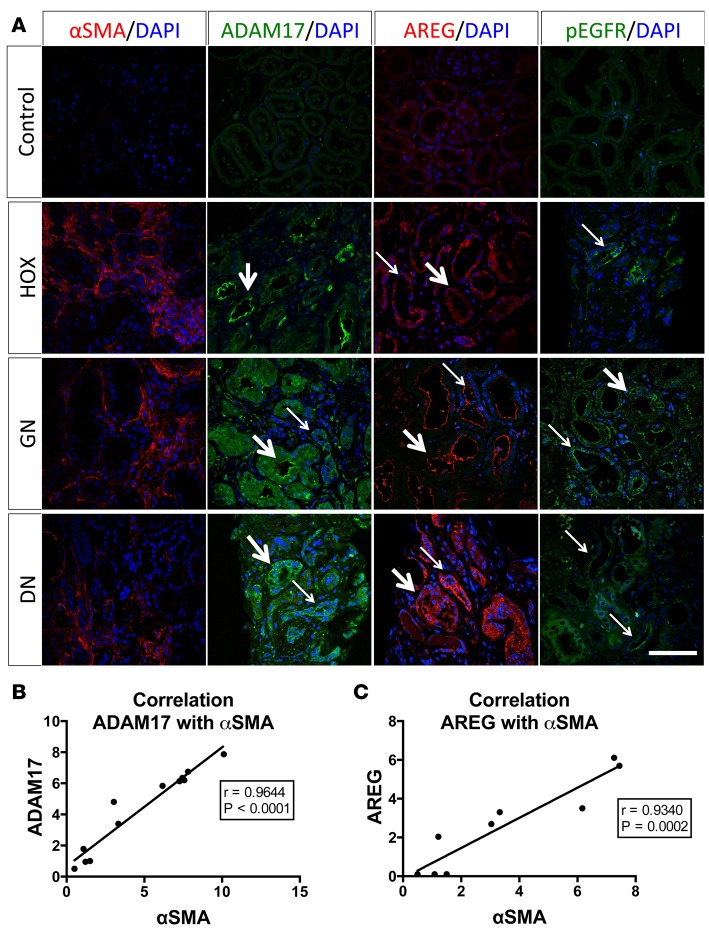
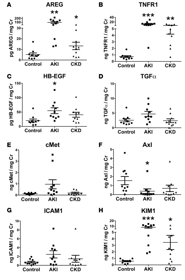
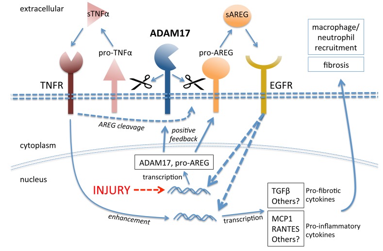
Kidney fibrosis following kidney injury is an unresolved health problem and causes significant morbidity and mortality worldwide. In a study into its molecular mechanism, we identified essential causative features. Acute or chronic kidney injury causes sustained elevation of a disintegrin and metalloprotease 17 (ADAM17); of its cleavage-activated proligand substrates, in particular of pro-TNFα and the EGFR ligand amphiregulin (pro-AREG); and of the substrates' receptors. As a consequence, EGFR is persistently activated and triggers the synthesis and release of proinflammatory and profibrotic factors, resulting in macrophage/neutrophil ingress and fibrosis. ADAM17 hypomorphic mice, specific ADAM17 inhibitor-treated WT mice, or mice with inducible KO of ADAM17 in proximal tubule (Slc34a1-Cre) were significantly protected against these effects. In vitro, in proximal tubule cells, we show that AREG has unique profibrotic actions that are potentiated by TNFα-induced AREG cleavage. In vivo, in acute kidney injury (AKI) and chronic kidney disease (CKD, fibrosis) patients, soluble AREG is indeed highly upregulated in human urine, and both ADAM17 and AREG expression show strong positive correlation with fibrosis markers in related kidney biopsies. Our results indicate that targeting of the ADAM17 pathway represents a therapeutic target for human kidney fibrosis.


Conflict of interest statement

The authors have declared that no conflict of interest exists.

Figures

Figure 1
Figure 1. Global targeting of ADAM17 results in similar initial IRI-induced injury but in reduced IRI-induced fibrosis.
ADAM17ex/ex mice and their WT littermates were subjected to bilateral ischemia for 30 minutes followed by reperfusion. (A and B) Time-course of changes in serum creatinine and BUN (n = 6–15 per time point). (C) Time-course of urinary KIM1 levels over time measured by ELISA (n = 5–10 per time point). (D) Tubular damage was scored in kidney cortex at day 1 after ischemia (n = 6–7). (E) Time course of cortical CD31+ area quantified after immunostaining (n = 4). (F) The induction of fibronectin and αSMA in kidney cortex was examined by immunostaining 21 days after injury (top: representative images; bottom: quantification, n = 6; scale bar: 50 μm). (G) Fibronectin protein expression at day 21 after injury was examined by Western blot (left: sample blots; right: quantification; GAPDH was used as loading control; n = 5). (H and I) Masson’s trichrome staining at day 21 after injury in kidney cortex (H: representative images; I: quantification; n = 6; scale bar: 100 μm). (J) Total interstitial area (n = 6) and (K) dilated tubules were quantified at day 21 after injury (n = 6). (L) Soluble TNFα was measured by ELISA in control (WT/WT), hypomorphic (ex/ex), and WT mice injected with recombinant ADAM17 prodomain (WT/WT+A17pro) 2 days after injury (n = 4). (M) The induction of fibronectin and αSMA was examined in kidney cortex by immunostaining in vehicle or ADAM17 prodomain inhibitor–injected WT mice 5 days after injury (left: representative images, right: quantification; n = 4; scale bar: 50 μm). *P < 0.05; **P < 0.01; ***P < 0.001 as determined by an unpaired 2-tailed Student’s t test. Diamond symbol denotes position of kidney capsule. ADAM17, a disintegrin and metalloprotease 17; BUN, blood urea nitrogen; IRI, ischemia reperfusion injury; KIM1, kidney injury molecule 1; MT, Masson’s trichrome; αSMA, alpha smooth muscle actin; TNFα, tumor necrosis factor alpha; WB, Western blot.
Figure 2
Figure 2. Proximal tubular-specific ADAM17 KO confers protection against fibrosis after IRI.
Control (SLC34a1GCE/+ ADAM17WT/WT, Cre WT/WT) and ADAM17 PTC-KO (SLC34a1GCE/+ ADAM17fl/fl, Cre fl/fl) mice were subjected to bilateral ischemia for 29 minutes followed by reperfusion. (A) KO specificity was examined in healthy kidneys (left: control, right: PTC-KO mouse) by immunostaining of ADAM17 (red color) together with LTL staining (green color) as a proximal tubular marker (thick arrows, LTL-positive tubules; thin arrows, LTL-negative tubules; scale bar: 50 μm). (BD) Time-course of BUN, urinary NGAL, and urinary KIM1 (n = 6–15). (E) The induction of fibronectin and αSMA was examined in kidney cortex by immunostaining at day 21 (left: representative images, right: quantification, n = 10; scale bar: 50 μm). (F) Fibronectin protein expression at day 21 after injury was examined by Western blot (top: sample blots; bottom: quantification; GAPDH was used as loading control; n = 4). (G and H) Masson’s trichrome staining in kidney cortex at day 21 after injury (G: representative images; H: quantification; n = 6; scale bar: 100 μm). (I) Total interstitial area (n = 6) and (J) dilated tubules (n = 6) were quantified in kidney cortex at day 21 after injury. *P < 0.05; **P < 0.01; ***P < 0.001 as determined by an unpaired 2-tailed Student’s t test. Diamond symbol denotes position of kidney capsule. ADAM17, a disintegrin and metalloprotease 17; BUN, blood urea nitrogen; IRI, ischemia reperfusion injury; KIM1, kidney injury molecule 1; LTL, Lotus Tetragonolobus lectin; MT, Masson’s trichrome; NGAL, neutrophil gelatinase–associated lipocalin; PTC-KO, proximal tubule cell KO; αSMA, alpha smooth muscle actin; WB, Western blot.
Figure 3
Figure 3. ADAM17ex/ex and ADAM17 PTC-KO mice are protected from UUO-induced fibrosis.
Mice were subjected to UUO, and their kidneys were examined 7 or 14 days after ureteral ligation, as noted. (A) qPCR analysis of fibronectin or αSMA mRNA expression levels in whole kidneys of ex/ex or WT/WT mice at day 7, expressed as fold over respective sham-injured mice (n = 6). (B) The induction of fibronectin and αSMA in the injured kidney cortex of UUO-subjected WT/WT or ex/ex mice was examined by immunostaining (left: representative images; right: quantification; n = 6; scale bar: 50 μm). (C) Fibronectin protein expression at day 7 after ligation was examined by Western blot (top: sample blots; bottom: quantification; GAPDH was used as loading control; n = 3). (D) Masson’s trichrome staining in kidney cortex at day 7 or day 14 after ligation (left: representative images day 14; right: quantification; n = 5–6; scale bar: 100 μm). (E) qPCR analysis of fibronectin or αSMA mRNA expression levels in whole kidneys of UUO-subjected control (Cre WT/WT) or ADAM17 PTC-KO (Cre fl/fl) at day 7, expressed as fold over respective sham-injured mice (n = 3). (F) The induction of fibronectin and αSMA in the injured kidney cortex of UUO-subjected control (Cre WT/WT) or ADAM17 PTC-KO (Cre fl/fl) mice was examined at day 7 by immunostaining (left: representative images, right: quantification; n = 5; scale bar: 50 μm). (G) Fibronectin protein expression at day 7 after ligation was examined by Western blot (top: sample blots; bottom: quantification; GAPDH was used as loading control; n = 3). (H) Masson’s trichrome staining in kidney cortex at day 7 after ligation in control (Cre WT/WT) or ADAM17 PTC-KO (Cre fl/fl) mice (left: representative images; right: quantification; n = 6; scale bar: 100 μm). *P < 0.05; **P < 0.01; ***P < 0.001 as determined by an unpaired 2-tailed Student’s t test. ADAM17; a disintegrin and metalloprotease 17; FN1, fibronectin; MT, Masson’s trichrome; PTC-KO, proximal tubule cell KO; αSMA, alpha smooth muscle actin; UUO, unilateral ureteral obstruction; WB, Western blot.
Figure 4
Figure 4. IRI-induced ADAM17 pathway components are reduced in ADAM17ex/ex mice.
The expression levels of ADAM17 and its pathway components were examined at different time points after ischemic injury in whole kidneys. (A) qPCR analysis of ADAM17 mRNA expression levels in ex/ex or WT/WT mice, expressed as fold over respective sham-injured mice (n = 3–5). (B) Western blot analysis of ADAM17 protein levels in ex/ex or WT/WT mice subjected to sham or IRI (day 5 after ischemia) (left: sample blots; right: quantification; GAPDH was used as loading control; n = 5). (CH) qPCR analysis of EGFR ligands, EGFR, TNFα, and TNFR1/2 mRNA expression levels in ex/ex or WT/WT mice, expressed as fold over respective sham-injured mice (time points as indicated; n = 3–6). (I) Soluble ectodomains of the ADAM17 substrates TNFα, TGFα, and AREG were measured in whole kidney lysates of WT/WT or ex/ex mice at day 2 after ischemia by ELISA (ADAM10 substrate cMET is used as control; n = 4). (J) Time-course of EGFR phosphorylation (Y1068) as measured by Western blot in whole kidney lysates of WT/WT or ex/ex mice (n = 5). (K) Representative images of EGFR phosphorylation (Y1173) examined in kidney cortex by immunostaining in WT/WT or ex/ex mice at day 2 after injury (left: representative images; right: quantification; n = 3; scale bar: 50 μm). *P < 0.05; **P < 0.01; ***P < 0.001 as determined by an unpaired 2-tailed Student’s t test. ADAM17; a disintegrin and metalloprotease 17; EGFR, epidermal growth factor receptor; IRI, ischemia reperfusion injury; NS, nonspecific band; TNFα, tumor necrosis factor α; TNFR, tumor necrosis factor receptor; AREG, amphiregulin; ADAM10, a disintegrin and metalloprotease 10; cMet, met proto-oncogene; WB, Western blot.
Figure 5
Figure 5. Amphiregulin (AREG) and TNFα crosstalk causes sustained EGFR activation and strongly enhanced cytokine production in human proximal tubular cells.
(A and B) qPCR analysis of ADAM17 pathway components (A) and proinflammatory/profibrotic cytokines (B) in human proximal tubular cells treated with AREG, TNFα, or both (AREG+TNFα) for 24 hours (n = 4). (C) Time course of TNFα-induced AREG cleavage producing soluble AREG (sAREG) measured by ELISA in HPTC culture medium (n = 3). (DF) Time course of HB-EGF–induced (D) or AREG-induced (E and F) Y1068-EGFR and ERK1/2 phosphorylation (analyzed by Western blot) in the absence (D and E) or presence (F) of the metalloprotease inhibitor BB94. Tubulin is used as loading control for quantification (G; n = 3). *P < 0.05; **P < 0.01 as determined by an unpaired 2-tailed Student’s t test. ADAM17, a disintegrin and metalloprotease 17; AREG, amphiregulin; BB94, batimastat; EGF, epidermal growth factor; EGFR, epidermal growth factor receptor; EREG, epiregulin; ERK, extracellular regulated mitogen activated protein kinase; HB-EGF, heparin binding epidermal growth factor-like growth factor; HPTC, human proximal tubule cells; MCP1, monocyte chemoattractant protein 1; MIP1A, macrophage inflammatory protein 1 alpha; MIP1B, macrophage inflammatory protein 1 beta; RANTES, regulated on activation normal T cell expressed and secreted; TNFα, tumor necrosis factor alpha; TNFR, tumor necrosis factor receptor.
Figure 6
Figure 6. Injury-induced cytokine expression and kidney ingress of macrophages and neutrophils is reduced in vivo in ADAM17ex/ex and ADAM17 PTC-KO mice.
(AC) The mRNA expression of TGFβ, MCP1, and PDGFRβ was examined by qPCR in whole kidney lysates of WT/WT or ex/ex mice at different time points after ischemia (n = 4). (DG) Macrophage and neutrophil infiltration in the kidney of ADAM17ex/ex mice (D and E; n = 5–6) or PTC-KO (Cre fl/fl) mice (F and G; n = 4–5) and their WT/WT littermates was examined by immunostaining at different time points after ischemia or 7 days after UUO (n = 5). *P < 0.05; **P < 0.01;***P < 0.001 as determined by an unpaired 2-tailed Student’s t test. ADAM17, a disintegrin and metalloprotease 17; IRI, ischemia reperfusion injury; MCP1, monocyte chemoattractant protein 1; PDGFRβ, platelet derived growth factor receptor beta; PTC-KO, proximal tubule cell KO; UUO, unilateral ureteral obstruction.
Figure 7
Figure 7. Induction of ADAM17 and AREG protein expression and EGFR phosphorylation in human kidney disease.
(A) The presence of the fibrotic marker αSMA and of ADAM17, AREG, and phospho-EGFR were examined in human kidney biopsies by immunostaining. Representative images of hyperoxaluria (HOX; intrarenal tubular obstruction by crystals not shown in image), glomerulonephritis (GN), and diabetic nephropathy (DN) cases are shown (4 controls, 4 HOX, 4 GN, and 4 diabetic patients were examined; representative images are shown) (thick arrows: positive staining in proximal tubular-like structures; thin arrows: positive staining in distal tubular-like structures; scale bar: 50 μm). (B and C) Correlation of ADAM17 (B) or AREG (C) protein expression levels with αSMA levels across all patients studied (n = 9–12). The Pearson correlation coefficient (R) and P value are provided in each graph. ADAM17; a disintegrin and metalloprotease 17; AREG, amphiregulin; EGFR, epidermal growth factor receptor; αSMA, alpha smooth muscle actin.
Figure 8
Figure 8. Soluble AREG and TNFR1 are significantly increased in the urine of AKI and CKD patients.
The urinary levels of the soluble ADAM17 substrates (AREG [A], TNFR1 [B], HB-EGF [C], TGFα [D], Axl [F], ICAM1 [G], and KIM1 [H]) and of the ADAM10 substrate cMET (E) were measured by ELISA in control, AKI, and CKD patients (n = 10; 50% of CKD patients were diabetics; patient demographics and details in Supplemental Table 1). *P < 0.05; **P < 0.01; ***P < 0.001 as determined by an unpaired 2-tailed Student’s t test. AREG, amphiregulin; TNFR, tumor necrosis factor receptor; AKI, acute kidney injury; CKD, chronic kidney disease; ADAM17, a disintegrin and metalloprotease 17; HB-EGF, heparin binding epidermal growth factor-like growth factor; ICAM1, intercellular adhesion molecule 1; KIM1, kidney injury molecule 1; cMET, met.
Figure 9
Figure 9. Mechanisms of injury-induced ADAM17-dependent sustained EGFR activation in proximal tubule cells.
Injury drives upregulation of ADAM17 and of its substrates, in particular pro-AREG. Increased ADAM17 activity on the cell surface leads to enhanced release of soluble AREG and TNFα, which activate their respective receptors. In a positive feedback loop, AREG-mediated EGFR activation increases ADAM17 and AREG expression. TNFα-mediated TNFR activation strengthens this feedback loop further by enhancing soluble AREG release (pathway crosstalk). Both pathways, AREG/EGFR and TNFα/TNFR, potentiate each other in inducing profibrotic and proinflammatory cytokines that drive kidney fibrosis. pro-TNFα, transmembrane pro–tumor necrosis factor α; sTNFα, soluble tumor necrosis factor α; ADAM17, a disintegrin and metalloprotease 17; EGFR, epidermal growth factor receptor; pro-AREG, proamphiregulin; sAREG, soluble amphiregulin; TNFR, tumor necrosis factor receptor; MCP1, monocyte chemoattractant protein 1.

Similar articles

Cited by

References

    1. Parr SK, Siew ED. Delayed consequences of acute kidney injury. Adv Chronic Kidney Dis. 2016;23(3):186–194. doi: 10.1053/j.ackd.2016.01.014. - DOI - PMC - PubMed
    1. Rewa O, Bagshaw SM. Acute kidney injury-epidemiology, outcomes and economics. Nat Rev Nephrol. 2014;10(4):193–207. doi: 10.1038/nrneph.2013.282. - DOI - PubMed
    1. Declèves AE, Sharma K. Novel targets of antifibrotic and anti-inflammatory treatment in CKD. Nat Rev Nephrol. 2014;10(5):257–267. doi: 10.1038/nrneph.2014.31. - DOI - PMC - PubMed
    1. Bonventre JV, Yang L. Cellular pathophysiology of ischemic acute kidney injury. J Clin Invest. 2011;121(11):4210–4221. doi: 10.1172/JCI45161. - DOI - PMC - PubMed
    1. Grgic I, et al. Targeted proximal tubule injury triggers interstitial fibrosis and glomerulosclerosis. Kidney Int. 2012;82(2):172–183. doi: 10.1038/ki.2012.20. - DOI - PMC - PubMed

Publication types