Review
. 2016 Oct;16(10):635-49.
doi: 10.1038/nrc.2016.77. Epub 2016 Sep 16.

Reprogramming glucose metabolism in cancer: can it be exploited for cancer therapy?

Affiliations
Review

Reprogramming glucose metabolism in cancer: can it be exploited for cancer therapy?

Nissim Hay. Nat Rev Cancer. 2016 Oct.

Abstract

In recent years there has been a growing interest among cancer biologists in cancer metabolism. This Review summarizes past and recent advances in our understanding of the reprogramming of glucose metabolism in cancer cells, which is mediated by oncogenic drivers and by the undifferentiated character of cancer cells. The reprogrammed glucose metabolism in cancer cells is required to fulfil anabolic demands. This Review discusses the possibility of exploiting the reprogrammed glucose metabolism for therapeutic approaches that selectively target cancer cells.


Conflict of interest statement

Competing interests statement

The author declares no competing interests.

Figures

Figure 1
Figure 1. Changes that occur in glucose metabolism of cancer cells
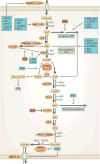
Compared with normal cells (left), the flux of glucose metabolism and glycolysis is accelerated in cancer cells (right) by preferential expression of transporters and enzyme isoforms that drive glucose flux forward and to adapt to the anabolic demands of cancer cells. Enzymes that catalyse the metabolic reactions are shown in ovals. Enzymes that are predominant in cancer cells are shown in bold. The thickness of the arrows indicates relative flux. 1,3BPG, 1,3-bisphosphoglycerate; 2PG, 2-phosphoglycerate; 3PG, 3-phosphoglycerate; α-KG, α-ketoglutarate; AcCoA, acetyl-CoA; ALDO, aldolase; DHAP, dihydroxyacetone-phosphate; ENO, enolase; F1,6BP, fructose-1,6-bisphosphate; F2,6BP, fructose-2,6-bisphosphate; F6P, fructose-6-phosphate; FAS, fatty acid synthesis; G3P, glyceraldehyde-3-phosphate; G6P, glucose-6-phosphate; HK, hexokinase; LDH, lactate dehydrogenase; GAPDH, glyceraldehyde-3-phosphate dehydrogenase; GCK, glucokinase; GLUT, glucose transporter; glycerol-3P, glycerol-3-phosphate; GPI, glucose-6-phosphate isomerase; MCT, monocarboxylate transporter; OAA, oxaloacetate; PEP, phosphoenolpyruvate; PFK1, phosphofructokinase 1; PFKFB, 6-phosphofructo 2-kinase/fructose-2,6-bisphosphatase; PGAM1, phosphoglycerate mutase 1; PGK1, phosphoglycerate kinase 1; PK, pyruvate kinase; PPP, pentose phosphate pathway; TCA, tricarboxylic acid; TPI, triosephosphate isomerase.
Figure 2
Figure 2. Branching pathways from glucose-6-phosphate
Mitochondrial hexokinase 1 (HK1) and HK2 phosphorylate glucose to glucose-6-phosphate (G6P) by preferentially using ATP derived from oxidative phosphorylation (OXPHO) in mitochondria. a | The pentose phosphate pathway (PPP), which generates NADPH and pentose phosphates. b | The hexosamine pathway that generates metabolites for glycosylation. c | Glycogenesis, which stores glycogen as an intracellular source of G6P. 6PG, 6-phosphogluconate; 6PGDH, 6-phosphogluconate dehydrogenase; 6PGL, 6-phosphogluconolactonase; AcCoA, acetyl-CoA; ANT, adenine nucleotide translocator; F1,6BP, fructose-1,6-bisphosphate; F6P, fructose-6-phosphate; FAS, fatty acid synthesis; G1P, glucose-1-phosphate; G3P, glyceraldehyde-3-phosphate; G6PDH, glucose-6-phosphate dehydrogenase; GFAT, glutamine:fructose-6-phosphate amidotransferase; GlcN-6P, glucosamine-6-phosphate; GSH, reduced glutathione; NAcGlcN-1P, N-acetyl D-glucosamine-1-phosphate; NAcGlcN-6P, N-acetyl D-glucosamine-6-phosphate; PPi, inorganic phosphate; Rib-5P, ribose-5-phosphate; UDP-Glc, UDP-glucose; UDP-GlcNAc, UDP-N-acetylglucosamine; VDAC, voltage-dependent anion channel.
Figure 3
Figure 3. The serine biosynthesis pathway and extensions to the one-carbon metabolism, the methionine cycle, the purine biosynthesis pathway and the generation of glutathione
3-phosphoglycerate (3PG) generated by glycolysis provides the initial substrate for serine biosynthesis. In the first step in the serine biosynthesis pathway, 3PG is oxidized by phosphoglycerate dehydrogenase (PHGDH) in a reaction that consumes NAD+. The second step is catalysed by phosphoserine aminotransferase (PSAT1) in a reaction that is coupled to deamination of glutamate (Glu) to α-ketoglutarate (α-KG). The last step is catalysed by phosphoserine phosphatase (PSP). The conversion of serine to glycine generates 5,10-methylenetetrahydrofolate (CH2-THF), which is then used in folate metabolism and in the methionine cycle. Glycine is used to generate glutathione (GSH), and together with ribose-5-phosphate (Rib-5P) to generate purines. The folate pathway can generate NADPH, and 10-formyl-THF (10-CHO-THF). 10-CHO-THF together with Rib-5P and glycine participates in the generation of purines. Demethylation of 5-methyl-THF (CH3-THF) contributes one carbon to the methionine cycle by the methylation of homocysteine (Hcys) to generate methionine (Met). Methionine is converted into S-adenosylmethionine (SAM) and is used by methyltransferases. Demethylation of SAM generates S-adenosylhomocysteine (SAH), which is converted back into Hcys by deadenylation. The thickness of the arrows indicatesrelative flux. 1,3BPG, 1,3-bisphosphoglycerate; 3P-HydPyr, 3-phosphohydroxypyruvate; 3P-serine, 3-phosphoserine; CH-THF, 5,10 methenyl-THF; F1,6BP, fructose-1,6-bisphosphate; F6P, fructose-6-phosphate; G3P, glyceraldehyde-3-phosphate; G6P, glucose-6-phosphate; MTHFD1, methylenetetrahydrofolate dehydrogenase 1; PPP, pentose phosphate pathway; SHMT1, serine hydroxymethyltransferase 1.
Figure 4
Figure 4. Positive and negative regulation of enzymes in glucose metabolism
Metabolites can either positively or negatively regulate the activities of enzymes in glucose metabolism. Reactive oxygen species (ROS) are known to oxidize and inhibit glyceraldehyde-3-phosphate dehydrogenase (GAPDH) and pyruvate kinase M2 (PKM2) activities. PKM2 is also inhibited by tyrosine phosphorylation (Ptyr) or by acetylation (Acetyl). Positive regulators are in green boxes and negative regulators are in blue boxes. 2PG, 2-phosphoglycerate; 3PG, 3-phosphoglycerate; 6PGDH, 6-phosphogluconate dehydrogenase; F1,6BP, fructose-1,6-bisphosphate; F2,6BP, fructose-2,6-bisphosphate; G6P, glucose-6-phosphate; GlcNAc, N-acetylglucosamine; HK, hexokinase; PEP, phosphoenolpyruvate; PFK1, phosphofructokinase 1; PHGDH, phosphoglycerate dehydrogenase; SAICAR, phosphoribosylaminoimidazolesuccinocarboxamide.
Figure 5
Figure 5. Regulation of glucose metabolism by oncoproteins and tumour suppressors
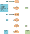
Several oncoproteins (in blue boxes) are known to either elevate the expression or induce the activity of enzymes and transporters that facilitate a high rate of glucose metabolism in cancer cells. The tumour suppressor p53 (in dark red boxes) is known to inhibit certain glucose metabolism pathways. Enzymes that are predominant in cancer cells are shown in bold. The thickness of the arrows indicates relative flux. 1,3BPG, 1,3-bisphosphoglycerate; 2PG, 2-phosphoglycerate; 3PG, 3-phosphoglycerate; ALDO, aldolase; DHAP, dihydroxyacetone phosphate; ENO, enolase; F1,6BP, fructose-1,6-bisphosphate; F2,6BP, fructose-2,6-bisphosphate; F6P, fructose-6-phosphate; FAS, fatty acid synthesis; G3P, glyceraldehyde-3-phosphate; G6P, glucose-6-phosphate; GAPDH, glyceraldehyde-3-phosphate dehydrogenase; GLUT, glucose transporter; glycerol-3P, glycerol-3-phosphate; GPI, glucose-6-phosphate isomerase; HK, hexokinase; LDH, lactate dehydrogenase; MCT, monocarboxylate transporter; mTORC1, mTOR complex 1; mtp53, mutant p53; PEP, phosphoenolpyruvate; PFK1, phosphofructokinase 1; PFKFB, 6-phosphofructo 2-kinase/fructose-2,6-bisphosphatase; PGAM1, phosphoglycerate mutase 1; PGK1, phosphoglycerate kinase 1; PK, pyruvate kinase; PPP, pentose phosphate pathway; TPI, triosephosphate isomerase.
Figure 6
Figure 6. Reprogramming of glucose metabolism in hepatocellular carcinoma
Differentiated hepatocytes use the reversal glucose transporter, GLUT2, for the uptake and export of glucose. The first committed step in glucose metabolism is attenuated because it is catalysed by the low-affinity hexokinase, HK4 (also known as glucokinase). The three committed steps in glucose metabolism could be reversed by the gluconeogenic enzymes (dark red). Glucose-6-phosphatase (G6Pase) dephosphorylates glucose-6-phosphate (G6P) back to glucose. Fructose-1,6-bisphosphatase (FBP1) dephosphorylates fructose-1,6-bisphosphate (F1,6BP) back to fructose-6-phosphate (F6P) and phosphoenolpyruvate carboxykinase (PEPCK) reverses the last committed step in glycolysis by converting oxaloacetate (OAA) to phosphoenolpyruvate (PEP), both in the mitochondria by the mitochondrial enzyme, PEPCK-M, and in the cytoplasm by PEPCK-C. In hepatocellular carcinoma the suppression of HK4 expression and the induction of the high-affinity hexokinase HK2 and by suppressing the expression of gluconeogenic (HCC), glucose metabolism is accelerated by the expression of GLUT1, enzymes. Unlike differentiated hepatocytes, HCC cells express relatively high levels of aldolase A (ALDOA), they express pyruvate kinase M2 (PKM2) instead of PKL, and they elevate the expression of lactate dehydrogenase A (LDHA; shown in bold). The thickness of the arrows indicates relative flux. 1,3BPG, 1,3-bisphosphoglycerate; 2PG, 2-phosphoglycerate; 3PG, 3-phosphoglycerate; α-KG, α-ketoglutarate; AcCoA, acetyl-CoA; F2,6BP, fructose-2,6-bisphosphate; F6P, fructose-6-phosphate; FAS, fatty acid synthesis; G3P, glyceraldehyde-3-phosphate; GPI, glucose-6-phosphate isomerase; PFK1, phosphofructokinase 1; PFKFB, 6-phosphofructo 2-kinase/fructose-2,6-bisphosphatase; PPP, pentose phosphate pathway; TCA, tricarboxylic acid.
Figure 7
Figure 7. Energetic and oxidative stress during solid tumour formation
During migration to the lumen, tumour cells suppress glucose uptake. Consequently NADPH and ATP levels decline. The decline in NADPH level increases the intracellular level of reactive oxygen species (ROS), which can cause cell death. The decline in ATP level induces the activation of AMP-activated protein kinase (AMPK), which in turn inhibits fatty acid synthesis (FAS) and induces fatty acid oxidation (FAO). The inhibition of FAS reduces NADPH consumption, and the induction of FAO could generate malate independently of pyruvate. The conversion of malate into pyruvate regenerates NADPH. Therefore NADPH homeostasis is maintained to reduce the elevated ROS. A similar scenario may occur in circulating metastatic cells. ANT, adenine nucleotide translocator; CPT1, carnitine O-palmitoyltransferase; G6P, glucose-6-phosphate; OXPHO, oxidative phosphorylation; PPP, pentose phosphate pathway; TCA, tricarboxylic acid; VDAC, voltage-dependent anion channel.

Similar articles

Cited by

References

    1. Warburg O, Wind F, Negelein E. The metabolism of tumors in the body. J Gen Physiol. 1927;8:519–530. - PMC - PubMed
    1. Warburg O. The Metabolism of Tumours: Investigations from the Kaiser Wilhelm Institute for Biology, Berlin-Dahlen. Constable; 1930.
    1. Warburg O. On the origin of cancer cells. Science. 1956;123:309–314. - PubMed
    1. Warburg O. On respiratory impairment in cancer cells. Science. 1956;124:269–270. - PubMed
    1. Weinhouse S. On respiratory impairment in cancer cells. Science. 1956;124:267–269. - PubMed