. 2016 Oct 28;90(22):10398-10413.
doi: 10.1128/JVI.01326-16. Print 2016 Nov 15.

The Presumed Polyomavirus Viroporin VP4 of Simian Virus 40 or Human BK Polyomavirus Is Not Required for Viral Progeny Release

Affiliations

The Presumed Polyomavirus Viroporin VP4 of Simian Virus 40 or Human BK Polyomavirus Is Not Required for Viral Progeny Release

Stian Henriksen et al. J Virol. .

Abstract

The minor capsid protein of human BK polyomavirus (BKPyV), VP2, and its N-terminally truncated form, VP3, are both important for viral entry. The closely related simian virus 40 (SV40) reportedly produces an additional truncated form of VP2/3, denoted VP4, apparently functioning as a viroporin promoting progeny release. The VP4 open reading frame is conserved in some polyomaviruses, including BKPyV. In this study, we investigated the role of VP4 in BKPyV replication. By transfecting viral genomes into primary human renal proximal tubule epithelial cells, we demonstrated that unaltered BKPyV and mutants with start codon substitutions in VP4 (VP2M229I and VP2M229A) abolishing putative VP4 production were released at the same level to supernatants. However, during infection studies, VP2M229I and VP2M229A exhibited 90% and 65% reduced infectivity, respectively, indicating that isoleucine substitution inadvertently disrupted VP2/3 function to the detriment of viral entry, while inhibition of VP4 production during late infection was well tolerated. Unexpectedly, and similarly to BKPyV, wild-type SV40 and the corresponding VP4 start codon mutants (VP2M228I and VP2M228A) transfected into monkey kidney cell lines were also released at equal levels. Upon infection, only the VP2M228I mutant exhibited reduced infectivity, a 43% reduction, which also subsequently led to delayed host cell lysis. Mass spectrometry analysis of nuclear extracts from SV40-infected cells failed to identify VP4. Our results suggest that neither BKPyV nor SV40 require VP4 for progeny release. Moreover, our results reveal an important role in viral entry for the amino acid in VP2/VP3 unavoidably changed by VP4 start codon mutagenesis.

Importance: Almost a decade ago, SV40 was reported to produce a late nonstructural protein, VP4, which forms pores in the nuclear membrane, facilitating progeny release. By performing transfection studies with unaltered BKPyV and SV40 and their respective VP4-deficient mutants, we found that VP4 is dispensable for progeny release, contrary to the original findings. However, infection studies demonstrated a counterintuitive reduction of infectivity of certain VP4-deficient mutants. In addition to the isoleucine-substituted SV40 mutant of the original study, we included alanine-substituted VP4-deficient mutants of BKPyV (VP2M229A) and SV40 (VP2M228A). These revealed that the reduction in infectivity was not caused by a lack of VP4 but rather depended on the identity of the single amino acid substituted within VP2/3 for VP4 start codon mutagenesis. Hopefully, our results will correct the longstanding misconception of VP4's role during infection and stimulate continued work on unraveling the mechanism for release of polyomavirus progeny.


Figures

FIG 1
FIG 1
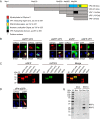
BKPyV minor capsid proteins are recognized by SV40 VP2/3 antiserum. (A) Schematic overview of BKPyV minor capsid protein VP2 and its confirmed and putative truncated variants. Annotated regions and motifs are derived from the UniProtKB database, accession numbers P03093 and P03094, and the VP4 hydrophobic domain is annotated by similarity to SV40 VP4. (B) Immunofluorescence labeling of RPTECs at 1 dpt with the eGFP-VP4 expression plasmid, using the SV40 VP2/3 antisera (red) and the DNA stain Draq5 (blue). The level of eGFP expression is indicated in parentheses as weak (w) or strong (s). As a positive and negative control for the SV40 VP2/3 antiserum, RPTECs were transfected with eGFP-VP3 and eGFP, respectively. In addition, cells were transfected with eGFP-VP1 plasmid and immunofluorescence labeling was performed using the BKPyV VP1 antisera (red). Images were acquired by confocal microscopy with a 40× objective and with a 2× zoom. (C) Western blot of HeLa cell lysate harvested at 1 dpt with the indicated expression plasmids. The membrane was probed with a GFP antibody (red) and the SV40 VP2/3 antisera (green). M, molecular mass marker. (D) Immunofluorescence labeling of HeLa cells at 1 dpt with the SV40 VP4-eGFP expression plasmid, using the SV40 VP2/3 antisera (red) and the DNA stain Draq5 (blue). VP4-eGFP is shown in green. Images were acquired by confocal microscopy with a 40× objective and a 2× zoom. (E) Western blot of nuclear and cytoplasmic cell fractions of mock-infected and BKPyV-infected RPTECs harvested at 3 dpi. The membrane was probed with the SV40 VP2/3 antisera. The empty arrowhead indicates a band with the expected size of BKPyV VP4, and the asterisk indicates a nonspecific band in the mock-infected nuclear fraction. Nuc, nuclear fraction; Cyt, cytoplasmic fraction.
FIG 2
FIG 2
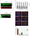
BKPyV VP4 is not required for viral progeny release, but VP4 start codon substitution affects infectivity. (A) Western blot of RPTEC cell lysates harvested at 2 dpt with the indicated BKPyV genomes. The membrane was probed with rabbit BKPyV VP1 antisera (green) and rabbit SV40 VP2/3 antisera (green). Mouse GAPDH antibody was used as a loading control (red). The asterisk indicates binding to a nonspecific cellular protein by the VP1 or VP2/3 antisera. M, molecular mass marker. (B) BKPyV load in DNase I-treated supernatants from RPTECs at 1 and 3 dpt, determined by real-time quantitative PCR and presented as GEq/ml. The figure represents data from three independent experiments, each performed in duplicate wells, and the error bars indicate the standard deviations. (C) Western blot of capsid proteins pelleted by ultracentrifugation from Vero cell supernatants harvested 3 dpt. A volume corresponding to 400 μl supernatant was loaded on the gel. The membrane was probed with rabbit BKPyV VP1 antisera (upper) and rabbit SV40 VP2/3 antisera (lower). The signal intensity in the lower panel was increased for better visualization of the VP2 and VP3 proteins. (D) Immunofluorescence staining of RPTECs at 3 dpi with the indicated BKPyV variants at similar BKPyV load (approximately 8 log10 GEq/ml), using the rabbit agno antisera (green) and the mouse monoclonal LTag antibody (Ab-2; red). Representative images of the immunofluorescence staining are shown in the upper panel, and quantification in relation to the WT infection is displayed in the graph in the lower panel. Each experiment was done in duplicate and repeated twice. Approximately 3,000 to 5,000 Draq5-positive cells per well were analyzed for virus protein expression.
FIG 3
FIG 3
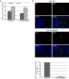
BKPyV VP4 is not required for viral progeny release of the WW strain, but the VP4 start codon substitution M229I affects infectivity. (A) BKPyV load in DNase I-treated supernatants from RPTECs at 3 and 6 dpt, determined by real-time quantitative PCR and presented as GEq/ml. The figure represents data from two independent experiments, each performed in duplicate wells, and the error bars indicate the standard deviations. (B) Immunofluorescence staining of COS-7 cells 4 dpi with BKPyV WW WT and BKPyV WW VP2M229I at similar viral loads (approximately 8 log10 GEq/ml) using a BKPyV VP1 antibody (red) and the agno antisera (green). Representative images of the immunofluorescence staining are shown in the upper panels, and quantification in relation to WT infection is displayed in the graph in the lower panel. Each experiment was performed twice in duplicate wells. All agno-positive cells present in 0.75-cm2 wells were counted.
FIG 4
FIG 4
SV40 VP4 is not required for viral progeny release, but the VP4 start codon substitution affects infectivity. (A) SV40 load in DNase I-treated supernatants from BS-C-1 cells and CV-1 cells at 1, 2, 4, and 5 dpt, determined by real-time quantitative PCR and presented as GEq/ml. The figure represents data from three independent experiments (except BS-C-1 cells at 5 dpt and CV-1 cells at 4 dpt, which were performed twice). Each experiment was performed in duplicate wells, and the error bars indicate the standard deviations. (B) Western blot of capsid proteins pelleted by ultracentrifugation of CV-1 cell supernatants harvested 2 dpt. A volume corresponding to 400 μl supernatant was loaded on the gel. The membrane was probed with SV40 VP1 and VP2/3 antisera. (C) Quantification of infectivity in CV-1 cells at 2 dpi with SV40 WT and VP4-deficient mutants. CV-1 cells were infected with the indicated SV40 variants at similar viral load (approximately 10 log10 GEq/ml). Quantification of LTag- and VP1-positive cells in relation to the SV40 WT infection, determined by immunofluorescence microscopy, is displayed in the graph. Immunofluorescence staining was performed using the SV40 VP1 antisera and the LTag antibody (Pab419), and the quantification is related to the number of SV40 WT-infected cells. Each experiment was done in duplicate and repeated twice. Approximately 3,000 to 5,000 Draq5-positive cells per well were analyzed for virus protein expression. (D) Western blot of nuclear and cytoplasmic fractions from CV-1 cells that were mock infected or SV40 infected with the indicated SV40 variants and harvested at 2 dpi. The membrane was probed with SV40 VP2/3 antisera. The empty arrowhead indicates a band with a size similar to that of the putative VP4 protein. Nuc, nuclear fraction; Cyt, cytoplasmic fraction; M, molecular mass marker.
FIG 5
FIG 5
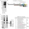
SV40 VP4 is not detected upon immunoprecipitation (IP) using SV40 VP2/3 antiserum. (A) Western blot (WB) of immunoprecipitated proteins from nuclear extracts of SV40-infected CV-1 cells at 6 dpi. Immunoprecipitation was performed using the SV40 VP2/3 antisera. The membrane was probed with SV40 VP2/3 antisera. M, molecular mass marker. (B) Schematic presentation of the location in VP2 of the peptides identified by LC-MS/MS from visible bands below 20 kDa excised from a Coomassie-stained SDS-PAGE gel after immunoprecipitation. Note that the SV40 VP5 ORF is similar to the BKPyV VP6 ORF, as shown in Fig. 1A. (C) Coomassie-stained SDS-PAGE gel of immunoprecipitated proteins from nuclear extracts of SV40-infected CV-1 cells at 6 dpi. The location of the excised bands and the sequence of VP2/3/4 peptides detected in these bands are listed. The initial methionine residues for both VP3 and VP4 are highlighted in red. The gel pieces were fragmented in gel by chymotrypsin. ND, not detected; M, molecular mass marker.
FIG 6
FIG 6
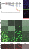
SV40 VP2M228I shows a delayed and reduced host cell lysis compared to WT SV40. (A) Electrical impedance in SV40-transfected CV-1 monolayers was measured using the xCELLigence system from 6 to ∼120 hpt. The cell index was normalized to the cell index of mock-transfected cells. The experiment was repeated 2 times, and a representative experiment where the cell index of 4 parallel wells was measured and shown as means with standard deviations is presented. At the end of this experiment, immunofluorescence staining was performed using the SV40 VP1 antisera (green), an LTag antibody (Pab419; red), and the DNA stain Draq5 (blue). (B) Time-lapse microscopy was used to monitor SV40-infected CV-1 monolayers from 6 hpi until 120 hpi (5 dpi). A green cyanine dye staining the DNA of dead cells green was added directly after infection. Photos were taken every 15 min, and selected time points (6 hpi, 2 dpi, 3 dpi, and 5 dpi) are shown. (C) Immunofluorescence staining of SV40-infected CV-1 cells at the end of the time-lapse experiment described for panel B, using the SV40 VP1 antisera (red). A Nikon Biostation was used to take immunofluorescence and phase contrast images, and merged images are shown.

Similar articles

Cited by

References

    1. Drachenberg CB, Papadimitriou JC, Wali R, Cubitt CL, Ramos E. 2003. BK polyoma virus allograft nephropathy: ultrastructural features from viral cell entry to lysis. Am J Transplant 3:1383–1392. doi:10.1046/j.1600-6135.2003.00237.x. - DOI - PubMed
    1. Hirsch HH. 2010. Polyoma and papilloma virus infections after hematopoietic stem cell or solid organ transplantation, p 465–482. In Bowden P, Ljungman P, Snydman DR (ed), Transplant infections, 3rd ed Lippincott Williams & Wilkins, Philadelphia, PA.
    1. Hirsch HH, Kardas P, Kranz D, Leboeuf C. 2013. The human JC polyomavirus (JCPyV): virological background and clinical implications. APMIS 121:685–727. doi:10.1111/apm.12128. - DOI - PubMed
    1. Mazlo M, Tariska I. 1980. Morphological demonstration of the first phase of polyomavirus replication in oligodendroglia cells of human brain in progressive multifocal leukoencephalopathy (PML). Acta Neuropathol 49:133–143. doi:10.1007/BF00690753. - DOI - PubMed
    1. Liddington RC, Yan Y, Moulai J, Sahli R, Benjamin TL, Harrison SC. 1991. Structure of simian virus 40 at 3.8-A resolution. Nature 354:278–284. doi:10.1038/354278a0. - DOI - PubMed

Substances

Grants and funding

The funders had no role in study design, data collection and interpretation, or the decision to submit the work for publication.

LinkOut - more resources