Review
. 2016 Oct;68(4):954-1013.
doi: 10.1124/pr.115.011395.

Glucagon-Like Peptide-1 and Its Class B G Protein-Coupled Receptors: A Long March to Therapeutic Successes

Affiliations
Review

Glucagon-Like Peptide-1 and Its Class B G Protein-Coupled Receptors: A Long March to Therapeutic Successes

Chris de Graaf et al. Pharmacol Rev. 2016 Oct.

Abstract

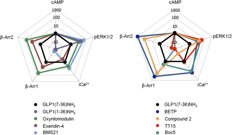
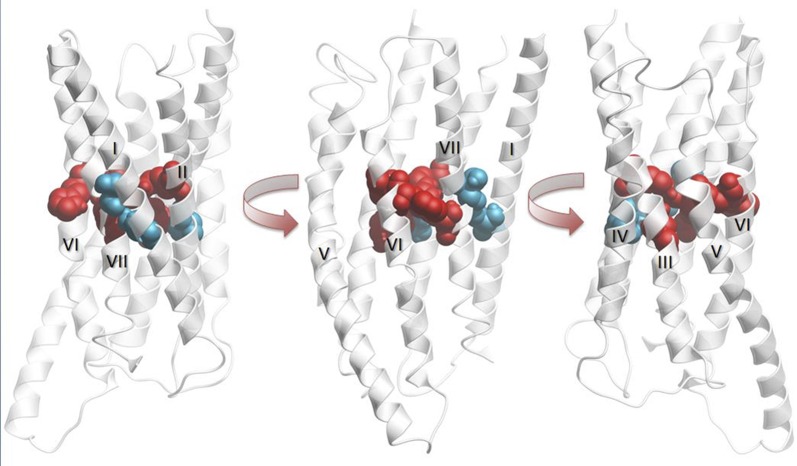
The glucagon-like peptide (GLP)-1 receptor (GLP-1R) is a class B G protein-coupled receptor (GPCR) that mediates the action of GLP-1, a peptide hormone secreted from three major tissues in humans, enteroendocrine L cells in the distal intestine, α cells in the pancreas, and the central nervous system, which exerts important actions useful in the management of type 2 diabetes mellitus and obesity, including glucose homeostasis and regulation of gastric motility and food intake. Peptidic analogs of GLP-1 have been successfully developed with enhanced bioavailability and pharmacological activity. Physiologic and biochemical studies with truncated, chimeric, and mutated peptides and GLP-1R variants, together with ligand-bound crystal structures of the extracellular domain and the first three-dimensional structures of the 7-helical transmembrane domain of class B GPCRs, have provided the basis for a two-domain-binding mechanism of GLP-1 with its cognate receptor. Although efforts in discovering therapeutically viable nonpeptidic GLP-1R agonists have been hampered, small-molecule modulators offer complementary chemical tools to peptide analogs to investigate ligand-directed biased cellular signaling of GLP-1R. The integrated pharmacological and structural information of different GLP-1 analogs and homologous receptors give new insights into the molecular determinants of GLP-1R ligand selectivity and functional activity, thereby providing novel opportunities in the design and development of more efficacious agents to treat metabolic disorders.


Figures

Fig. 1.
Fig. 1.
Gene structure, expression, processing, degradation, and elimination of proglucagon. The proglucagon gene is located in human chromosome 2 and transcribed as one single mRNA in three major tissues, namely, the pancreas, the intestine, and the CNS. The mRNA is first translated into one single protein and then processed by prohormone convertase (PC) in different tissues. In the pancreatic α cells, proglucagon protein is processed by PC2 into glicentin-related polypeptide (GRPP), glucagon (Gluc), intervening peptide-1 (IP-1), and major proglucagon fragment, whereas in L cells of the small intestine and the brain, proglucagon is processed by PC1/3 into oxyntomodulin, intervening peptide-2 (IP-2), GLP-1, and GLP-2. GLP-1 is degraded by DPP-4 via cleavage of two amino acids from the N terminus, or by NEP-24.11 through cleavage of the C terminus in vivo. The cleaved products are eventually eliminated in the kidney. UTR, untranslated region.
Fig. 2.
Fig. 2.
Structural characteristics of GLP-1 and its cognate receptor. (A) GLP-1–bound full-length GLP-1R homology model based on a previously published full-length glucagon-bound GCGR model combining structural and experimental information from the GCGR 7TMD crystal structure (PDB: 4L6R), the GCGR ECD structure (PDB: 4ERS), and the ECD structure of GLP-1–bound GLP-1R (PDB: 3IOL), complemented by site-directed mutagenesis, electron microscopy, hydrogen-deuterium exchange, and cross-linking studies (Siu et al., 2013; Yang et al., 2015b, 2016). The C-terminal helix of GLP-1 bound to the ECD region of GLP-1R is depicted as cartoon, whereas the atoms of the flexible N-terminal region of GLP-1 predicted to be bound to the 7TMD of GLP-1R are depicted as spheres. GLP-1 is color coded according to mutation effects (blue: 10-fold effect IC50; see II. Glucagon-Like Peptide-1); mutation effects of GLP-1R are reported in Fig. 3 and Table 1. The Cα/Cβ atoms of GLP-1/GLP-1R residue pairs identified in photo cross-linking studies (Chen et al., 2009, 2010b; Miller et al., 2011) are depicted as green-colored spheres. (B) Structural alignment of the ECD structures of GLP-1 and exendin9–39–bound GLP-1R (PDB: 3IOL, 3C59), GIP-bound GIPR (PDB: 2QKH), and the mAb23-bound GCGR ECD structure (PDB: 4ERS). Comparison of the crystal structure binding modes of (C) GLP-1 and (D) exendin9–39. The surfaces of GLP-1R residues involved in important apolar interactions with GLP-1/apolar are colored pink, whereas residues involved in polar interactions described in II. Glucagon-Like Peptide-1 are also depicted as sticks (and their H-bond interaction networks are depicted as dashed lines). (E) Structure-based sequence alignment of GLP-1, exendin9–39, glucagon, GIP, and GLP-2. The regions of the peptide ligands solved in ECD–ligand complex crystal structures are marked above the amino acid sequences using the same color coding as in (B). Amino acids of GLP-1 are marked according to mutation study effects, as indicated in (A). The residues that are boxed are found in an α-helical conformation in the crystal structure complex (solid lines: GLP-1, exendin9–39, GIP) or in NMR studies in micelle DPC (dashed lines: glucagon, GLP-2), as described in II. Glucagon-Like Peptide-1.
Fig. 3.
Fig. 3.
Summary of GLP-1R mutagenesis. A snake plot of GLP-1R from http://www.gpcrdb.org that has been colored (see Key) to highlight the location of signal peptide, glycosylation, and phosphorylation sites, as well as the mutated residues in Table 1. It should be noted that the color coding of 74 of the 195 mutated residues (W39, W72, W87, W91, W110, F169-C174, R176, N177, H180, N182, A200-Q213, W214-G225, R227-F230, L232, M233, E262-L268, F321-I332, K334-K336, and R348-K351) reflects the effects of rat GLP-1R mutations projected on the hGLP-1R amino acid sequence. The color coding of 27 of the 185 residues (K202, W203, S206-Q211, Q213, W214, G216-Q221, S223-G225, R227-F230, L332, M233, I325, and F326) reflects the effect of double mutations, not single-point mutations. Information on the fold change in ligand affinity and potency as well as expression levels of the GLP-1R mutants is reported in Table 1.
Fig. 4.
Fig. 4.
Summary of the main characterized pathways of glucose and GLP-1 signaling in the pancreatic β cell. Glucose enters the cell through glucose transporter 2 and undergoes glycolysis to produce pyruvate that enters the mitochondria for oxidative metabolism and ATP production. This increase in cytosolic ATP closes the KATP channels, depolarizing the membrane and opening the voltage-dependent calcium channels, increasing calcium influx into the cell, causing insulin exocytosis. GLP-1 increases insulin exocytosis through a number of mechanisms. GLP-1R couples to Gαs, activates adenylate cyclase that converts ATP to cAMP, and mobilizes two downstream effectors, PKA and Epac. These have a range of effects, including closing KATP channels, enhancing fusion of insulin secretory granules with the membrane, whereas PKA also closes Kv channels, inhibiting membrane repolarization. PKA and Epac also increase intracellular calcium by facilitating CICR through the opening of inositol 1,4,5-trisphosphate receptor and ryanodine receptor calcium channels, respectively. This increase in calcium has also been proposed to upregulate mitochondrial ATP production and activate calcineurin, so nuclear factor of activated T cells promotes insulin gene transcription to increase insulin stores. Alongside increasing insulin synthesis and exocytosis, GLP-1 signals through a number of pathways to increase β cell mass. PKA reduces ER stress through ATF-Gadd34 signaling, increases β cell neogenesis by activating cyclin D, and elevates the expression of insulin receptor substrate 2 (IRS2), a β cell survival factor, as well as anti-apoptotic proteins Bcl-2 and Bcl-xL through CREB. PI3K is activated by IRS2 and transactivation of epidermal growth factor receptor, and this further promotes increased β cell mass through upregulation of PDX-1 and nuclear factor κB, which upregulates anti-apoptotic Bcl-2 /Bcl-xL and inhibitor of apoptosis protein-2.
Fig. 5.
Fig. 5.
Web of bias illustrating distinctions in the pattern of signaling of different peptide agonists (left panel) or nonpeptidic modulators (right panel) at GLP-1R. The web of bias plots ΔΔτ/KA values on a logarithmic scale for each ligand and for every signaling pathway tested. Formation of these values included normalization to the reference ligand GLP-17–36 amide and the reference pathway, cAMP accumulation. The plots do not provide information on absolute potency, but on relative efficacy for signaling of individual pathways in comparison with that for cAMP. Data are from Koole et al., 2010; Willard et al., 2012a; and Wootten et al., 2013b.
Fig. 6.
Fig. 6.
Homology model of GLP-1R illustrating the relative position of key residues involved in the receptor-signaling bias. The modeling indicates that these residues reside at a fulcrum position of the receptor transmembrane bundle, where the splayed helices of the open extracellular face of the receptor converge, with the residues that contribute to ligand-dependent signaling forming a central interaction network (space fill, red) and the smaller polar residues that are globally important for signaling external to this core (space fill, blue). The receptor is displayed in three views at different horizontal rotation. Transmembrane helices are numbered with Roman numerals.
Fig. 7.
Fig. 7.
Nonpeptidic GLP-1R modulators and peptide mimetics. Liraglutide (Novo Nordisk) was the first approved human GLP-1 analog to teat diabetes (European Union, 2009; United States, 2010) and obesity (United States, 2014; European Union, 2015). Liraglutide has 97% sequence homology to human GLP-1 and was designed to reversibly bind to albumin by attachment of palmatic acid via a L-γ-glutamic linker to lys26 in Arg34 GLP-17–37. The modification of Lys34 to Arg34 made it possible to produce Arg34 GLP-17–37 in yeast, followed by acylation of Lys26. The native GLP-1 peptide has a half-life of approximately 2 minutes due to rapid cleavage of GLP-17–37 to GLP-19–37 by DPP-4 (Deacon et al., 1995a,b; Vilsboll et al., 2003). Liraglutide comprises the natural GLP-1 N terminus, but has a half-life of about 11 hours after s.c. dosing to humans combined with a delayed absorption from sub cutis that gives a pharmacokinetic profile applicable for once-daily administration. The reason for extended circulation is due to reversible albumin binding that protects from DPP-4 degradation and glomerular filtration, whereas the delayed absorption is explained by the ability of liraglutide to form heptamers by self-assemble controlled by the fatty acid side-chain at position 26. Liraglutide is well tolerated and capable of substantially improving glycemic control with low risk of hypoglycemia and weight loss benefit (Knudsen et al., 2000; Knudsen, 2004; Madsen et al., 2007; Steensgaard et al., 2008; Dharmalingam et al., 2011; Wang et al., 2015).

Similar articles

Cited by

References

    1. Abrahamsen N, Lundgren K, Nishimura E. (1995) Regulation of glucagon receptor mRNA in cultured primary rat hepatocytes by glucose and cAMP. J Biol Chem 270:15853–15857. - PubMed
    1. Adelhorst K, Hedegaard BB, Knudsen LB, Kirk O. (1994) Structure-activity studies of glucagon-like peptide-1. J Biol Chem 269:6275–6278. - PubMed
    1. Ahn J-M, Murage EN, Lo S-T, Lin M, and Sun X (2011) Highly constrained GLP-1 analogues as non-invasive PET imaging agents for the assessment of pancreatic beta-cell mass, in Peptides: Building Bridges, Proceedings of the 22nd American Peptide Symposium (Lebl M ed); 2011 June 25-30; San Diego, CA. pp 220-221, American Peptide Society.
    1. Al-Sabah S, Donnelly D. (2003a) A model for receptor-peptide binding at the glucagon-like peptide-1 (GLP-1) receptor through the analysis of truncated ligands and receptors. Br J Pharmacol 140:339–346. - PMC - PubMed
    1. Al-Sabah S, Donnelly D. (2003b) The positive charge at Lys-288 of the glucagon-like peptide-1 (GLP-1) receptor is important for binding the N-terminus of peptide agonists. FEBS Lett 553:342–346. - PubMed

Publication types

MeSH terms

Substances