Monocytes and B cells support active replication of Chandipura virus
- PMID: 27628855
- PMCID: PMC5024506
- DOI: 10.1186/s12879-016-1794-6
Monocytes and B cells support active replication of Chandipura virus
Abstract
Background: Interaction between immune system and Chandipura virus (CHPV) during different stages of its life cycle remain poorly understood. The exact route of virus entry into the blood and CNS invasion has not been clearly defined. The present study was undertaken to assess the population in PBMC that supports the growth of virus and to detect active virus replication in PBMC as well as its subsets.
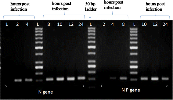
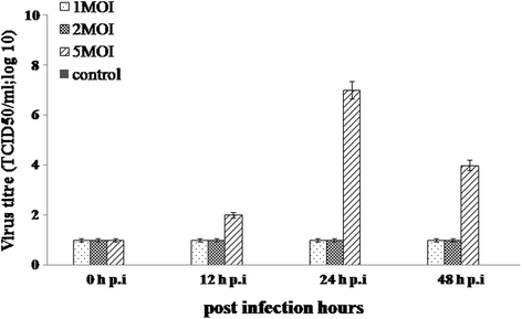
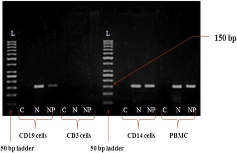
Methods: PBMC subsets viz.: CD3(+), CD14(+), CD19(+), CD56(+)cells were separated and infected with CHPV. The infected cells were then assessed for transcription (N gene primer) and replication (NP gene primer) of CHPV by PCR. The supernatant collected from infected cells were titrated in Baby Hamster Kidney (BHK) cells to assess virus release. The cytokine and chemokine expression was quantified by flow cytometry.
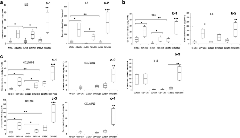
Results: Amplification of N and NP gene was detected in CD14(+) (monocyte) and CD19(+) (B cell), significant increase in virus titre was also observed in these subsets. It was observed that, although the levels of IL-6 and IL-10 were elevated in CD14(+) cells as compared to CD19(+)cells, the differences were not significant. However the levels of TNFα and IL-8 were significantly elevated in CD14(+) cells than in CD19(+)cells. The levels of chemokine (CXCL9, CCL5, CCL2, CXCL10) were significantly elevated in CHPV infected PBMC as compared to uninfected cells. CCL2 and CXCL9 were significantly increased in CHPV infected CD14(+)cells as compared to CD19(+) cells.
Conclusion: CD14(+)and CD19(+)cells support active replication of CHPV. High viral load was detected in CD14(+) cells infected with CHPV hence it might be the primary target cells for active replication of CHPV. An elevated levels of cytokines and chemokines observed in CD14(+) cells may help in predicting the pathogenecity of CHPV and possible entry into the central nervous system.
Keywords: B cells; Chandipura virus; Chemokine; Cytokine; Monocytes; PBMC.
Figures
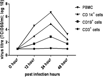
 ) PBMC, (
 ) CD14+cells, (
 ) CD19+cells and (
 ) CD3+cells infected with MOI = 5 of CHPV. The culture supernatant was collected at different time interval. The release of progeny virus was quantitated by calculating TCID50 titre in BHK cells. The assay was performed in triplicates
Similar articles
-
Detection of hepatitis C virus (HCV) negative strand RNA and NS3 protein in peripheral blood mononuclear cells (PBMC): CD3+, CD14+ and CD19+.Virol J. 2013 Nov 26;10:346. doi: 10.1186/1743-422X-10-346. Virol J. 2013. PMID: 24279719 Free PMC article.
-
Neuroinvasion by Chandipura virus.Acta Trop. 2014 Jul;135:122-6. doi: 10.1016/j.actatropica.2014.03.028. Epub 2014 Apr 5. Acta Trop. 2014. PMID: 24713200 Review.
-
Hepatitis C virus (HCV) infection of peripheral blood mononuclear cells in patients with type II cryoglobulinemia.Hum Immunol. 2013 Dec;74(12):1559-62. doi: 10.1016/j.humimm.2013.08.273. Epub 2013 Aug 28. Hum Immunol. 2013. PMID: 23993984
-
Borrelia burgdorferi stimulation of chemokine secretion by cells of monocyte lineage in patients with Lyme arthritis.Arthritis Res Ther. 2010;12(5):R168. doi: 10.1186/ar3128. Epub 2010 Sep 9. Arthritis Res Ther. 2010. PMID: 20828409 Free PMC article.
-
Neuropathogenesis by Chandipura virus: An acute encephalitis syndrome in India.Natl Med J India. 2017 Jan-Feb;30(1):21-25. Natl Med J India. 2017. PMID: 28731002 Review.
Cited by
-
Gammaherpesviruses and B Cells: A Relationship That Lasts a Lifetime.Viral Immunol. 2020 May;33(4):316-326. doi: 10.1089/vim.2019.0126. Epub 2020 Jan 8. Viral Immunol. 2020. PMID: 31913773 Free PMC article. Review.
-
Chandipura Viral Encephalitis: A Brief Review.Open Virol J. 2018 Aug 31;12:44-51. doi: 10.2174/1874357901812010044. eCollection 2018. Open Virol J. 2018. PMID: 30288194 Free PMC article. Review.
-
Age-dependent alterations in serum cytokines, peripheral blood mononuclear cell cytokine production, natural killer cell activity, and prostaglandin F2α.Immunol Res. 2017 Oct;65(5):1009-1016. doi: 10.1007/s12026-017-8940-0. Immunol Res. 2017. PMID: 28762199
-
Alternative pathway of complement activation has a beneficial role against Chandipura virus infection.Med Microbiol Immunol. 2020 Apr;209(2):109-124. doi: 10.1007/s00430-019-00648-z. Epub 2019 Nov 28. Med Microbiol Immunol. 2020. PMID: 31781935 Free PMC article.
References
-
- Bhatt PN, Rodrigues FM. Chandipura: a new Arbovirus isolated in India from patients with febrile illness. Indian J Med Res. 1967;55:1295–1305. - PubMed
-
- Rao BL, Basu A, Wairagkar NS, Gore MM, Arankalle VA, Thakare JP, Jadi RS, Rao KA, Mishra AC. A large outbreak of acute encephalitis with high fatality rate in children in Andrapradesh, India, in 2003, associated with Chandipura virus. Lancet. 2004;364:869–874. doi: 10.1016/S0140-6736(04)16982-1. - DOI - PMC - PubMed
-
- Chadha MS, Arankalle VA, Jadi RS, Joshi MV, Thakare JP, Mahadev PVM, Mishra AC. An outbreak of Chandipura virus encephalitis in the eastern districts of Gujarat state, India. Am J Trop Med. 2005;73:566–570. - PubMed
MeSH terms
Substances
LinkOut - more resources
Full Text Sources
Other Literature Sources
Research Materials
Miscellaneous

