. 2016 Dec 15;44(22):10603-10618.
doi: 10.1093/nar/gkw788. Epub 2016 Sep 12.

A feedback loop comprising PRMT7 and miR-24-2 interplays with Oct4, Nanog, Klf4 and c-Myc to regulate stemness

Affiliations

A feedback loop comprising PRMT7 and miR-24-2 interplays with Oct4, Nanog, Klf4 and c-Myc to regulate stemness

Sung-Hun Lee et al. Nucleic Acids Res. .

Abstract

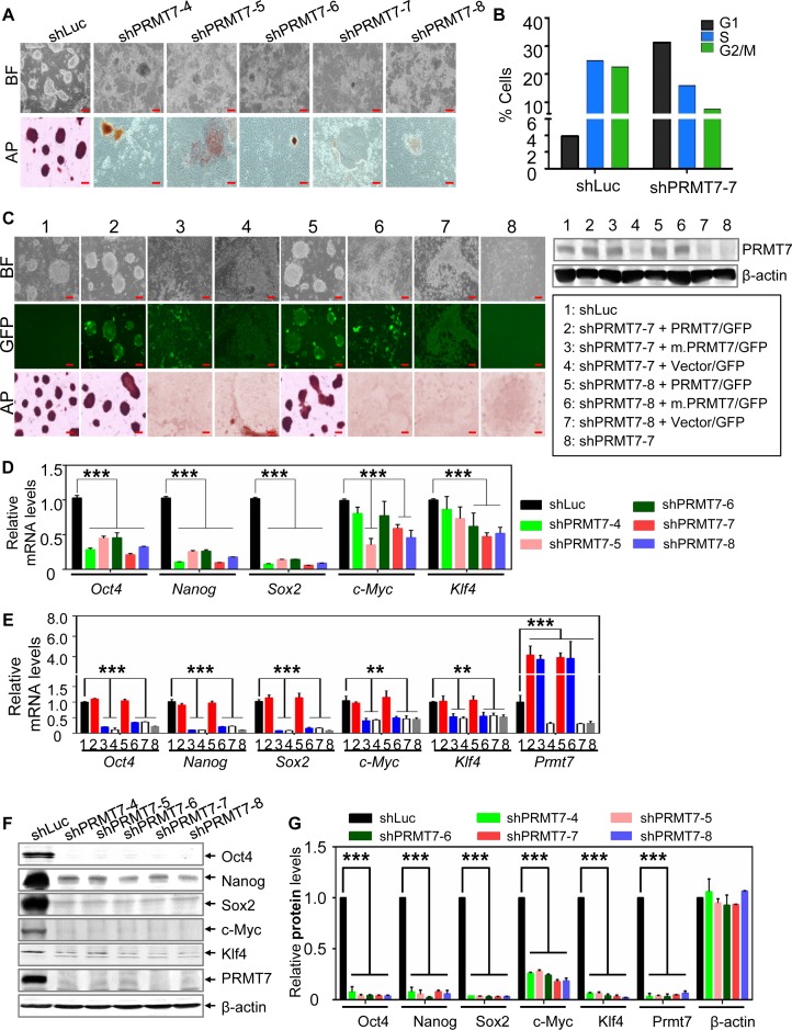
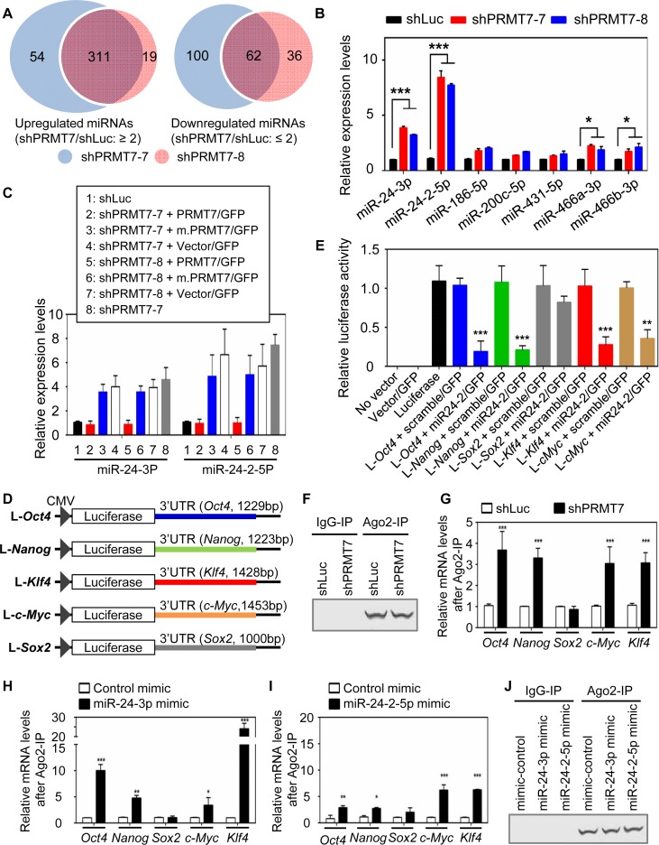
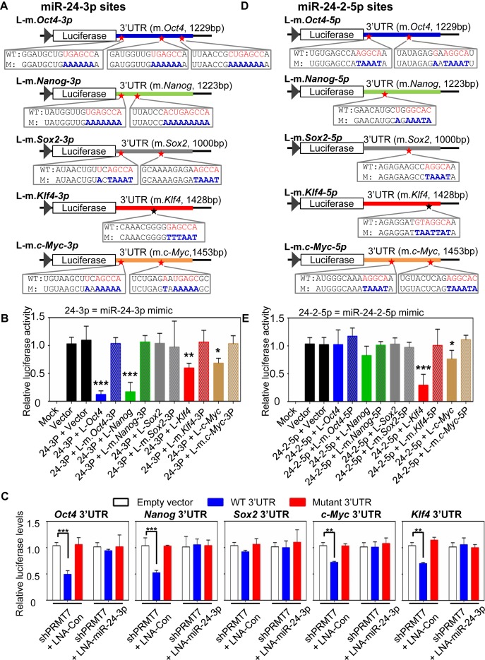
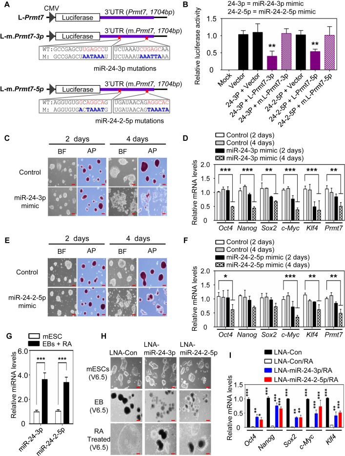
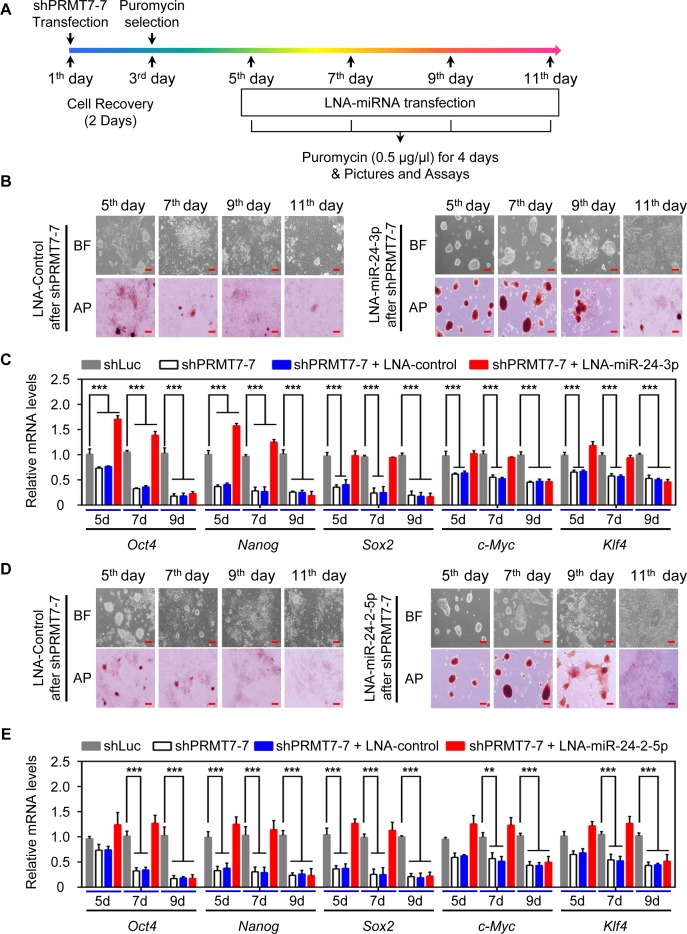
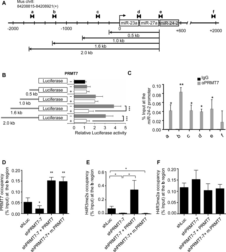
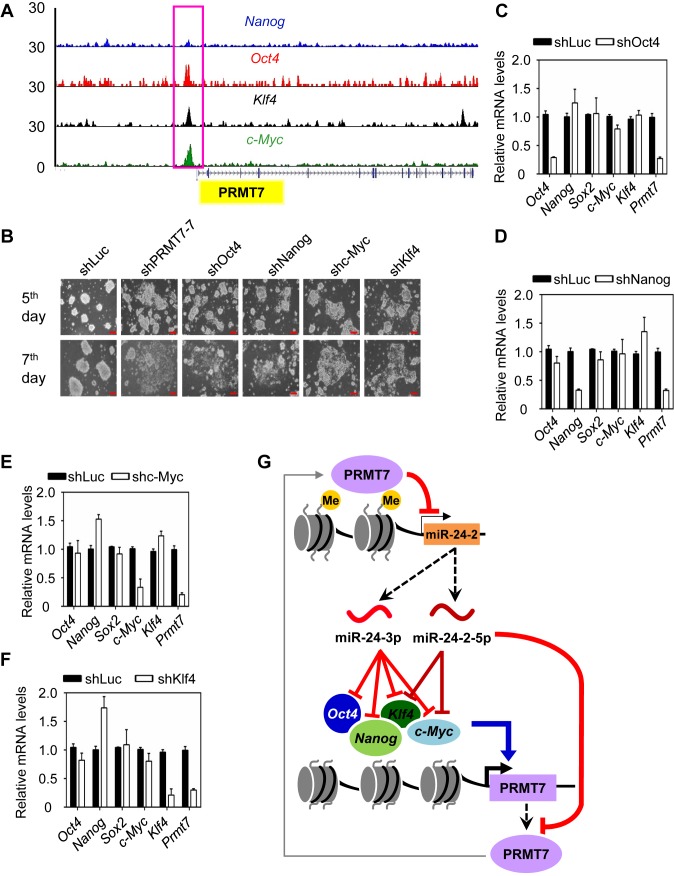
Self-renewal and pluripotency are two fundamental characteristics of embryonic stem cells (ESCs) and are controlled by diverse regulatory factors, including pluripotent factors, epigenetic regulators and microRNAs (miRNAs). Although histone methyltransferases are key epigenetic regulators, whether and how a histone methyltransferase forms a network with miRNAs and the core pluripotent factor system to regulate ESC stemness is little known. Here, we show that the protein arginine methyltransferase 7 (PRMT7) is a pluripotent factor essential for the stemness of mouse ESCs. PRMT7 repressed the miR-24-2 gene encoding miR-24-3p and miR-24-2-5p by upregulating the levels of symmetrically dimethylated H4R3. Notably, miR-24-3p targeted the 3' untranslated regions (UTRs) of the major pluripotent factors Oct4, Nanog, Klf4 and c-Myc, whereas miR-24-2-5p silenced Klf4 and c-Myc expression. miR-24-3p and miR-24-2-5p also targeted the 3'UTR of their repressor gene Prmt7 miR-24-3p and miR-24-2-5p induced mouse ESC differentiation, and their anti-sense inhibitors substantially reversed spontaneous differentiation of PRMT7-depleted mouse ESCs. Oct4, Nanog, Klf4 and c-Myc positively regulated Prmt7 expression. These findings define miR-24-3p and miR-24-2-5p as new anti-pluripotent miRNAs and also reveal a novel epigenetic stemness-regulatory mechanism in which a double-negative feedback loop consisting of PRMT7 and miR-24-3p/miR24-2-5p interplays with Oct4, Nanog, Klf4 and c-Myc to control ESC stemness.


Figures

Figure 1.
Figure 1.
PRMT7 and its catalytic activity are indispensable for the stemness of mESCs, and Oct4, Nanog, Klf4 and c-Myc protein levels are more reduced than their mRNA levels in PRMT7-depleted mESCs. (A) Microscopic and AP-staining images of shLuc-treated and PRMT7-depleted V6.5 mESCs. Five shPRMT7s (shPRMT7-4, -5, -6, -7 and -8) were used to deplete PRMT7. BF, bright field; AP, alkaline phosphatase; red scale bar, 100 μm. (B) The percentages of shLuc- or shPRMT7-treated V6.5 mESCs in G1, S and G2/M phase of the cell cycle. shLuc-treated cells were used as a control. (C) Rescue experiments of PRMT7-depleted mESCs with PRMT7 or its catalytic mutant (m.PRMT7). shPRMT7-7 or shPRMT7-8 plasmids were co-electroporated with expression plasmids encoding both GFP and either PRMT7 or its catalytic mutant. The GFP-expressing vector pCDH1-EF1-IRES-GFP (vector/GFP) was used to clone PRMT7 cDNA. Microscopic, green-fluorescent and AP staining images of indicated group of cells are shown (left panel). Western blotting was used to analyze the expression levels of PRMT7 or its mutant in eight different experimental conditions (right panel). (D) Analysis of Oct4, Nanog, Sox2, c-Myc and Klf4 mRNA levels in shLuc-treated and PRMT7-depleted V6.5 mESCs using quantitative RT-PCR. (E) The effect of ectopic expression of PRMT7 or its catalytic mutant (m.PRMT7) on Oct4, Nanog, Sox2, c-Myc, Klf4 and Prmt7 mRNA levels in PRMT7-depleted mESCs. Relative Oct4, Nanog, Sox2, c-Myc, Klf4 and Prmt7 mRNA levels were examined in eight different experimental conditions (see panel C for lane description). Red scale bar, 100 μm. (F and G) Western blot analysis of Oct4, Nanog, Sox2, c-Myc, Klf4 and PRMT7 levels in shLuc-treated and PRMT7-depleted V6.5 mESCs. Oct4, Nanog, Sox2, c-Myc, Klf4 and PRMT7 protein levels in shLuc-treated and PRMT7-depleted V6.5 mESCs were quantified (G). Data are presented as the mean ± SD of three independent experiments. P < 0.05 (*), P < 0.01 (**) and P < 0.001 (***) indicate statistically significant changes.
Figure 2.
Figure 2.
PRMT7 downregulates the levels of miR-24-3p and miR-24-2-5p, which collectively target the 3′UTRs of Oct4, Nanog, Klf4 and c-Myc mRNAs. (A) Venn diagrams of miRNAs that were significantly upregulated or downregulated by two shPRMT7s (shPRMT7-7 and shPRMT7-8). (B) Comparison of cellular levels of multiple miRNAs between shLuc-treated and PRMT7-depleted V6.5 mESCs using quantitative, miRNA-specific PCR. (C) The effect of ectopic expression of PRMT7 or its catalytic mutant (m.PRMT7) on miR-24-3p and miR24-2-5p levels in PRMT7-depleted mESCs. (D) Schematic representation of luciferase reporter constructs containing Oct4-3′UTR, Nanog-3′UTR, Sox2-3′UTR, Klf4-3′UTR or c-Myc-3′UTR. (E) Relative luciferase activities of reporter constructs containing Oct4-3′UTR, Nanog-3′UTR, Sox2-3′UTR, Klf4-3′UTR or c-Myc-3′UTR in the absence or presence of miR24-2 expression. The reporter constructs, alone or together with a miR-24-2 expression plasmid encoding miR-24-3p and miR24-2-5p, were transfected into HEK293T cells. Firefly luciferase activities were normalized to the internal transfection control Renila luciferase. (F and G) Comparison of endogenous association of Ago2 with Oct4, Nanog, Sox2, Klf4 and c-Myc mRNAs between shLuc-treated cells and PRMT7-depleted cells. Ago2 IP was performed (F) and IP eluates were analyzed using quantitative RT-PCR (G). (HJ) Analysis of the association of Ago2 with Oct4, Nanog, Sox2, Klf4 and c-Myc mRNAs after transient transfection of control mimic, miR-24-3p mimic (H) or miR-24-2-5p mimic (I) in mESCs. Following transfection of mimic RNAs, cells were incubated for 48 h, lysed and used for Ago2 IP (J). IP eluates were analyzed using quantitative RT-PCR (H and I). Data are presented as the mean ± SD of three independent experiments. P < 0.05 (*), P < 0.01 (**) and P <0.001 (***) indicate statistically significant changes.
Figure 3.
Figure 3.
miR-24-3p's targeting sites are located at Oct4, Nanog, Klf4 and c-Myc 3′UTRs, whereas miR24-2-5p's sites are localized at Klf4 and c-Myc 3′UTRs. (A) Schematic representation of luciferase reporter constructs containing mutations in miR-24-3p target sites in Oct4-3′UTR, Nanog-3′UTR, Sox2-3′UTR, Klf4-3′UTR and c-Myc-3′UTR. WT, wild-type; M, mutant. (B) The effect of miR-24-3p mimic on reporter activities of WT Oct4-3′UTR, Nanog-3′UTR, Sox2-3′UTR, Klf4-3′UTR, c-Myc-3′UTR and their mutants. The reporter constructs, together with miR-24-3p mimic, were transfected into HEK293T cells. Mock indicates non-transfected cells, and vector denotes a reporter plasmid without any 3′UTR. Firefly luciferase activities were normalized to an internal transfection control (Renila luciferase). (C) The effect of endogenous miR-24-3p on the reporter activities of Oct4-3′UTR, Nanog-3′UTR, Sox2-3′UTR, Klf4-3′UTR and c-Myc-3′UTR. mESCs (5 × 10⁵ cells) were first electroporated with 30 μg of the shPRMT7-7 plasmid. Twelve days later, PRMT7-depleted mESCs were co-transfected with both WT luciferase-3′UTR (or luciferase-mutant 3′UTR) reporters and control LNA (or LNA-miR-24-3p [a strong anti-sense inhibitor of miR-24-3p]). Cells were harvested 2 days after transfection. (D) Schematic representation of luciferase reporter constructs bearing mutations in miR-24-2-5p target sites in Oct4-3′UTR, Nanog-3′UTR, Sox2-3′UTR, Klf4-3′UTR and c-Myc-3′UTR. WT, wild-type; M, mutant. (E) The effect of miR-24-2-5p mimic on reporter activities of WT Oct4-3′UTR, Nanog-3′UTR, Sox2-3′UTR, Klf4-3′UTR, c-Myc-3′UTR and their mutants. Data are presented as the mean ± SD of three independent experiments. P < 0.05 (*), P < 0.01 (**) and P < 0.001 (***) indicate statistically significant changes.
Figure 4.
Figure 4.
miR-24-3p and miR-24-2-5p target Prmt7 3′UTR and impede the stemness of mESCs. (A) Schematic representation of luciferase reporter constructs containing WT Prmt7-3′UTR or its mutants. (B) Relative luciferase activities of reporter constructs containing Prmt7-3′UTR or its mutants in HEK293T cells after transfection of miR-24-3p and miR-24-2-5p mimics. (C and E) Microscopic and AP staining images of V6.5 mESCs after treatment with miR-24-3p (C) or miR-24-2-5p (E) mimic. V6.5 mESCs were treated with miR-24-3p and miR-24-2-5p mimics and incubated for 2d or 4d. (D and F) Quantitative analysis of Oct4, Nanog, Sox2, Klf4, c-Myc and Prmt7 mRNA levels in V6.5 mESCs after treatment with miR-24-3p (D) or miR-24-2-5p (F) mimic. (G) Quantitative RT-PCR analysis of miR-24-3p and miR-24-2-5p levels in WT and differentiated V6.5 mESCs (EBs + RA). (H and I) The effect of LNA-miRNAs (a type of anti-sense microRNAs) against miR-24-3p or miR-24-2-5p on RA-induced mESC differentiation. mESCs were transfected with LNA-miRNAs (H). Oct4, Nanog, Sox2, c-Myc and Klf4 mRNA levels were measured using quantitative RT-PCR (I). In G−I, mESCs were induced to form EBs for 5 days and then treated with RA for another 5 days. Data are presented as the mean ± SD of three independent experiments. P < 0.05 (*), P < 0.01 (**) and P < 0.001 (***) indicate statistically significant changes.
Figure 5.
Figure 5.
PRMT7-mediated repression of miR-24-3p/miR-24-2-5p levels is required for maintaining self-renewal and pluripotency. (A) The outline for treatment of PRMT7-depleted cells with LNA-control and LNA-miRNAs. (B and D) Microscopic and AP staining images of PRMT7-depleted V6.5 mESCs at the 5th, 7th, 9th, or 11th day after treatment with LNA-control or LNA-miR-24-3p (B) (LNA-miR-24-2-5p in [D]). Red scale bar, 100 μm. (C and E) Quantitative analysis of Oct4, Nanog, Sox2, c-Myc and Klf4 mRNA levels in PRMT7-depleted V6.5 mESCs at the 5th, 7th, 9th, or 11th day after treatment with LNA-control or LNA-miR-24-3p (C) (LNA-miR-24-2-5p in [E]). Data are presented as the mean ± SD of three independent experiments. P < 0.05 (*), P < 0.01 (**) and P < 0.001 (***) indicate statistically significant changes.
Figure 6.
Figure 6.
PRMT7 occupies the miR-24-2 gene and negatively regulates its expression via H4R3 methylation. (A) Schematic representation of the miR-24-2 gene. A set of arrow heads indicate a ChIP PCR amplicon. (B) Luciferase activities of the miR-24-2 promoter and its deletion mutants with or without ectopic expression of PRMT7 in V6.5 mESCs. V6.5 mESCs were transfected using Lipofectamine 3000 and incubated for 48 h. (C) Analysis of PRMT7 occupancy at the miR-24-2 promoter using quantitative ChIP. (D−F) Analysis of PRMT7 (D), H4R3me2s (E) and H4R3me2a (F) levels at the region ‘b’ in the miR-24-2 promoter after rescue experiments of PRMT7-depleted mESCs. Quantitative ChIP assays were performed using four groups of V6.5 mESCs: (i) shLuc-treated cells, (ii) PRMT7-depleted cells, (iii) PRMT7-depleted cells with ectopic expression of PRMT7 and (iv) PRMT7-depleted cells with ectopic expression of a catalytic mutant of PRMT7 (m.PRMT7).
Figure 7.
Figure 7.
The Prmt7 gene is activated by Oct4, Nanog, c-Myc and Klf4. (A) Analysis of Oct4, Nanog, c-Myc and Klf4 levels at the Prmt7 promoter using the publicly available ChIP-Seq database. (B) Microscopic images of V6.5 mESCs treated with shPRMT7-7, shOct4, shc-Myc, shKlf4 or shNanog. Red scale bar, 100 μm. (C−F) Effects of individual knockdown of Oct4 (C), Nanog (D), c-Myc (E), and Klf4 (F) on Prmt7 mRNA levels. V6.5 mESCs were electroporated with shOct4, shc-Myc, shKlf4, or shNanog. Four days later, cells were harvested. Expression levels were analyzed using quantitative RT-PCR. (G) A proposed model depicting a role for PRMT7 in maintaining the self-renewal and pluripotency of mESCs. miR-24-3p can silence the expression of Oct4, Nanog, Klf4 and c-Myc, whereas miR-24-2-5p can target Klf4 and c-Myc 3′UTRs. In addition, miR-24-3p and miR-24-2-5p target Prmt7 3′UTR. In mESCs, PRMT7 represses the miR-24-2 gene encoding miR-24-3p and miR-24-2-5p via H4R3 methylation. Therefore, PRMT7 antagonizes the anti-pluripotent effects of miR-24-3p and miR-24-2-5p against Oct4, Nanog, Klf4 and c-Myc in mESCs and positively regulates Oct4, Nanog, Klf4, and c-Myc levels to maintain mESC stemness. During differentiation, increased miR-24-3p and miR-24-2-5p levels may reduce PRMT7, Oct4, Nanog, Klf4, and c-Myc levels, facilitating mESC differentiation. The regulatory loop involving PRMT7 and miR-24-3p/miR-24-2-5p is interactive with the major pluripotent system containing Oct4, Nanog, Klf4, and c-Myc to fine-tune mESC stemness.

Similar articles

Cited by

References

    1. Orkin S.H., Hochedlinger K. Chromatin connections to pluripotency and cellular reprogramming. Cell. 2011;145:835–850. - PMC - PubMed
    1. Meissner A. Epigenetic modifications in pluripotent and differentiated cells. Nat. Biotechnol. 2010;28:1079–1088. - PubMed
    1. Spivakov M., Fisher A.G. Epigenetic signatures of stem-cell identity. Nat. Rev. Genet. 2007;8:263–271. - PubMed
    1. Gu B., Lee M.G. Histone H3 lysine 4 methyltransferases and demethylases in self-renewal and differentiation of stem cells. Cell Biosci. 2013;3:39–53. - PMC - PubMed
    1. Jenuwein T., Allis C.D. Translating the histone code. Science. 2001;293:1074–1080. - PubMed

MeSH terms