. 2016 Oct 18;7(42):67966-67985.
doi: 10.18632/oncotarget.11817.

Human cytomegalovirus encoded chemokine receptor US28 activates the HIF-1α/PKM2 axis in glioblastoma cells

Affiliations

Human cytomegalovirus encoded chemokine receptor US28 activates the HIF-1α/PKM2 axis in glioblastoma cells

Raymond H de Wit et al. Oncotarget. .

Abstract

The human cytomegalovirus (HCMV) encoded chemokine receptor US28 promotes tumorigenesis through activation of various proliferative and angiogenic signaling pathways. Upon infection, US28 displays constitutive activity and signals in a G protein-dependent manner, hijacking the host's cellular machinery. In tumor cells, the hypoxia inducible factor-1α/pyruvate kinase M2 (HIF-1α/PKM2) axis plays an important role by supporting proliferation, angiogenesis and reprogramming of energy metabolism. In this study we show that US28 signaling results in activation of the HIF-1α/PKM2 feedforward loop in fibroblasts and glioblastoma cells. The constitutive activity of US28 increases HIF-1 protein stability through a Gαq-, CaMKII- and Akt/mTOR-dependent mechanism. Furthermore, we found that VEGF and lactate secretion are increased and HIF-1 target genes, glucose transporter type 1 (GLUT1) and glyceraldehyde-3-phosphate dehydrogenase (GAPDH), involved in glucose metabolism, are upregulated in US28 expressing cells. In addition, PKM2 is phosphorylated and found to be in a tumor-associated dimeric state upon US28 expression. Also in HCMV-infected cells HIF-1 activity is enhanced, which in part is US28-dependent. Finally, increased proliferation of cells expressing US28 is abolished upon inhibition of the HIF-1α/PKM2 cascade. These data highlight the importance of HIF-1α and PKM2 in US28-induced proliferation, angiogenesis and metabolic reprogramming.

Keywords: G protein-coupled receptor (GPCR); chemokine; glioblastoma; human cytomegalovirus (HCMV); hypoxia-inducible factor (HIF).


Conflict of interest statement

CONFLICTS OF INTEREST

The authors declare no conflicts of interest in relation to this manuscript.

Figures
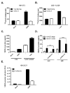
Figure 1
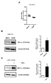
Figure 1. US28 constitutively induces promoter activation and secretion of VEGF via HIF-1
NIH-3T3 cells stably expressing US28 A. or U251 cells with inducible expression of US28 B. were assayed for binding of radiolabeled 125I-CCL5 (200 pM) and displacement with unlabeled US28 ligand CX3CL1 (100 nM). C. 48 hour secretion of VEGF-A in the supernatant of synchronized mock and US28-expressing NIH-3T3 or U251 cells. D. VEGF-A secretion after 24/48 hour incubation with or without HIF-1 dimerization inhibitor acriflavine (1 μM) treatment. Significant differences between conditions are depicted by asterisks (*** = P < 0.001, **** = P < 0.0001). E. HEK293T cells transfected with US28 and VEGF promoter reporter gene were treated with HIF-1 dimerization inhibitor acriflavine (1 μM), VEGF promoter activity was measured after 24 hours.
Figure 2
Figure 2. US28 constitutively induces the transcriptional activity of the HIF-1 complex by increasing the HIF-1α protein level
A. Stably transfected mock and US28 NIH-3T3 cells were used for RNA isolation. Quantitative RT-PCR is performed to determine HIF1A relative mRNA expression levels, using ACTB as normalization control. Lysates were prepared from stably transfected NIH-3T3 cells with US28 or control vector B. or U251-iUS28 cells with inducible US28 expression C., expression of HIF-1α protein was evaluated with a specific antibody by Western blotting, followed by signal quantification using β-actin expression as a loading control. Significant differences between conditions are depicted by asterisks (** = P < 0.01, *** = P < 0.001)
Figure 3
Figure 3. US28 stimulates HIF-1 transcriptional activity through concerted activation of Gαq, CaMKII and the Akt/mTOR signaling cascade
A.-D. HEK293T cells were transiently co-transfected with US28-encoding DNA and 500 ng of hypoxia response element (HRE) HIF-1 reporter gene, transcriptional activity was measured 24 hours after transfection and treatment. A. Increasing amounts of US28-WT DNA were transfected with a fixed amount of reporter gene B. Transfection of US28-WT or the G protein-uncoupled mutant US28 R129A and treatment with cobalt chloride (150 μM) to mimic hypoxia C. Treatment with HIF-1 dimerization inhibitor acriflavine (1 μM). D. A panel of pharmacological inhibitors is applied to inhibit US28-dependent HIF-1 activation in a HIF-1 reporter gene assay using HEK293T cells. US28-expressing and mock (reporter only) cells were treated with inhibitors for 24 hours before activity was measured. The results normalized to the vehicle condition of the reporter only control. The dotted lines represent the basal level of US28-expressing or mock cell reportergene activity. Significant (α=0.05, P < 0.05) reporter gene activity inhibition compared to the vehicle controls is represented by an asterisk (*) above the bar.
Figure 4
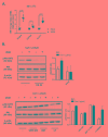
Figure 4. US28 promotes HIF-1 target gene transcription and reprograms activation of Akt and PKM2 which are involved in proliferation, angiogenesis and glucose metabolism through a Gαq/CaMKII/Akt/HIF-1 dependent fashion in fibroblasts and glioma cells
A. HIF-1 target genes GAPDH, GLUT1 and VEGFA relative mRNA levels were determined by quantitative RT-PCR using RNA isolated from US28 or mock transfected NIH-3T3 cells and gene-specific primers. Significant differences in HIF-1 target gene expression are depicted by asterisks (* = P < 0.05, **** = P < 0.0001). B. Lysates were prepared from synchronized U251-iUS28 cells with/without 48 hours of doxycycline-induced US28 expression and treatment with signaling pathway inhibitors. SDS/PAGE Western blot analysis for phosphorylated Akt (Ser 473), total Akt and the loading control β-actin. Relative Akt activity is semi-quantified by pAkt/Akt ratios C. Lysates were prepared from synchronized, stably transfected NIH-3T3 or U251-iUS28 cells and phosphorylation status of PKM2 were assessed by Western blotting. Relative PKM2 phosphorylation is semi-quantified by pPKM2/PKM2 ratios D. Native lysates were prepared from synchronized US28 or mock NIH-3T3 and U251-iUS28 cells. For U251-iUS28, native lysates were prepared after 48 hours of US28 expression with/without vehicle, DMSO or UBO-QIC treatment. PKM2 protein was cross-linked using 0.25% formaldehyde/PBS solution (+), lysates without cross-linking treatment (−) were included as loading and total PKM2 expression controls. PKM2 and β-actin expression levels were assessed by Western blotting.
Figure 5
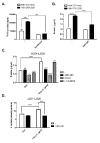
Figure 5. US28 signaling results in increased cell proliferation and metabolic reprogramming
A. Stably transfected NIH-3T3 cells with US28 or empty vector were treated with acriflavine for 48 hours. Proliferation of the cells was determined by 3H- thymidine incorporation assay. B. Mock and US28 NIH-3T3 cells were synchronized and subsequently treated with UBO-QIC for 48 hours before lysates were prepared and protein content was determined using a BCA assay C., D. Synchronized U251 cells with inducible US28 expression were cultured for 48 hours with/without doxycycline and pharmacological inhibitors. (D) The lysate protein content was determined using a BCA assay, E. whereas cell culture medium supernatant was analyzed for L-Lactate levels using a cell-based glycolysis assay. The L-Lactate levels are normalized to the total protein content. Significant differences between conditions are depicted by asterisks (***= P < 0.001, **** = P < 0.0001).
Figure 6
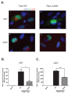
Figure 6. US28 contributes to HCMV-dependent HIF-1 activation in U251 glioblastoma and HFF cells
A. Immunofluorescence of HCMV Titan WT and ΔUS28 infected U251 or HFF cells 48 hours post-infection at MOI2. Cells were stained with US28 antibody (red fluorescence), anti-immediate early antigen (IEA) antibody (green fluorescence) and DAPI (blue fluorescence). Additionally, since a GFP tag is incorporated in the viral genome, the cytosol of infected cells can also be detected with green fluorescence. Panel I-IV display IEA-specific staining in the nuclei and GFP in the cytosol of HCMV Titan-infected cells. Panels I and II display US28-specific staining in primarily the perinuclear region of Titan WT infected cells, which cannot be detected in cells infected with the US28 deletion mutant (ΔUS28). B. HFF or C. U251 cells were infected with HCMV Titan WT or ΔUS28 at MOI of 2. Mock and infected cells were transfected with the HRE reporter gene 24 hours post-infection, followed by HIF-1 activity measurement 48 hours post-infection. Significant differences between conditions are depicted by asterisks (** = P < 0.01, **** = P < 0.0001).
Figure 7
Figure 7. Proposed mechanism of US28 induced activation of the HIF-1α/PKM2 feedforward loop
US28 constitutively stimulates Gαq-dependent signaling cascades resulting in CaMKII activity and subsequent stimulation of the Akt/mTOR cascade to increase HIF-1α translation and/or stability. HIF-1α translocates to the nucleus and dimerizes with HIF-1β to form a functional HIF-1 transcription factor that binds HRE regions to regulate HIF-1 target genes supporting proliferation, angiogenesis and metabolic reprogramming. US28 signaling modulates PKM2 phosphorylation and rearrangement into enzymatically less active, but transcriptional active dimers. Dimeric PKM2 is linked with glycolysis and thus increased levels lactate production and secretion. PKM2 dimers and HIF-1α engage in a feedforward loop supporting a proliferative, angiogenic and glycolytic phenotype.

Similar articles

Cited by

References

    1. Gandhi MK, Khanna R. Human cytomegalovirus: clinical aspects, immune regulation, and emerging treatments. The Lancet Infectious diseases. 2004;4:725–738. - PubMed
    1. White MK, Gorrill TS, Khalili K. Reciprocal transactivation between HIV-1 and other human viruses. Virology. 2006;352:1–13. - PubMed
    1. Fischer SA. Emerging viruses in transplantation: there is more to infection after transplant than CMV and EBV. Transplantation. 2008;86:1327–1339. - PubMed
    1. Nakase H, Honzawa Y, Toyonaga T, Yamada S, Minami N, Yoshino T, Matsuura M. Diagnosis and treatment of ulcerative colitis with cytomegalovirus infection: importance of controlling mucosal inflammation to prevent cytomegalovirus reactivation. Intestinal research. 2014;12:5–11. - PMC - PubMed
    1. Vischer HF, Siderius M, Leurs R, Smit MJ. Herpesvirus-encoded GPCRs: neglected players in inflammatory and proliferative diseases? Nat Rev Drug Discov. 2014;13:123–139. - PubMed

MeSH terms