Effects of aging on the architecture of the ileocecal junction in rats
- PMID: 27602243
- PMCID: PMC4986394
- DOI: 10.4292/wjgpt.v7.i3.416
Effects of aging on the architecture of the ileocecal junction in rats
Abstract
Aim: To evaluate the structural organization of the elastic and collagen fibers in the region of the ileocecal transition in 30 young and old male Wistar rats.
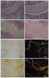
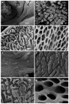
Methods: Histology, immunohistochemistry (IHC), transmission electron microscopy and scanning electron microscopy were employed in this study. The results demonstrated that there was a demarcation of the ileocecal region between the ileum and the cecum in both groups.
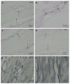
Results: The connective tissue fibers had different distribution patterns in the two groups. IHC revealed the presence of nitric oxide synthase, enteric neurons and smooth muscle fibers in the ileocecal junctions (ICJs) of both groups. Compared to the young group, the elderly group exhibited an increase in collagen type I fibers, a decrease in collagen type III fibers, a decreased linear density of oxytalan elastic fibers, and a greater linear density of elaunin and mature elastic fibers.
Conclusion: The results revealed changes in the patterns of distribution of collagen and elastic fibers that may lead to a possible decrease in ICJ functionality.
Keywords: Aging; Collagen fibers; Elastic fibers; Ileocecal junction; Rats.
Figures


Similar articles
-
A comparative study of aging of the elastic fiber system of the diaphragm and the rectus abdominis muscles in rats.Braz J Med Biol Res. 2000 Dec;33(12):1449-54. doi: 10.1590/s0100-879x2000001200008. Braz J Med Biol Res. 2000. PMID: 11105097
-
Abnormal elastic system fibers in fibrotic human liver.Med Electron Microsc. 2000;33(3):135-42. doi: 10.1007/s007950000013. Med Electron Microsc. 2000. PMID: 11810471
-
Elastic system fibers (oxytalan, elaunin and elastic fibers) in the skin of a freshwater teleost : optical and electron microscopy study.Arch Anat Microsc Morphol Exp. 1980;69(4):259-66. Arch Anat Microsc Morphol Exp. 1980. PMID: 7212696
-
Oxytalan connective tissue fibers: a review.J Oral Pathol. 1974;3(6):291-316. doi: 10.1111/j.1600-0714.1974.tb01724.x. J Oral Pathol. 1974. PMID: 4142890 Review.
-
[Elastic system fibers of healthy human gingiva].J Parodontol. 1990 Feb;9(1):29-34. J Parodontol. 1990. PMID: 2200871 Review. French.
Cited by
-
Metabolomic Profiles of Mouse Tissues Reveal an Interplay between Aging and Energy Metabolism.Metabolites. 2021 Dec 26;12(1):17. doi: 10.3390/metabo12010017. Metabolites. 2021. PMID: 35050139 Free PMC article.
References
-
- Ferraz de Carvalho CA, Rodrigues de Souza R, Henrique A, Henrique A, Nogueira de Lima MA. Functional anatomy of the tela submucosa of the valva ileocecalis in the adult man. Anat Anz. 1987;164:63–76. - PubMed
-
- Shafik AA, Shafik A, Asaad S, Wahdan M. A study of an anatomic-physiological cecocolonic sphincter in humans. Clin Anat. 2010;23:851–861. - PubMed
-
- Shafik AA, Ahmed IA, Shafik A, Wahdan M, Asaad S, El Neizamy E. Ileocecal junction: anatomic, histologic, radiologic and endoscopic studies with special reference to its antireflux mechanism. Surg Radiol Anat. 2011;33:249–256. - PubMed
-
- Pollard MF, Thompson-Fawcett MW, Stringer MD. The human ileocaecal junction: anatomical evidence of a sphincter. Surg Radiol Anat. 2012;34:21–29. - PubMed
-
- Kumar D, Phillips SF. The contribution of external ligamentous attachments to function of the ileocecal junction. Dis Colon Rectum. 1987;30:410–416. - PubMed
LinkOut - more resources
Full Text Sources
Other Literature Sources

