. 2016 Oct 4;7(40):65389-65402.
doi: 10.18632/oncotarget.11700.

Activation of AKT pathway by Nrf2/PDGFA feedback loop contributes to HCC progression

Affiliations

Activation of AKT pathway by Nrf2/PDGFA feedback loop contributes to HCC progression

Danyang Liu et al. Oncotarget. .

Abstract

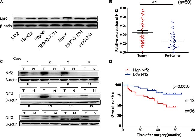
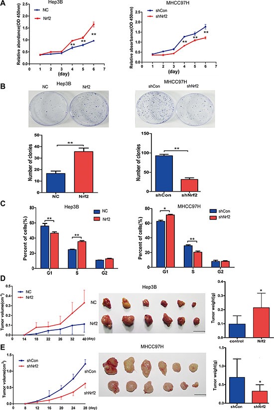
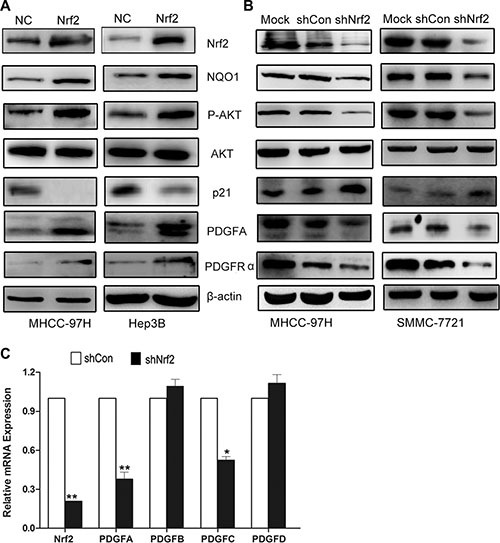
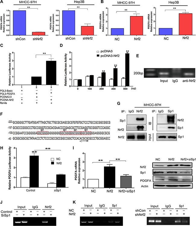
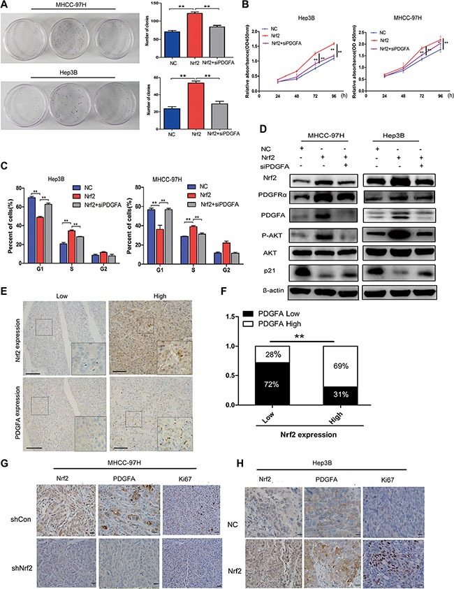
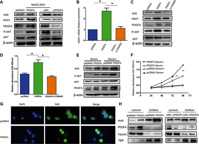
Nuclear factor erythroid-2-related factor 2 (Nrf2), a master transcription factor in the antioxidant response, has been found to be ubiquitously expressed in various cancer cells and in the regulation tumor proliferation, invasion, and chemoresistance activities. The regulatory roles of Nrf2 in controlling Hepatocellular carcinoma (HCC) progression remain unclear. In this study, we demonstrated that Nrf2 was significantly elevated in HCC cells and tissues and was correlated with poor prognosis of HCCs. Consistently, Nrf2 significantly promoted HCC cell growth both in vitro and in vivo. Further investigation suggested a novel association of Nrf2 with Platelet-Derived Growth Factor-A (PDGFA). Nrf2 promoted PDGFA transcription by recruiting specificity protein 1 (Sp1) to its promoter, resulting in increased activation of the AKT/p21 pathway and cell cycle progression of HCC cells. As a feedback loop, PDGFA enhanced Nrf2 expression and activation in an AKT dependent manner. In line with these findings, expression of Nrf2 and PDGFA were positively correlated in HCC tissues. Taken together, this study uncovers a novel mechanism of the Nrf2/PDGFA regulatory loop that is crucial for AKT-dependent HCC progression, and thereby provides potential targets for HCC therapy.

Keywords: AKT; Nrf2; PDGFA; hepatocellular carcinoma; tumorigenesis.


Conflict of interest statement

CONFLICTS OF INTEREST

The authors declare that they have no competing interests.

Figures

Figure 1
Figure 1. Nrf2 is significantly up-regulated in HCC
(A) Expression of Nrf2 was higher in a panel of HCC cell lines. The expression of Nrf2 in indicated HCC cell lines was subjected to western blot analysis using anti-Nrf2 antibody. (B) The mRNA levels of Nrf2 were elevated in HCC tissues. The mRNA transcripts of Nrf2 were measured by qRT-PCR using specific primers of Nrf2 in HCC tissues and normal counterparts. (C) The protein levels of Nrf2 were up-regulated in HCC tissues. The whole cell lysates of HCC tissues and normal counterparts were extracted and measured by western blot analysis using anti-Nrf2 antibody. (D) Overall survival (OS) differences between patients with high and low levels of Nrf2 protein expression. IHC results were independently graded by two experienced pathologists. Tissue samples with grades 0 (no staining) and 1(weak staining) were grouped as ‘low’ expression, and samples with grade 2 (strong staining) were grouped as ‘high’ expression. Survival curve was generated by using Graphpad software Prism 5 and Log Rank Mantel-Cox Test analysis. *P < 0.05; **P < 0.01. Student's-t test was used for the statistical analysis.
Figure 2
Figure 2. Nrf2 promotes HCC cell proliferation by up-regulating cell cycle progression both in vitro and in vivo
(A) Nrf2 triggered HCC cell growth in Hep3B and MHCC-97H cells. Equal numbers of the cells infected with indicated lentivirus were plated into 96-well plates. The proliferation activity was measured by CCK8 assay. (B) Nrf2 promoted the colony formation ability of HCC cells. 500–1000 cells that infected with indicated lentivirus was plated into 35 cm dish for 2 weeks. The number of colonies was counted and calculated. (C) Nrf2 controlled HCC cell cycle progression. 106 cells that infected with indicated lentivirus were analysed by FACS for the relative percentages of cell cycle phases. Means ± SD from three independent experiments are presented. *P < 0.05; **P < 0.01. Student's-t test was used for the statistical analysis. (D) Ectopic expression of Nrf2 promoted tumor growth in vivo. Left, growth curve of tumor volumes at indicated time. Middle, demonstration of tumor volumes of xenograft tumors removed from indicated group. Right, Statistical analysis of tumor weight of the resected tumors. (E) Depletion of Nrf2 repressed tumor growth in vivo. Left, growth curve of tumor volumes at indicated time. Middle, demonstration of tumor volumes of xenograft tumors removed from indicated group. Right, Statistical analysis of tumor weight of the resected tumors. Scale bars, 1 cm *P < 0.05; **P < 0.01. Student's-t test was used for the statistical analysis.
Figure 3
Figure 3. Nrf2 possibly modulates cell cycle progression by upregulating PDGFA and activation of AKT/p21 pathway
(A) In Hep3B and MHCC-97H cells with Nrf2 overexpression, the protein expression levels of PDGFA, PDGFRα, phosphor-AKT (p-AKT) were significantly increased while the p21 expression decreased compared with the control. (B) Depletion of Nrf2 decreased PDGFA expression. The expression of indicated proteins was measured in MHCC-97H and MHCC-7721 cells by western blotting using indicated antibodies. (C) The PDGF family genes in MHCC-97H in response to Nrf2 stable knockdown were performed by Real-time PCR. Means ± SD from three independent experiments are presented. *P < 0.05; **P < 0.01. Student's-t test was used for the statistical analysis.
Figure 4
Figure 4. Nrf2 enhances the mRNA transcription of PDGFA by binding to PDGFA promoter via Sp1
(A) Ablation of Nrf2 downregulated PDGFA mRNA expression. MHCC-97H and Hep3B cells were infected with indicated lentivirus and the mRNA levels of PDGFA in indicated group were measured by qRT-PCR. (B) Ectopic expression of Nrf2 increased PDGFA mRNA expression. The mRNA levels of PDGFA in indicated group were measured by qRT-PCR. Means ± SD from three independent experiments are presented as relative ratio to the control whose value was taken as 1.0. (C) Nrf2 activated PDGFA promoter. MHCC-97H cells were co-transfected with indicated plasmids for 48 h. Means ± SD of normalized luciferase activity from three independent experiments are presented. (D) Nrf2 dose-dependently activated PDGFA promoter. HEK-293T cells cotransfected with PGL3-PDGFA, pRL-SV40, and various amounts (0, 100, 200, 400, 800 ng/well) of pcDNA-Nrf2 or pcDNA3.0 vectors. Means ± SD of normalized luciferase activity from three independent experiments are presented. (E) Nrf2 could bind to PDGFA promoter. ChIP assays were performed by immunoprecipitation chromatin fragments from MHCC-97H cells using anti-Nrf2 mAb or IgG control. 5% cell lysate was used as input. (F) Characterization of the region −223/+20 in PDGFA promoter revealed four binding sites for Sp1 (marked in red box). (G) Nrf2 interacts with Sp1 in the nucleus. Nucleoproteins were then extracted and subjected to Co-IP assay analysis. In this way we further revealed the interactions between Nrf2 and Sp1. 5% cell lysate was used as input. IB, immunoblot. IP, immunoprecipitation. (H) PDGFA luciferase activities were detected under the NC and Nrf2 overexpression group, respectively. Cells were transfected with no siRNAs (control), or Sp1 siRNA (siSp1). Means ± s.d. of normalized luciferase activity from three independent experiments are presented. (I) The upregulation of PDGFA by Nrf2 was dependent on Sp1. Cells that were infected with the indicated lentivirus were transfected with the indicated siSp1. The expression of PDGFA was measured by qRT-PCR and western blotting. (J) Sp1 is essential for Nrf2 binding to PDGFA promoter. ChIP assays were performed by immunoprecipitation chromatin fragments from MHCC-97H cells with Mock group or Sp1 depletion group using anti-Nrf2 mAb or IgG control. 5% cell lysate was used as input. (K) Nrf2 is required for Sp1 enrichment on PDGFA promoter. ChIP assays were performed with antibody against Sp1 or control IgG in MHCC-97H cells expressed high Nrf2 level or low Nrf2 level. Left, Nrf2 expression promotes Sp1 association with PDGFA promoter. Right, Nrf2 ablation attenuates Sp1 interaction with PDGFA promoter. *P < 0.05; **P < 0.01. Student's-t test was used for the statistical analysis.
Figure 5
Figure 5. The tumor-promoting effect of Nrf2 is dependent on PDGFA
(A) Colony formation (B) Growth curve (C) Cell cycle distribution. Depletion of PDGFA in Nrf2 over-expressed cells abolished the oncogenic activity of Nrf2. Cells infected with indicated lentivirus was transfected with siPDGFA. CCK8 assay, clony formation and cell cycle analysis were evaluated. (D) Depletion of PDGFA in Nrf2 expression cells reduced AKT/p21 pathway activation. Cells infected with indicated lentivirus was transfected with siPDGFA. The expression of protein levels were measured by western blotting using indicated antibodies. (E) IHC staining of Nrf2 and PDGFA. IHC results were independently graded by two experienced pathologists. Tissue samples with grades 0(no staining) and 1(weak staining) were grouped as ‘low’ expression, and samples with grade 2 (strong staining) were grouped as ‘high’ expression. Representative IHC staining images of Nrf2 and PDGFA are shown. (magnification ×400; Scale bars, 100 μm). (F) The expression of Nrf2 and PDGFA was positively correlated in HCC tissues. The correlation was analysed by Low-high bar graph using Graphpad software Prism 5. (G) and (H) Expression of Nrf2, PDGFA and Ki67 were positively correlated. Immunohistochemistry staining for indicated proteins in tumor tissues from mice with subcutaneous HCC implantation. (magnification ×400; ×100; scale bars, 50 μm). *P < 0.05; **P < 0.01. Student's-t test was used for the statistical analysis.
Figure 6
Figure 6. Nrf2 forms a feedback loop with PDGFA in the activation of the AKT pathway
(A) Overexpression of PDGFA increased Nrf2 protein levels. MHCC97H cells infected with the indicated PDGFA plasmid and PDGFA siRNA were lysed and subjected to western blotting using indicated antibodies. (B) PDGFA up-regulated the mRNA expression of the Nrf2 downstream target gene NQO1 through the AKT pathway. MHCC97H cells infected with the indicated PDGFA plasmid and then treated with LY294002 (30 μM) for 24 hours were subjected to qRT-PCR using specific primers. (C) The role of PDGFA in promoting Nrf2 expression was dependent on the AKT pathway. Cells infected with the indicated plasmid was treated with LY294002 (30 μM) for 24 hours. The expression of Nrf2 was measured by western blotting using indicated antibodies. (D) MHCC-97H cell proliferation was analyzed by a CCK8 assay after cells were infected with PDGFA or PDGFA plus LY294002. (E) Active AKT signaling in cultured cells enhanced PDGFA-mediated modulation of Nrf2. MHCC-97H cells infected with indicated plasmids were maintained in cell culture medium containing serum or serum-free medium, followed by western blotting analysis of Nrf2 expression using the indicated antibody. (F) AKT activation enhanced PDGFA-mediated cell survival by Nrf2 upregulation. MHCC-97H cells infected with indicated plasmids were maintained in cell culture medium containing serum or serum-free medium. Cell viability was assessed by CCK8 assays. Means ± SD of relative absorbance from three independent experiments are presented. (G) Immunofluorescence revealed that exogenous PDGFA expression promoted Nrf2 translocation to the nucleus in MHCC-97H cells. (H) Cell fractionation assays revealed that PDGFA increased the nuclear Nrf2 protein levels in MHCC-97H cells. Tubulin and TATA box binding protein (TBP) were used as loading control.*P < 0.05; **P < 0.01. Student's-t test was used for the statistical analysis.

Similar articles

Cited by

References

    1. Tang ZY, Ye SL, Liu YK, Qin LX, Sun HC, Ye QH, Wang L, Zhou J, Qiu SJ, Li Y, Ji XN, Liu H, Xia JL, et al. A decade's studies on metastasis of hepatocellular carcinoma. J Cancer Res Clin Oncol. 2004;130:187–196. - PubMed
    1. Forner A, Llovet JM, Bruix J. Hepatocellular carcinoma. Lancet. 2012;379:1245–1255. - PubMed
    1. Kaspar JW, Niture SK, Jaiswal AK. Nrf2:INrf2 (Keap1) signaling in oxidative stress. Free Radic Biol Med. 2009;47:1304–1309. - PMC - PubMed
    1. Mitsuishi Y, Taguchi K, Kawatani Y, Shibata T, Nukiwa T, Aburatani H, Yamamoto M, Motohashi H. Nrf2 redirects glucose and glutamine into anabolic pathways in metabolic reprogramming. Cancer Cell. 2012;22:66–79. - PubMed
    1. Zipper LM, Mulcahy RT. The Keap1 BTB/POZ dimerization function is required to sequester Nrf2 in cytoplasm. J Biol Chem. 2002;277:36544–36552. - PubMed

MeSH terms

Substances