miR-223 increases gallbladder cancer cell sensitivity to docetaxel by downregulating STMN1
- PMID: 27577078
- PMCID: PMC5308733
- DOI: 10.18632/oncotarget.11634
miR-223 increases gallbladder cancer cell sensitivity to docetaxel by downregulating STMN1
Abstract
Background: MicroRNAs (miRs) are involved in cancer carcinogenesis, and certain regulatory miRs could provide promising therapeutic methods for refractory malignancies, such as gallbladder cancer (GBC). miR-223 was found to play a pivotal role in enhancing chemotherapeutic effects, therefore evoking interest in the role of miR-223 in GBC.
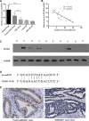
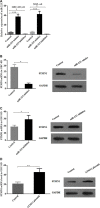
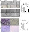
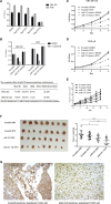
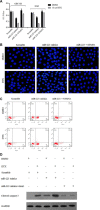
Results: miR-223 was decreased in GBC tissues and cell lines, and ectopic miR- 223 expression exhibited multiple anti-tumorigenic effects in GBC cells, including decreased proliferation, migration and invasion in vitro. However, treatment with a miR-223 inhibitor increased cell viability. We determined that STMN1 was negatively correlated with and regulated by miR-223 in GBC. miR-223 increased GBC sensitivity to docetaxel in vitro and in vivo, and the induced sensitivity to docetaxel was suppressed by the restoration of STMN1 expression.
Methods: We examined miR-223 expression in GBC tissue and GBC cell lines using qRT-PCR. The effects of modulated miR-223 expression in GBC cells were assayed using Cell Counting Kit-8 (CCK8), flow cytometry, and wound-healing and invasion assays. Susceptibility to docetaxel was evaluated in miR-223/STMN1-modulated GBC cells and xenograft tumor models. The protein expression of relevant genes was examined by Western blotting.
Conclusions: These findings indicated that miR-223 might serve as an onco-suppressor that enhances susceptibility to docetaxel by downregulating STMN1 in GBC, highlighting its promising therapeutic value.
Keywords: STMN1; gallbladder cancer; malignancy; miR-223.
Conflict of interest statement
All authors declare no conflicts of interest. All authors declare no previous presentation of the manuscript.
Figures

Similar articles
-
MiR-31 regulates the cisplatin resistance by targeting Src in gallbladder cancer.Oncotarget. 2016 Dec 13;7(50):83060-83070. doi: 10.18632/oncotarget.13067. Oncotarget. 2016. PMID: 27825112 Free PMC article.
-
Downregulation of stathmin 1 in human gallbladder carcinoma inhibits tumor growth in vitro and in vivo.Sci Rep. 2016 Jun 28;6:28833. doi: 10.1038/srep28833. Sci Rep. 2016. PMID: 27349455 Free PMC article.
-
Long non-coding RNA PVT1 promotes tumor progression by regulating the miR-143/HK2 axis in gallbladder cancer.Mol Cancer. 2019 Mar 2;18(1):33. doi: 10.1186/s12943-019-0947-9. Mol Cancer. 2019. PMID: 30825877 Free PMC article.
-
MicroRNA aberrations: An emerging field for gallbladder cancer management.World J Gastroenterol. 2016 Feb 7;22(5):1787-99. doi: 10.3748/wjg.v22.i5.1787. World J Gastroenterol. 2016. PMID: 26855538 Free PMC article. Review.
-
Genetic Factors and MicroRNAs in the Development of Gallbladder Cancer: The Prospective Clinical Targets.Curr Drug Targets. 2024;25(6):375-387. doi: 10.2174/0113894501182288240319074330. Curr Drug Targets. 2024. PMID: 38544392 Review.
Cited by
-
LINC01006 promotes cell proliferation and metastasis in pancreatic cancer via miR-2682-5p/HOXB8 axis.Cancer Cell Int. 2019 Dec 2;19:320. doi: 10.1186/s12935-019-1036-2. eCollection 2019. Cancer Cell Int. 2019. PMID: 31827394 Free PMC article.
-
Overview of current targeted therapy in gallbladder cancer.Signal Transduct Target Ther. 2020 Oct 7;5(1):230. doi: 10.1038/s41392-020-00324-2. Signal Transduct Target Ther. 2020. PMID: 33028805 Free PMC article. Review.
-
Low serum miR-223 expression predicts poor outcome in patients with acute myeloid leukemia.J Clin Lab Anal. 2020 Mar;34(3):e23096. doi: 10.1002/jcla.23096. Epub 2019 Nov 6. J Clin Lab Anal. 2020. PMID: 31691380 Free PMC article.
-
Changes of serum miR-223-3p in patients with oral cancer treated with TPF regimen and the prognosis.Oncol Lett. 2020 Mar;19(3):2527-2532. doi: 10.3892/ol.2020.11258. Epub 2020 Jan 24. Oncol Lett. 2020. PMID: 32194755 Free PMC article.
-
Reiterating the Emergence of Noncoding RNAs as Regulators of the Critical Hallmarks of Gall Bladder Cancer.Biomolecules. 2021 Dec 8;11(12):1847. doi: 10.3390/biom11121847. Biomolecules. 2021. PMID: 34944491 Free PMC article. Review.
References
-
- Alvarez H, Corvalan A, Roa JC, Argani P, Murillo F, Edwards J, Beaty R, Feldmann G, Hong SM, Mullendore M, Roa I, Ibanez L, Pimentel F, et al. Serial analysis of gene expression identifies connective tissue growth factor expression as a prognostic biomarker in gallbladder cancer. Clin Cancer Res. 2008;14:2631–8. doi: 10.1158/1078-0432.CCR-07-1991. - DOI - PubMed
MeSH terms
Substances
LinkOut - more resources
Full Text Sources
Other Literature Sources
Medical
Miscellaneous

