

Connexin 43 is overexpressed in human fetal membrane defects after fetoscopic surgery
- PMID: 27568096
- PMCID: PMC5082503
- DOI: 10.1002/pd.4917
Connexin 43 is overexpressed in human fetal membrane defects after fetoscopic surgery
Abstract
Objective: We examined whether surgically induced membrane defects elevate connexin 43 (Cx43) expression in the wound edge of the amniotic membrane (AM) and drives structural changes in collagen that affects healing after fetoscopic surgery.
Method: Cell morphology and collagen microstructure was investigated by scanning electron microscopy and second harmonic generation in fetal membranes taken from women who underwent fetal surgery. Immunofluoresence and real-time quantitative polymerase chain reaction was used to examine Cx43 expression in control and wound edge AM.
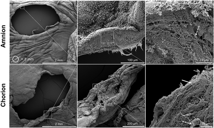
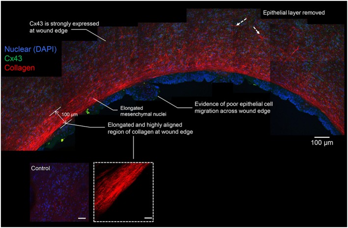
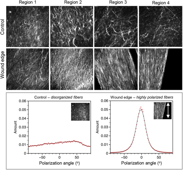
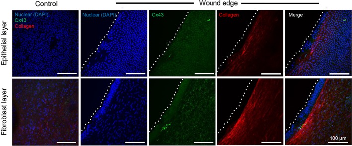
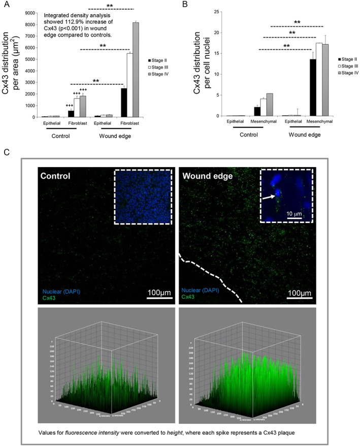
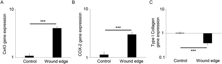
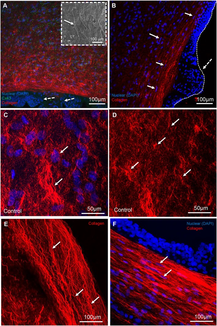
Results: Scanning electron microscopy showed dense, helical patterns of collagen fibrils in the wound edge of the fetal membrane. This arrangement changed in the fibroblast layer with evidence of collagen fibrils that were highly polarised along the wound edge but not in control membranes. Cx43 was increased by 112.9% in wound edge AM compared with controls (p < 0.001), with preferential distribution in the fibroblast layer compared with the epithelial layer (p < 0.01). In wound edge AM, mesenchymal cells had a flattened morphology, and there was evidence of poor epithelial migration across the defect. Cx43 and COX-2 expression was significantly increased in wound edge AM compared with controls (p < 0.001).
Conclusion: Overexpression of Cx43 in the AM after fetal surgery induces morphological and structural changes in the collagenous matrix that may interfere with normal healing mechanisms. © 2016 The Authors. Prenatal Diagnosis published by John Wiley & Sons, Ltd.
© 2016 The Authors. Prenatal Diagnosis published by John Wiley & Sons, Ltd.
Figures
Similar articles
-
Trauma induces overexpression of Cx43 in human fetal membrane defects.Prenat Diagn. 2017 Sep;37(9):899-906. doi: 10.1002/pd.5104. Epub 2017 Aug 1. Prenat Diagn. 2017. PMID: 28664994 Free PMC article.
-
Cx43 mediates changes in myofibroblast contraction and collagen release in human amniotic membrane defects after trauma.Sci Rep. 2021 Aug 18;11(1):16975. doi: 10.1038/s41598-021-94767-4. Sci Rep. 2021. PMID: 34408164 Free PMC article.
-
Tensile strain increased COX-2 expression and PGE2 release leading to weakening of the human amniotic membrane.Placenta. 2014 Dec;35(12):1057-64. doi: 10.1016/j.placenta.2014.09.006. Epub 2014 Sep 19. Placenta. 2014. PMID: 25280972
-
Minimally Invasive Fetal Surgery.Clin Perinatol. 2017 Dec;44(4):729-751. doi: 10.1016/j.clp.2017.08.001. Epub 2017 Sep 23. Clin Perinatol. 2017. PMID: 29127956 Review.
-
Obstetric outcomes after fetal intervention - a single-center descriptive review.J Matern Fetal Neonatal Med. 2022 Dec;35(25):7102-7108. doi: 10.1080/14767058.2021.1943658. Epub 2021 Jul 11. J Matern Fetal Neonatal Med. 2022. PMID: 36411675 Review.
Cited by
-
Perinatal Outcomes after Fetal Endoscopic Tracheal Occlusion for Isolated Congenital Diaphragmatic Hernia: Rapid Review.Rev Bras Ginecol Obstet. 2022 Jan;44(1):74-82. doi: 10.1055/s-0041-1740596. Epub 2022 Jan 29. Rev Bras Ginecol Obstet. 2022. PMID: 35092962 Free PMC article. Clinical Trial.
-
Comprehensive quantitative characterization of the human term amnion proteome.Matrix Biol Plus. 2021 Sep 21;12:100084. doi: 10.1016/j.mbplus.2021.100084. eCollection 2021 Dec. Matrix Biol Plus. 2021. PMID: 34765964 Free PMC article.
-
Trauma induces overexpression of Cx43 in human fetal membrane defects.Prenat Diagn. 2017 Sep;37(9):899-906. doi: 10.1002/pd.5104. Epub 2017 Aug 1. Prenat Diagn. 2017. PMID: 28664994 Free PMC article.
-
Tissuepatch is biocompatible and seals iatrogenic membrane defects in a rabbit model.Prenat Diagn. 2018 Jan;38(2):99-105. doi: 10.1002/pd.5191. Epub 2017 Dec 11. Prenat Diagn. 2018. PMID: 29178347 Free PMC article.
References
-
- Slaghekke F, Lopriore E, Lewi L, et al. Fetoscopic laser coagulation of the vascular equator versus selective coagulation for twin‐to‐twin transfusion syndrome: an open‐label randomised controlled trial. Lancet 2014;383(9935):2144–51. - PubMed
-
- Deprest J, Van Schoubroeck D, Van Ballaer P, et al. Alternative access for fetoscopic Nd:YAG laser in TTS with anterior placenta. US Obstet Gynecol 1998;347–55. - PubMed
-
- Gratacos E, Sanin‐Blair J, Lewi L, et al. A histological study of fetoscopic membrane defects to document membrane healing. Placenta 2006;27:452–6. - PubMed
-
- Mallik AS, Fichter MA, Rieder S, et al. Fetoscopic closure of punctured fetal membranes with acellular human amnion plugs in a rabbit model. Obstet Gynecol 2007;110(5):1121–9. - PubMed
MeSH terms
Substances
Grants and funding
LinkOut - more resources
Full Text Sources
Other Literature Sources
Research Materials
