. 2016 Aug 24;36(34):8815-25.
doi: 10.1523/JNEUROSCI.0345-16.2016.

Chimeric Glutamate Receptor Subunits Reveal the Transmembrane Domain Is Sufficient for NMDA Receptor Pore Properties but Some Positive Allosteric Modulators Require Additional Domains

Affiliations

Chimeric Glutamate Receptor Subunits Reveal the Transmembrane Domain Is Sufficient for NMDA Receptor Pore Properties but Some Positive Allosteric Modulators Require Additional Domains

Timothy J Wilding et al. J Neurosci. .

Abstract

NMDA receptors are ligand-gated ion channels that underlie transmission at excitatory synapses and play an important role in regulating synaptic strength and stability. Functional NMDA receptors require two copies of the GluN1 subunit coassembled with GluN2 (and/or GluN3) subunits into a heteromeric tetramer. A diverse array of allosteric modulators can upregulate or downregulate NMDA receptor activity. These modulators include both synthetic compounds and endogenous modulators, such as cis-unsaturated fatty acids, 24(S)-hydroxycholesterol, and various neurosteroids. To evaluate the structural requirements for the formation and allosteric modulation of NMDA receptor pores, we have replaced portions of the rat GluN1, GluN2A, and GluN2B subunits with homologous segments from the rat GluK2 kainate receptor subunit. Our results with these chimeric constructs show that the NMDA receptor transmembrane domain is sufficient to account for most pore properties, but that regulation by some allosteric modulators requires additional cytoplasmic or extracellular domains.

Significance statement: Glutamate receptors mediate excitatory synaptic transmission by forming cation channels through the membrane that open upon glutamate binding. Although many compounds have been identified that regulate glutamate receptor activity, in most cases the detailed mechanisms that underlie modulation are poorly understood. To identify what parts of the receptor are essential for pore formation and sensitivity to allosteric modulators, we generated chimeric subunits that combined segments from NMDA and kainate receptors, subtypes with distinct pharmacological profiles. Surprisingly, our results identify separate domain requirements for allosteric potentiation of NMDA receptor pores by pregnenolone sulfate, 24(S)-hydroxycholesterol, and docosahexaenoic acid, three endogenous modulators derived from membrane constituents. Understanding where and how these compounds act on NMDA receptors should aid in designing better therapeutic agents.

Keywords: carboxy terminal domain; docosahexaenoic acid; palmitoylation.


Figures

Figure 1.
Figure 1.
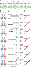
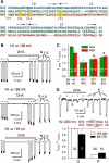
Heteromeric subunit chimeras form functional NMDA receptor pores. A, Protein sequence at the five joints used to construct the chimeric subunits in B. B, Simplified diagram of the four domains in two chimeric subunits of a heteromeric receptor highlighting the pore loop and α helical segments in the TMD derived from the GluN1 (blue) or GluN2 (red) subunit. Non-TMD portions of each chimera derived from the GluK2 kainate receptor subunit are shown in black. Subunits with N1 and N2 extracellular domains are presumed to be adjacent in functional tetramers. For clarity, the subunits are positioned opposite each other in the diagram. C, Mean peak whole-cell agonist-evoked current (± SEM) recorded at −80 mV in HEK 293 cells transiently transfected with chimeric subunit cDNAs and activated with either 10 μm kainate (LBD from GluK2) or 10 μm NMDA and 10 μm glycine (LBDs from NMDA receptor subunits). Agonist-evoked inward currents were significant (i.e., different from zero, t-statistic) for all of the coexpressed construct combinations, but not for any of the chimeric constructs expressed alone. D, I–V for agonist-evoked current recorded as the membrane potential was ramped from −110 to +60 mV at 0.75 mV ms −1 in the presence (red) or absence (black) of 1 mm external magnesium. All recordings from GluN1/2B chimeras. Voltage axis ticks, 50 mV; current axis ticks, 200 pA; except TMD, 500 pA.
Figure 2.
Figure 2.
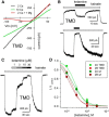
Calcium permeability and ketamine block of chimeric channels. A, Whole-cell current evoked by 10 μm NMDA plus 10 μm glycine as a function of voltage as membrane potential was ramped from −110 to +60 mV at 0.75 mV ms −1 in 160 mm NaCl, 10 mm HEPES with 2 mm CaCl2 (black), or with 10 mm CaCl2 (green), or with 2 mm CaCl2 and 1 mm MgCl2 (red). Note the rightward shift in reversal potential in elevated calcium. GluN1/2A TMD. B, Exposure to 30 μm ketamine (black bars) blocks current evoked by 10 μm kainate (open bars) at −80 mV, and to a lesser extent at +40 mV. GluN1/2A TMD. C, Whole-cell current evoked by 10 μm kainate is blocked progressively by 1, 3, 10, and 30 μm katamine. GluN1/2A TMD. D, Current in the presence of ketamine as a fraction of control (mean ± SEM) is plotted as a function of ketamine concentration. Smooth curves are best fits of 1/(1 + [(ketamine)/IC50]n) where IC50 is the concentration producing half-maximal inhibition and n is the slope factor. Values for chimeric constructs (N1/2A: IC50 = 2.6 ± 0.2, n = 1.9 ± 0.3, 7 cells; N1/2B: IC50 = 1.7 ± 0.1, n = 2.0 ± 0.3, 9 cells) were not significantly different from those of the respective wild-type subunits (N1/2A: IC50 = 2.0 ± 0.1 μm, n = 1.4 ± 0.2, 13 cells; N1/2B: IC50 = 1.6 ± 0.2, n = 1.7 ± 0.2, 10 cells) by t test.
Figure 3.
Figure 3.
Potentiation by 24(S)-HC only requires the NMDA receptor TMD. A, Exposure to 10 μm 24(S)-HC (shaded bar) potentiated whole-cell current evoked by 10 μm kainate (open bars) in an HEK cell transfected with the N1/N2B TMD chimera. B, Minimal effect of 24(S)-HC on the N1/N2B ATD+LBD chimera activated by 10 μm NMDA plus 10 μm glycine. C, Plot of current (mean ± SEM) evoked after exposure to 24(S)-HC as a fraction of control current before 24(S)-HC treatment. *, Significant difference from wild-type GluN1/2B was observed for homomeric GluK2 (Q or R) and for the N1/N2B ATD+LBD chimera, which has an unedited (Q) kainate receptor TMD but not for chimeric constructs with the NMDA receptor LBD+TMD or TMD alone (p < 0.001, ANOVA on ranks with post hoc Dunn's test).
Figure 4.
Figure 4.
PS potentiates chimeric channels that include the NMDA receptor LBD and TMD. A, B, Exposure to 100 μm PS (shaded bar) potentiates whole-cell current evoked by 10 μm NMDA plus 10 μm glycine in an HEK cell transfected with the N1/N2B LBD+TMD chimeric subunits (A), but not in a cell transfected with the N1/N2B TMD-alone chimera (B). C, Plot of current (mean ± SEM) evoked during exposure to PS as a fraction of control current before PS treatment. *, Significant difference from wild-type GluN1/2B was observed for homomeric GluK2 (Q or R), for heteromeric chimeras with the N1/2B TMD and linkers without the LBD and for the N1/N2B ATD+LBD chimera, which has an unedited (Q) kainate receptor TMD; however, potentiation for the chimeric construct with the NMDA receptor LBD+TMD was not significantly different from N1/2B. (p < 0.001, ANOVA on ranks with post hoc Dunn's test).
Figure 5.
Figure 5.
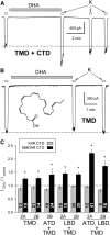
DHA potentiates chimeric channels that include the NMDA receptor TMD and CTD. A, Exposure to 15 μm DHA (shaded bar) potentiated whole-cell current evoked by 10 μm kainate (open bars) in an HEK cell transfected with the N1/N2B TMD+CTD chimera. B, Lack of effect of DHA on the N1/N2B TMD chimera. C, Plot of current (mean ± SEM) evoked immediately after exposure to DHA as a fraction of control current before DHA treatment. *, Significant potentiation for chimeric constructs with the NMDA receptor TMD+CTD, but not for chimeras that included the NMDA receptor TMD and linkers without the CTD (p < 0.05, Mann–Whitney rank-sum tests).
Figure 6.
Figure 6.
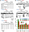
DHA potentiation requires the GluN2 CTD. A, Exposure to 15 μm DHA (shaded bars) did not potentiate whole-cell currents evoked by 10 μm NMDA plus 10 μm glycine (open bars) in HEK cells cotransfected with truncated GluN1840 and GluN2B846 (top trace), or for cotransfection of a wild-type and a truncated subunit: GluN1840/GluN2Bwt (middle trace), GluN1wt/GluN2B846 (bottom trace). B, Exposure to 15 μm DHA (shaded bar) potentiated whole-cell current evoked by 10 μm NMDA plus 10 μm glycine (open bars) in an HEK cell transfected with a GluN1 chimera bearing a GluK2 CTD and wild-type GluN2B (top); lack of effect of DHA on wild-type GluN1 with the GluN2B chimera bearing a GluK2 CTD (bottom). C, Plot of current (mean ± SEM) evoked immediately after exposure to DHA as a fraction of control current before DHA treatment for wild-type and truncated or chimeric NMDA receptor subunits. *, Significant difference from wild-type (p < 0.0001, ANOVA on ranks with post hoc Dunn's test) and not significantly different from no effect (i.e., IDHA/Icontrol = 1, t-statistic). # denotes that for the N1 (K2CTD)/N2 wild-type combinations IDHA/Icontrol was significantly >1 but also significantly less than that for wild-type receptors. Inset, Proximal CTD sequences of GluN1, GluN2A, and GluN2B. Shading denotes truncation after N1 H840, 2A L845, and 2B F846.
Figure 7.
Figure 7.
The GluN2 proximal CTD supports NMDA receptor TMD potentiation. A, Sequence of the GluN1.1 CTD (blue) noting joints for the C0, C1, and C2 splice cassettes; alignment of the membrane-proximal portions of GluN2A (green) and GluN2B (red) noting location of conserved Tyr (Y) and Cys (C1, 2, 3) residues. B, Exposure to 15 μm DHA (shaded bar) did not potentiate whole-cell current evoked by 10 μm NMDA plus 10 μm glycine (open bars) in HEK cells cotransfected with wild-type GluN1.1 and GluN2B truncated after S870 (top trace), but did potentiate for N2B truncation after E878 (middle trace) or V931 (bottom trace). C, Plot of current (mean ± SEM) evoked immediately after exposure to DHA as a fraction of control current before DHA treatment for wild-type and GluN2 truncated NMDA receptors. *, Significant difference from wild-type (p < 0.0001, ANOVA on ranks with post hoc Dunn's test) and not significant difference from no effect (IDHA/Icontrol = 1, t-statistic). # denotes that for the other N2 truncations IDHA/I control was significantly >1 but also significantly less than for wild-type receptors. D, Exposure to 15 μm DHA (shaded bar) inhibited whole-cell current evoked by 10 μm kainate (open bars) in an HEK cell transfected with chimeric GluK2(R) bearing a CTD from GluN2B. Plot of current (mean ± SEM) evoked at the end of DHA exposure as a fraction of control current before DHA treatment. *, DHA produced weak but significant (p = 0.030) inhibition of unedited (Q) chimeric GluK2 subunits and strong inhibition of edited (R) chimeric receptors that was not different from wild-type GluK2(R) (p = 0.148). Inset, Proximal CTD sequences of GluK2 and GluN2B indicating the position of the K2/2B joint.
Figure 8.
Figure 8.
Proximal GluN2 CTD substitutions prevent DHA potentiation. A, Plot of current (mean ± SEM) evoked immediately after exposure to DHA as a fraction of control current before DHA treatment for NMDA receptors with wild-type GluN1 and the indicated substitutions to full-length GluN2. Residues highlighted in yellow are wild-type. *, Significant difference from wild-type (p < 0.0001, ANOVA on ranks with post hoc Dunn's test) and not significant difference from no effect (IDHA/Icontrol = 1, t-statistic). # denotes that for the N1 wt/N2A Y842F combination IDHA/Icontrol was significantly >1 but also significantly less than for wild-type receptors. B, Traces show the quadruple FAAA mutant lacked potentiation by DHA (top trace). Potentiation was preserved for the Y842F substitution, in some cases reaching the level observed for wild-type receptors (bottom trace).
Figure 9.
Figure 9.
NMDA receptor model with GluN2A proximal CTD. Homology model for GluN1/GluN2A generated by Modeler (Eswar et al., 2008) using PDB files 4PE5 (Karakas and Furukawa, 2014) and 4TLM (Lee et al., 2014) as templates and with twofold symmetry constraint for the A/C and B/D subunits. Prediction for the proximal CTD generated by PEP-FOLD (Thévenet et al., 2012) with subsequent loop-refinement in Modeler. Palmitoylation at C1, 2, and 3 is expected to tether the proximal CTD to the membrane.

Similar articles

Cited by

References

    1. Ayalon G, Stern-Bach Y. Functional assembly of AMPA and kainate receptors is mediated by several discrete protein-protein interactions. Neuron. 2001;31:103–113. doi: 10.1016/S0896-6273(01)00333-6. - DOI - PubMed
    1. Burnashev N, Schoepfer R, Monyer H, Ruppersberg JP, Günther W, Seeburg PH, Sakmann B. Control by asparagine residues of calcium permeability and magnesium blockade in the NMDA receptor. Science. 1992;257:1415–1419. doi: 10.1126/science.1382314. - DOI - PubMed
    1. Casado M, Ascher P. Opposite modulation of NMDA receptors by lysophospholipids and arachidonic acid: common features with mechanosensitivity. J Physiol. 1998;513:317–330. doi: 10.1111/j.1469-7793.1998.317bb.x. - DOI - PMC - PubMed
    1. Cole C, Barber JD, Barton GJ. The Jpred 3 secondary structure prediction server. Nucleic Acids Res. 2008;36:W197–W201. doi: 10.1093/nar/gkn238. - DOI - PMC - PubMed
    1. Collingridge GL, Volianskis A, Bannister N, France G, Hanna L, Mercier M, Tidball P, Fang G, Irvine MW, Costa BM, Monaghan DT, Bortolotto ZA, Molnár E, Lodge D, Jane DE. The NMDA receptor as a target for cognitive enhancement. Neuropharmacology. 2013;64:13–26. doi: 10.1016/j.neuropharm.2012.06.051. - DOI - PMC - PubMed

Publication types

MeSH terms

LinkOut - more resources