

Foxp3 enhances HIF-1α target gene expression in human bladder cancer through decreasing its ubiquitin-proteasomal degradation
- PMID: 27557492
- PMCID: PMC5323164
- DOI: 10.18632/oncotarget.11395
Foxp3 enhances HIF-1α target gene expression in human bladder cancer through decreasing its ubiquitin-proteasomal degradation
Abstract
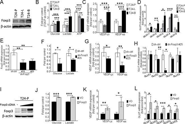
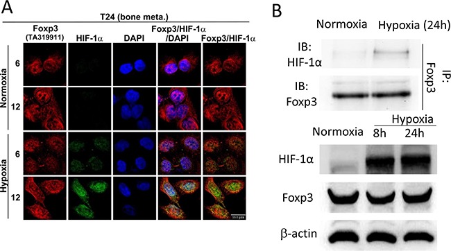
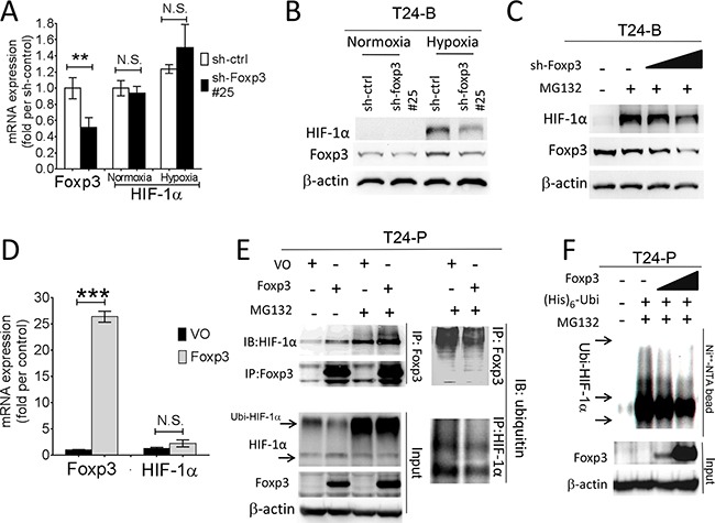
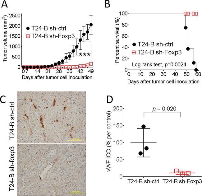
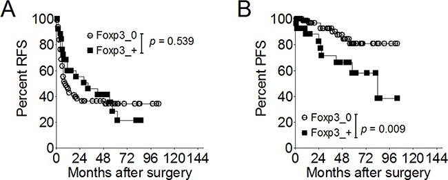
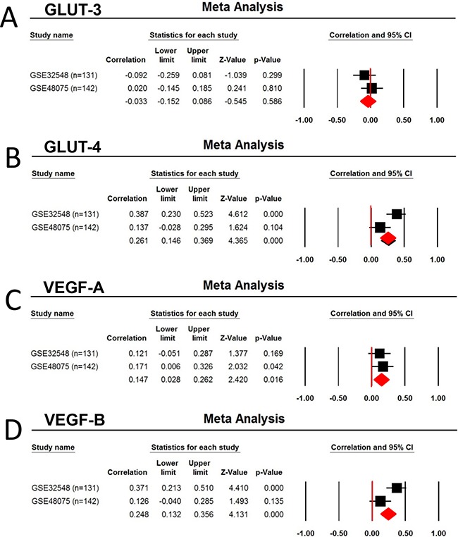
Hypoxia-inducible factor-1α (HIF-1α) can control a transcriptional factor forkhead box P3 (Foxp3) protein expression in T lymphocyte differentiation through proteasome-mediated degradation. In this study, we unveil a reverse regulatory mechanism contributing to bladder cancer progression; Foxp3 expression attenuates HIF-1α degradation. We first demonstrated that Foxp3 expression positively correlates with the metastatic potential in T24 cells and can increase the expression of HIF-1α-target genes, such as vascular endothelial growth factor (VEGF) and glucose transporter (GLUT). Foxp3 protein can bind with HIF-1α, particularly under hypoxia. In vivo ubiquination assay demonstrated that Foxp3 can decrease HIF-1α degradation in a dose-dependent manner. Knocking-down of Foxp3 expression blocks in vivo tumor growth in mice and prolongs mice's survival, which is associated with von Willebrand factor expression. Thirty-three of 145 (22.8 %) bladder tumors exhibit Foxp3 expression. Foxp3 expression is an independent predictor for disease progression in superficial bladder cancer patients (p = 0.032), associated with less number of intratumoral CD8+ lymphocyte. The metaanalysis from 2 published datasets showed Foxp3 expression is positively associated with GLUT-4,-9, and VEGF-A, B-, D expression. This reverse post-translational regulation of HIF-1α protein by Foxp3 provides a new potential target for developing new therapeutic strategy for bladder cancer.
Keywords: Foxp3; bladder neoplasms; glycolysis; immunohistochemistry; prognosis.
Conflict of interest statement
The authors declare no financial disclosure.
Figures

Similar articles
-
The Role of Hypoxia-inducible Factor-1 in Bladder Cancer.Curr Mol Med. 2024;24(7):827-834. doi: 10.2174/1566524023666230720163448. Curr Mol Med. 2024. PMID: 37475553 Free PMC article. Review.
-
Cyclin-dependent kinase inhibitor, P276-00, inhibits HIF-1α and induces G2/M arrest under hypoxia in prostate cancer cells.Prostate Cancer Prostatic Dis. 2012 Mar;15(1):15-27. doi: 10.1038/pcan.2011.51. Epub 2011 Nov 15. Prostate Cancer Prostatic Dis. 2012. PMID: 22083267
-
Effects of YC-1 on hypoxia-inducible factor 1 alpha in hypoxic human bladder transitional carcinoma cell line T24 cells.Urol Int. 2012;88(1):95-101. doi: 10.1159/000331881. Epub 2011 Oct 25. Urol Int. 2012. PMID: 22041818
-
TARBP2 Suppresses Ubiquitin-Proteasomal Degradation of HIF-1α in Breast Cancer.Int J Mol Sci. 2021 Dec 24;23(1):208. doi: 10.3390/ijms23010208. Int J Mol Sci. 2021. PMID: 35008634 Free PMC article.
-
Hypoxia-inducible factor 1α plays a predominantly negative role in regulatory T cell functions.J Leukoc Biol. 2018 Nov;104(5):911-918. doi: 10.1002/JLB.MR1217-481R. Epub 2018 Jun 14. J Leukoc Biol. 2018. PMID: 29901858 Review.
Cited by
-
FOXP3 Isoforms Expression in Cervical Cancer: Evidence about the Cancer-Related Properties of FOXP3Δ2Δ7 in Keratinocytes.Cancers (Basel). 2023 Jan 5;15(2):347. doi: 10.3390/cancers15020347. Cancers (Basel). 2023. PMID: 36672296 Free PMC article.
-
The Role of Hypoxia-inducible Factor-1 in Bladder Cancer.Curr Mol Med. 2024;24(7):827-834. doi: 10.2174/1566524023666230720163448. Curr Mol Med. 2024. PMID: 37475553 Free PMC article. Review.
-
Prediction of the Immune Phenotypes of Bladder Cancer Patients for Precision Oncology.IEEE Open J Eng Med Biol. 2022 Apr 15;3:47-57. doi: 10.1109/OJEMB.2022.3163533. eCollection 2022. IEEE Open J Eng Med Biol. 2022. PMID: 35519421 Free PMC article.
-
Upregulation of Cytotoxic T-Lymphocyte-Associated Protein 4 and Forkhead Box P3 Transcripts in Peripheral Blood of Patients with Bladder Cancer.Iran J Med Sci. 2021 Sep;46(5):339-346. doi: 10.30476/ijms.2020.84462.1426. Iran J Med Sci. 2021. PMID: 34539008 Free PMC article.
-
Advances in tumor microenvironment and underlying molecular mechanisms of bladder cancer: a systematic review.Discov Oncol. 2024 Apr 11;15(1):111. doi: 10.1007/s12672-024-00902-8. Discov Oncol. 2024. PMID: 38602556 Free PMC article. Review.
References
-
- Torre LA, Bray F, Siegel RL, Ferlay J, Lortet-Tieulent J, Jemal A. Global cancer statistics 2012. CA Cancer J Clin. 2015;65:87–108. - PubMed
-
- Wu XR. Urothelial tumorigenesis: a tale of divergent pathways. Nat Rev Cancer. 2005;5:713–25. - PubMed
-
- Roupret M, Zigeuner R, Palou J, Boehle A, Kaasinen E, Sylvester R, Babjuk M, Oosterlinck W. European guidelines for the diagnosis and management of upper urinary tract urothelial cell carcinomas: 2011 update. Eur Urol. 2011;59:584–594. - PubMed
-
- Stenzl A, Cowan NC, De Santis M, Kuczyk MA, Merseburger AS, Ribal MJ, Sherif A, Witjes J A. Treatment of muscle-invasive and metastatic bladder cancer: update of the EAU guidelines. Eur Urol. 2011;59:1009–1018. - PubMed
-
- Babjuk M, Oosterlinck W, Sylvester R, Kaasinen E, Bohle A, Palou-Redorta J, Roupret M. EAU guidelines on non-muscle-invasive urothelial carcinoma of the bladder the 2011 update. Eur Urol. 2011;59:997–1008. - PubMed
MeSH terms
Substances
LinkOut - more resources
Full Text Sources
Other Literature Sources
Medical
Research Materials
