. 2016 Aug 24;12(8):e1005825.
doi: 10.1371/journal.ppat.1005825. eCollection 2016 Aug.

The Prolyl Isomerase Pin1 Promotes the Herpesvirus-Induced Phosphorylation-Dependent Disassembly of the Nuclear Lamina Required for Nucleocytoplasmic Egress

Affiliations

The Prolyl Isomerase Pin1 Promotes the Herpesvirus-Induced Phosphorylation-Dependent Disassembly of the Nuclear Lamina Required for Nucleocytoplasmic Egress

Jens Milbradt et al. PLoS Pathog. .

Abstract

The nuclear lamina lines the inner nuclear membrane providing a structural framework for the nucleus. Cellular processes, such as nuclear envelope breakdown during mitosis or nuclear export of large ribonucleoprotein complexes, are functionally linked to the disassembly of the nuclear lamina. In general, lamina disassembly is mediated by phosphorylation, but the precise molecular mechanism is still not completely understood. Recently, we suggested a novel mechanism for lamina disassembly during the nuclear egress of herpesviral capsids which involves the cellular isomerase Pin1. In this study, we focused on mechanistic details of herpesviral nuclear replication to demonstrate the general importance of Pin1 for lamina disassembly. In particular, Ser22-specific lamin phosphorylation consistently generates a Pin1-binding motif in cells infected with human and animal alpha-, beta-, and gammaherpesviruses. Using nuclear magnetic resonance spectroscopy, we showed that binding of Pin1 to a synthetic lamin peptide induces its cis/trans isomerization in vitro. A detailed bioinformatic evaluation strongly suggests that this structural conversion induces large-scale secondary structural changes in the lamin N-terminus. Thus, we concluded that a Pin1-induced conformational change of lamins may represent the molecular trigger responsible for lamina disassembly. Consistent with this concept, pharmacological inhibition of Pin1 activity blocked lamina disassembly in herpesvirus-infected fibroblasts and consequently impaired virus replication. In addition, a phospho-mimetic Ser22Glu lamin mutant was still able to form a regular lamina structure and overexpression of a Ser22-phosphorylating kinase did not induce lamina disassembly in Pin1 knockout cells. Intriguingly, this was observed in absence of herpesvirus infection proposing a broader importance of Pin1 for lamina constitution. Thus, our results suggest a functional model of similar events leading to disassembly of the nuclear lamina in response to herpesviral or inherent cellular stimuli. In essence, Pin1 represents a regulatory effector of lamina disassembly that promotes the nuclear pore-independent egress of herpesviral capsids.


Conflict of interest statement

The authors have declared that no competing interests exist.

Figures

Fig 1
Fig 1. Site-specific phosphorylation of lamin A/C at Ser22 and Ser392 in herpesvirus-infected primary fibroblasts analysed by western blot.
HFFs were infected with different herpesviruses belonging to subfamily alpha (A), beta (B), and gamma (C). For HSV-1, HCMV, RhCMV, and MHV-68 infections, increasing MOIs were applied in a range between approx. 0.1–1. Cells were lysed at 24 hpi (HSV-1) or 72 hpi (HCMV, RhCMV, and MHV-68). Virus-positive carrier cells were cocultivated in serial dilutions with host HFFs for VZV and HHV-6A infections, producing ~60% and ~30% of infected cells, respectively. Cells were lysed at 72 h post-cocultivation. In all cases, total lysates were subjected to standard Western blot analysis for detection of lamin A/C phosphorylated at Ser22 (pSer22) or Ser392 (pSer392) (upper two panels), total lamin A/C (third panels), viral marker proteins (fourth and fifth panels), and loading control β-actin (lower panels). *, detection of a viral 17 kDa protein using a polyspecific MHV-68 post-infection murine antiserum. Ser22 and Ser392 phosphorylation signal intensities were quantified and related to the lamin A/C and β-actin signals by densitometry using AIDA image analyser.
Fig 2
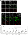
Fig 2. Ser22-specific phosphorylation of lamin A/C in herpesvirus-infected primary fibroblasts analysed by confocal imaging.
(A) HFFs were infected with different herpesviruses or remained uninfected (mock) as indicated. Cells were fixed at 24 hpi (HSV-1 and HSV-1 ΔUS3) or 72 hpi (VZV, HCMV AD, HCMV TB, HCMV ΔUL97, HHV-6A, and RhCMV) followed by immunofluorescence analysis using phospho-specific antibodies to detect lamin A/C phosphorylated at Ser22 in red. Staining of viral proteins or the green fluorescent protein (GFP) served as viral markers in green. Cell nuclei were counterstained with DAPI (4’,6-diamidino-2-phenylindole). Samples were analysed by confocal microscopy and a representative image of the focal plane is depicted for each setting. Filled arrows, nuclei of virus-positive cells; open arrows, nuclei of virus-positive cells showing increased Ser22 phosphorylation compared to virus-negative cells; scale bars, 30 μm. (B) Median intracellular intensities of lamin A/C phosphorylation. pSer22 signals were determined for infected (white boxes) and surrounding uninfected cells (grey-shaded boxes) as maximum projections of confocal z-series. One representative experiment out of three is depicted for each virus presenting the values of site-specific phosphorylation as box plots. Note, Table 2 contains the mean values ± standard deviation of three independent experiments. Centre lines show the medians with box limits indicating the 25th and 75th percentiles as determined by R software. Whiskers extend 1.5 times the interquartile range from the 25th and 75th percentiles, outliers are represented by circles, and the number of evaluated cells is depicted above each box in brackets. Statistical significance was determined by Student’s t-test (*, P < 0.05; **, P < 0.01; ***, P < 0.001; n.s., not significant, P ≥ 0.05).
Fig 3
Fig 3. Putative role of Pin1 in herpesviral replication.
(A) Conserved Pin1-binding motif in lamin A/C. Amino acid sequences of the lamin A/C precursor from humans (UniProt accession number: P02545), rhesus macaques (F7GLE9), and mice (P48678) were analysed by multiple sequence alignment. Note the 100% conservation of the lamin A/C N-terminus including the Pin1-binding motif. Depicted are the N-terminal 30 amino acids of each sequence. (B) Pin1 upregulation in herpesvirus-infected cells. HFFs were infected with HSV-1, VZV, HCMV, or RhCMV at increasing MOIs. Cells were lysed at 24 hpi (HSV-1) or 72 hpi (VZV, HCMV, and RhCMV). Lysates were subjected to standard Western blot analysis for detection of Pin1 (rabbit mAb-Pin1; upper panels), viral marker proteins (middle panels), and loading control β-actin (lower panels). Pin1 signal intensities were quantified by densitometry using AIDA image analyser. (C) Effect of pharmacological Pin1 inhibition on herpesviral replication efficiency. HFFs were infected with HCMV GFP at a MOI of 0.2 or remained uninfected (mock). The Pin1 inhibitor PiB and the anti-HCMV reference drug ganciclovir (GCV) were added immediately post-infection at 10 μM and 20 μM, respectively. Cells were lysed at 7 days post-infection to perform quantitative GFP fluorometry (n = 3; mean ± standard deviation; statistical significance was determined by Student’s t-test). Potential cytotoxic effects of these compounds in the concentrations used were excluded by microscopic evaluation before cell lysis.
Fig 4
Fig 4. Pin1-induced cis/trans isomerization of lamin A/C.
(A-B) NMR spectroscopy of lamin A/C peptides in the presence or absence of Pin1. Superimposed expanded HN-HN regions of the 2D 1H-1H NOESY spectra are depicted for phosphorylated and unphosphorylated versions of a lamin A/C peptide comprising amino acids 11–40. (A) Phosphorylated peptide prior to (red signals) and after addition of Pin1 (blue signals); note the appearance of exchange peaks originating from an enhanced prolyl cis/trans interconversion rate after addition of Pin1. (B) Unphosphorylated peptide prior to (red signals) and after addition of Pin1 (blue signals); note that no exchange peaks are observed for the unphosphorylated peptide. (C-D) Molecular dynamics (MD) simulation of lamin A/C peptides. (C) A histogram plot of the end-to-end distances for the lamin A/C (1–30) peptides that differ in the phosphorylation state of Ser22 and the isomerization state of Pro23. (D) Representative snapshots from the different MD simulations indicating the most populated conformation of the Ser22/Pro23 trans (left), pSer22/Pro23 trans (middle), and pSer22/Pro23 cis (right) peptide. Ser22 (green) and Pro23 (white) are highlighted as stick presentations.
Fig 5
Fig 5. Lamin phosphorylation by transiently expressed pUL97 kinase is not sufficient for lamina disassembly in Pin1 KO cells.
(A-B) Monitoring of the Pin1 KO in HeLa cells. (A) Total cell lysates of wt and Pin1 KO HeLa cells were subjected to standard Western blot analysis for detection of Pin1 (rabbit pAb-Pin1; upper panel) and loading control β-actin (lower panel). (B) Confocal microscopic images of fixed wt and Pin1 KO HeLa cells stained with rabbit pAb-Pin1 and DAPI. Scale bars, 50 μm. (C) Wt HeLa cells and Pin1 KO HeLa cells were transiently transfected with pcNDA3.1 (vector) or a plasmid coding for HCMV pUL97 fused to GFP. Cells were fixed at 24 h post-transfection followed by counterstaining of cell nuclei with DAPI. Samples were analysed by confocal microscopy. Scale bars, 10 μm. (D) Quantitation of lamin A/C signals. Signal intensities from raw images were measured along the nuclear rim (mean of ≥ 20 cells in each case, ± standard deviation; statistical significance was determined by Student’s t-test).
Fig 6
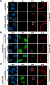
Fig 6. Effect of HCMV pUL97 coexpression on the localization of wild-type lamin A and phospho-mimetic and -deficient mutants.
Wt HeLa cells (A-B) and Pin1 KO HeLa cells (C) were transiently transfected with plasmids coding for HCMV pUL97 fused to GFP and wt or mutant lamin A fused to RFP as indicated. Cells were fixed at 24 h post-transfection followed by counterstaining of cell nuclei with DAPI. Samples were analysed by confocal microscopy. Insets show the magnification of dashed boxes. Scale bars, 10 μm.
Fig 7
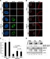
Fig 7. Reduced lamina disassembly upon inhibition of Pin1 activity in HCMV-infected cells.
HFFs were infected with HCMV AD at a MOI of 0.01 or remained uninfected (mock). At 48 hpi, cells were treated with DMSO, 10 μM PiB or 5 μM MBV as indicated. Cells were fixed at 72 hpi followed by immunofluorescence staining using rabbit mAb-lamin A/C to visualize the nuclear lamina in red. Staining of HCMV pUL44 served as a viral marker in green. (A) Representative images of confocal planes demonstrating the effect of inhibitory compounds on the distribution of lamin A/C (raw images). (B) Lamin A/C signals of images depicted in (A) were adjusted to levels of uninfected control cells treated with DMSO for qualitative analysis of lamin A/C distribution. Insets show the magnification of dashed boxes. Open arrows, lamina-depleted areas; scale bars, 10 μm. (C) Quantitation of lamin A/C signals. Signal intensities from raw images were measured along the nuclear rim (mean of 10 cells in each case, ± standard deviation; statistical significance was determined by Student’s t-test). (D) Effect of PiB on the HCMV-induced Ser22 phosphorylation. HFFs were infected with HCMV at MOIs of 0.1 and 1.0. At 48 hpi, cells were treated with DMSO, 10 μM PiB, and 5 μM MBV. Cells were lysed at 72 hpi and lysates were subjected to standard Western blot analysis for detection of lamin A/C phosphorylated at Ser22 (upper panel), total lamin A/C (second panel), viral protein kinase pUL97 (third panel), and loading control β-actin (lower panels).
Fig 8
Fig 8. Pharmacological inhibition of Pin1 results in the accumulation of phosphorylated lamins at the nuclear rim.
HFFs were infected with HCMV AD at a MOI of 0.01 (A-B) or remained uninfected (mock) (C-D). At 48 hpi, cells were treated with DMSO or 10 μM PiB as indicated. Cells were fixed at 72 hpi followed by immunofluorescence staining using a phospho-specific antibody to detect lamin A/C phosphorylated at Ser22. Staining of HCMV pUL44 served as a viral marker in green. (A,C) Representative confocal images illustrate partial relocalization of phosphorylated lamins to the nuclear envelope in cells treated with PiB. Insets show the magnification of dashed boxes. Filled arrows, accumulation of pSer22 lamin A/C at the nuclear envelope; scale bars, 10 μm. (B,D) Cells with lamina-associated pSer22 lamin A/C relative to cells with soluble pSer22 lamin A/C diffusely localized in the nucleoplasm. The percentage of cells showing these phenotypes was determined by scoring ≥ 39 cells for each setting.
Fig 9
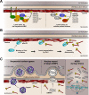
Fig 9. Putative conserved mechanism of Pin1-induced nuclear lamina disassembly.
(A) Site-specific lamin phosphorylation by herpesvirus-encoded or host cell protein kinases. NEC, nuclear egress complex; PKC, protein kinase C; CDK, cyclin-dependent kinase. (B) Lamina disassembly as a direct consequence of a Pin1-mediated conformational change of lamins. (C) Cellular processes which require disassembly of the nuclear lamina. mRNP, messenger ribonucleoprotein complexes; NEBD, nuclear envelope breakdown. Diagram not to scale.

Similar articles

Cited by

References

    1. Dechat T, Adam SA, Taimen P, Shimi T, Goldman RD. Nuclear lamins. Cold Spring Harb Perspect Biol. 2010;2: a000547 10.1101/cshperspect.a000547 - DOI - PMC - PubMed
    1. Gruenbaum Y, Foisner R. Lamins: nuclear intermediate filament proteins with fundamental functions in nuclear mechanics and genome regulation. Annu Rev Biochem. 2015;84: 131–164. 10.1146/annurev-biochem-060614-034115 - DOI - PubMed
    1. Güttinger S, Laurell E, Kutay U. Orchestrating nuclear envelope disassembly and reassembly during mitosis. Nat Rev Mol Cell Biol. 2009;10: 178–191. 10.1038/nrm2641 - DOI - PubMed
    1. Speese SD, Ashley J, Jokhi V, Nunnari J, Barria R, Li Y, et al. Nuclear envelope budding enables large ribonucleoprotein particle export during synaptic Wnt signaling. Cell. 2012;149: 832–846. 10.1016/j.cell.2012.03.032 - DOI - PMC - PubMed
    1. Montpetit B, Weis K. An alternative route for nuclear mRNP export by membrane budding. Science. 2012;336: 809–810. 10.1126/science.1222243 - DOI - PubMed

Publication types

Substances

Grants and funding

This study was funded by grants from Deutsche Forschungsgemeinschaft SFB796 (C3, A2) and MA 1289/8-1, Wilhelm Sander-Stiftung (no. 2011.085.2), Research Foundation Medicine of Univ. Erlangen-Nürnberg (JM-2010), and ELAN program of Univ. Erlangen-Nürnberg (Vi-12-10-29-1-Milbradt; Vi-15-04-07-1-Hutterer). The funders had no role in study design, data collection and analysis, decision to publish, or preparation of the manuscript.

LinkOut - more resources