Identification of Multiple QTLs Linked to Neuropathology in the Engrailed-1 Heterozygous Mouse Model of Parkinson's Disease
- PMID: 27550741
- PMCID: PMC4994027
- DOI: 10.1038/srep31701
Identification of Multiple QTLs Linked to Neuropathology in the Engrailed-1 Heterozygous Mouse Model of Parkinson's Disease
Abstract
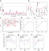
Motor symptoms in Parkinson's disease are attributed to degeneration of midbrain dopaminergic neurons (DNs). Heterozygosity for Engrailed-1 (En1), one of the key factors for programming and maintenance of DNs, results in a parkinsonian phenotype featuring progressive degeneration of DNs in substantia nigra pars compacta (SNpc), decreased striatal dopamine levels and swellings of nigro-striatal axons in the SwissOF1-En1+/- mouse strain. In contrast, C57Bl/6-En1+/- mice do not display this neurodegenerative phenotype, suggesting that susceptibility to En1 heterozygosity is genetically regulated. Our goal was to identify quantitative trait loci (QTLs) that regulate the susceptibility to PD-like neurodegenerative changes in response to loss of one En1 allele. We intercrossed SwissOF1-En1+/- and C57Bl/6 mice to obtain F2 mice with mixed genomes and analyzed number of DNs in SNpc and striatal axonal swellings in 120 F2-En1+/- 17 week-old male mice. Linkage analyses revealed 8 QTLs linked to number of DNs (p = 2.4e-09, variance explained = 74%), 7 QTLs linked to load of axonal swellings (p = 1.7e-12, variance explained = 80%) and 8 QTLs linked to size of axonal swellings (p = 7.0e-11, variance explained = 74%). These loci should be of prime interest for studies of susceptibility to Parkinson's disease-like damage in rodent disease models and considered in clinical association studies in PD.
Conflict of interest statement
The authors declare no competing financial interests.
Figures


Similar articles
-
Nigral transcriptomic profiles in Engrailed-1 hemizygous mouse models of Parkinson's disease reveal upregulation of oxidative phosphorylation-related genes associated with delayed dopaminergic neurodegeneration.Front Aging Neurosci. 2024 Feb 5;16:1337365. doi: 10.3389/fnagi.2024.1337365. eCollection 2024. Front Aging Neurosci. 2024. PMID: 38374883 Free PMC article.
-
Progressive nigrostriatal terminal dysfunction and degeneration in the engrailed1 heterozygous mouse model of Parkinson's disease.Neurobiol Dis. 2015 Jan;73:70-82. doi: 10.1016/j.nbd.2014.09.012. Epub 2014 Oct 2. Neurobiol Dis. 2015. PMID: 25281317 Free PMC article.
-
A WNT1-regulated developmental gene cascade prevents dopaminergic neurodegeneration in adult En1(+/-) mice.Neurobiol Dis. 2015 Oct;82:32-45. doi: 10.1016/j.nbd.2015.05.015. Epub 2015 Jun 3. Neurobiol Dis. 2015. PMID: 26049140
-
Progressive loss of dopaminergic neurons in the ventral midbrain of adult mice heterozygote for Engrailed1: a new genetic model for Parkinson's disease?Parkinsonism Relat Disord. 2008;14 Suppl 2:S107-11. doi: 10.1016/j.parkreldis.2008.04.007. Epub 2008 Jun 27. Parkinsonism Relat Disord. 2008. PMID: 18585951 Review.
-
Dissecting the role of Engrailed in adult dopaminergic neurons--Insights into Parkinson disease pathogenesis.FEBS Lett. 2015 Dec 21;589(24 Pt A):3786-94. doi: 10.1016/j.febslet.2015.10.002. Epub 2015 Oct 13. FEBS Lett. 2015. PMID: 26459030 Free PMC article. Review.
Cited by
-
Nigral transcriptomic profiles in Engrailed-1 hemizygous mouse models of Parkinson's disease reveal upregulation of oxidative phosphorylation-related genes associated with delayed dopaminergic neurodegeneration.Front Aging Neurosci. 2024 Feb 5;16:1337365. doi: 10.3389/fnagi.2024.1337365. eCollection 2024. Front Aging Neurosci. 2024. PMID: 38374883 Free PMC article.
-
Astrocytic Expression of GSTA4 Is Associated to Dopaminergic Neuroprotection in a Rat 6-OHDA Model of Parkinson's Disease.Brain Sci. 2017 Jun 26;7(7):73. doi: 10.3390/brainsci7070073. Brain Sci. 2017. PMID: 28672859 Free PMC article.
-
Loss of One Engrailed1 Allele Enhances Induced α-Synucleinopathy.J Parkinsons Dis. 2019;9(2):315-326. doi: 10.3233/JPD-191590. J Parkinsons Dis. 2019. PMID: 30932894 Free PMC article.
References
-
- Hornykiewicz O. Dopamine (3-hydroxytyramine) and brain function. Pharmacological reviews 18, 925–964 (1966). - PubMed
-
- Riederer P. & Wuketich S. Time course of nigrostriatal degeneration in parkinson’s disease. A detailed study of influential factors in human brain amine analysis. J Neural Transm. 38, 277–301 (1976). - PubMed
Publication types
MeSH terms
Substances
LinkOut - more resources
Full Text Sources
Other Literature Sources
Medical
Miscellaneous

