

Ins and Outs of Multipartite Positive-Strand RNA Plant Viruses: Packaging versus Systemic Spread
- PMID: 27548199
- PMCID: PMC4997590
- DOI: 10.3390/v8080228
Ins and Outs of Multipartite Positive-Strand RNA Plant Viruses: Packaging versus Systemic Spread
Abstract
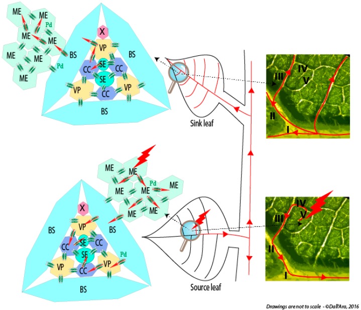
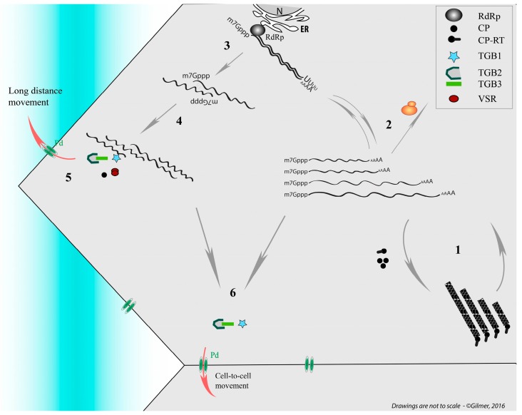
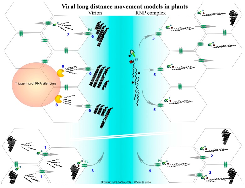
Viruses possessing a non-segmented genome require a specific recognition of their nucleic acid to ensure its protection in a capsid. A similar feature exists for viruses having a segmented genome, usually consisting of viral genomic segments joined together into one viral entity. While this appears as a rule for animal viruses, the majority of segmented plant viruses package their genomic segments individually. To ensure a productive infection, all viral particles and thereby all segments have to be present in the same cell. Progression of the virus within the plant requires as well a concerted genome preservation to avoid loss of function. In this review, we will discuss the "life aspects" of chosen phytoviruses and argue for the existence of RNA-RNA interactions that drive the preservation of viral genome integrity while the virus progresses in the plant.
Keywords: RNA-RNA interaction; genome integrity; phytovirus; segmented genome; systemic movement.
Figures
Similar articles
-
Long-distance movement of helical multipartite phytoviruses: keep connected or die?Curr Opin Virol. 2018 Dec;33:120-128. doi: 10.1016/j.coviro.2018.07.016. Epub 2018 Sep 7. Curr Opin Virol. 2018. PMID: 30199788 Review.
-
Integration of replication and assembly of infectious virions in plant RNA viruses.Curr Opin Virol. 2014 Dec;9:61-6. doi: 10.1016/j.coviro.2014.09.008. Epub 2014 Oct 10. Curr Opin Virol. 2014. PMID: 25308094 Review.
-
Molecular and biological factors regulating the genome packaging in single-strand positive-sense tripartite RNA plant viruses.Curr Opin Virol. 2018 Dec;33:113-119. doi: 10.1016/j.coviro.2018.07.019. Epub 2018 Aug 27. Curr Opin Virol. 2018. PMID: 30165268 Review.
-
Genome packaging by spherical plant RNA viruses.Annu Rev Phytopathol. 2006;44:61-87. doi: 10.1146/annurev.phyto.44.070505.143334. Annu Rev Phytopathol. 2006. PMID: 16480335 Review.
-
Adaptation of positive-strand RNA viruses to plants.Arch Virol Suppl. 1994;9:87-97. doi: 10.1007/978-3-7091-9326-6_10. Arch Virol Suppl. 1994. PMID: 8032285 Review.
Cited by
-
The Strange Lifestyle of Multipartite Viruses.PLoS Pathog. 2016 Nov 3;12(11):e1005819. doi: 10.1371/journal.ppat.1005819. eCollection 2016 Nov. PLoS Pathog. 2016. PMID: 27812219 Free PMC article. Review.
-
Nanovirus Disease Complexes: An Emerging Threat in the Modern Era.Front Plant Sci. 2020 Nov 19;11:558403. doi: 10.3389/fpls.2020.558403. eCollection 2020. Front Plant Sci. 2020. PMID: 33329624 Free PMC article. Review.
-
An Evolved 5' Untranslated Region of Alfalfa Mosaic Virus Allows the RNA Transport of Movement-Defective Variants.J Virol. 2022 Nov 23;96(22):e0098822. doi: 10.1128/jvi.00988-22. Epub 2022 Oct 31. J Virol. 2022. PMID: 36314818 Free PMC article.
-
Collective properties of viral infectivity.Curr Opin Virol. 2018 Dec;33:1-6. doi: 10.1016/j.coviro.2018.06.001. Epub 2018 Jul 14. Curr Opin Virol. 2018. PMID: 30015082 Free PMC article. Review.
-
Endemicity and prevalence of multipartite viruses under heterogeneous between-host transmission.PLoS Comput Biol. 2019 Mar 18;15(3):e1006876. doi: 10.1371/journal.pcbi.1006876. eCollection 2019 Mar. PLoS Comput Biol. 2019. PMID: 30883545 Free PMC article.
References
-
- Guu T.S., Zheng W., Tao Y.J. Bunyavirus: Structure and replication. Adv. Exp. Med. Biol. 2012;726:245–266. - PubMed
Publication types
MeSH terms
LinkOut - more resources
Full Text Sources
Other Literature Sources
