. 2016 Aug 1;57(10):4084-99.
doi: 10.1167/iovs.16-19949.

Tropomodulin 1 Regulation of Actin Is Required for the Formation of Large Paddle Protrusions Between Mature Lens Fiber Cells

Affiliations

Tropomodulin 1 Regulation of Actin Is Required for the Formation of Large Paddle Protrusions Between Mature Lens Fiber Cells

Catherine Cheng et al. Invest Ophthalmol Vis Sci. .

Abstract

Purpose: To elucidate the proteins required for specialized small interlocking protrusions and large paddle domains at lens fiber cell tricellular junctions (vertices), we developed a novel method to immunostain single lens fibers and studied changes in cell morphology due to loss of tropomodulin 1 (Tmod1), an F-actin pointed end-capping protein.

Methods: We investigated F-actin and F-actin-binding protein localization in interdigitations of Tmod1+/+ and Tmod1-/- single mature lens fibers.

Results: F-actin-rich small protrusions and large paddles were present along cell vertices of Tmod1+/+ mature fibers. In contrast, Tmod1-/- mature fiber cells lack normal paddle domains, while small protrusions were unaffected. In Tmod1+/+ mature fibers, Tmod1, β2-spectrin, and α-actinin are localized in large puncta in valleys between paddles; but in Tmod1-/- mature fibers, β2-spectrin was dispersed while α-actinin was redistributed at the base of small protrusions and rudimentary paddles. Fimbrin and Arp3 (actin-related protein 3) were located in puncta at the base of small protrusions, while N-cadherin and ezrin outlined the cell membrane in both Tmod1+/+ and Tmod1-/- mature fibers.

Conclusions: These results suggest that distinct F-actin organizations are present in small protrusions versus large paddles. Formation and/or maintenance of large paddle domains depends on a β2-spectrin-actin network stabilized by Tmod1. α-Actinin-crosslinked F-actin bundles are enhanced in absence of Tmod1, indicating altered cytoskeleton organization. Formation of small protrusions is likely facilitated by Arp3-branched and fimbrin-bundled F-actin networks, which do not depend on Tmod1. This is the first work to reveal the F-actin-associated proteins required for the formation of paddles between lens fibers.


Figures
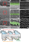
Figure 1
Figure 1
(AD) Scanning electron microscopy (SEM) at various depths in 3-month-old wild-type (WT) lenses. Boxed regions in (A) indicate the approximate location where (BD) higher-magnification images were obtained. (BD) Cortical newly formed fiber cells (B) are straight, with balls and sockets along the broad sides and small protrusions along the vertices. As fiber cells differentiate (C), the cells remain straight and more small protrusions are formed along the cell vertices. Mature lens fibers (D) form large interlocking paddle domains decorated by small protrusions along the cell vertices. Single fibers are highlighted in green to show the changes in cell morphology as the fibers mature. Note that (D) is a different orientation of the same fiber cell shown in Figure 3B of Blankenship et al. (EG) Confocal fluorescence microscopy of phalloidin staining of fiber cells in 6-week-old Tmod1+/+ lens fiber cell bundles (located at depths comparable to those in [BD]) reveals that F-actin is enriched in large paddle domains and small interlocking protrusions at the vertices of fiber cells. (H) Diagram (not to scale) of mature lens fiber cells with large paddles (light blue shading), valleys between large paddles (red lines), small protrusions (green shading), and bases of small protrusions (yellow lines) along the short sides of the cell. Scale bars: 50 μm (A); 6 μm (BD); 4 μm (EG).
Figure 2
Figure 2
Confocal fluorescence microscopy images (2D single optical planes, extended focus flattened Z-stack, 3D reconstruction and cross section) of F-actin in neighboring mature fiber cells in 6-week-old Tmod1+/+ lenses. Single fiber cells are pseudocolored green or pink or uncolored. 3D reconstruction of Z-stacks through three neighboring fibers shows interlocking structures between adjacent cells (A) and coordinated paddle domains between cells of neighboring layers (B) (red arrows). Dashed boxes show the level where single XZ planes in (C, D) are derived. A single 2D XZ plane from the 3D reconstruction shows normal hexagonal fiber cell morphology (C, D) (hexagonal cell body outlined with blue dashed lines) with paddles and protrusions along the short side (D) (red asterisk). The F-actin staining pattern in dissociated fiber cells is similar to what is typically observed in cross sections through hexagonal mature lens fibers that have undergone denucleation (E) (hexagonal cell body outlined with blue dashed lines), demonstrating enriched F-actin along the short sides of these fibers and in regions of paddles and protrusions (red asterisk) with less intense F-actin signals along the broad sides. Extended focus image of the flattened Z-stack shows F-actin–rich interlocking paddles decorated by small protrusions along the short sides of the cells, creating a 3D zipper between adjacent cells (F). Single XY optical sections along the anterior–posterior axis through the fiber cells (G, H), where the dashed lines are drawn through the XZ plane in (C) show interlocking domains between fibers with some separation between the cells. Scale bars: 4 μm.
Figure 3
Figure 3
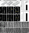
Confocal fluorescence microscopy images (2D, single optical plane) of F-actin in fiber cells at various depths and SEM in 6-week-old (A) and 2-month-old (C) Tmod1+/+ and Tmod1−/− lenses. (A) Diagrams of normal cortical, differentiating, and mature fiber cells are shown along the top. Tmod1+/+ and Tmod1−/− cortical and differentiating fiber cells are straight with small F-actin protrusions along their vertices. Protrusions in differentiating fibers have more pronounced head and neck regions (arrows). Tmod1+/+ mature fibers have large paddle domains (asterisks) with F-actin–rich small protrusions, while Tmod1−/− fiber cells have F-actin–positive protrusions, but very few paddles. (B) Tmod1−/− mature fiber cells display a significant decrease in tortuosity (n = 9, *P < 0.01), but the cell neck width was unaffected (double-headed arrows in [A]) between Tmod1+/+ and Tmod1−/− lenses (n = 9). (C) Low-magnification SEM with boxed regions indicating the location of higher-magnification images. Comparable regions are identified by measuring outward from the nucleus at the center of the lens, since some peripheral cortical fibers are lost in the sample preparation procedure. Tmod1+/+ lenses have rows of mature fiber cells with coordinated paddle protrusions that are decorated by smaller protrusions of equal size and spacing. In contrast, Tmod1−/− mature lens fibers have disorganized paddles with irregular protrusions. Scale bars: 4 μm (A); 1 mm (C) (low magnification left); 4 μm (C) (high magnification).
Figure 4
Figure 4
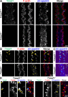
Immunostaining of single mature fiber cells from 6-week-old Tmod1+/+ and Tmod1−/− lenses for Tmod1 (green), F-actin (red), and β2-spectrin (blue). (A) Extended focus of Z-stacks through mature fiber cells. The Tmod1+/+ fiber has F-actin–rich large paddle domains and small protrusions along cell vertices. Tmod1 and β2-spectrin are enriched in numerous puncta in the control fiber cell. While the Tmod1−/− fiber has very few paddles, F-actin–rich small protrusions are still present. (B, D) Single optical section (2D) from a Z-stack, showing a section through the cytoplasm of the fiber cells, with enlargements. Tmod1 and β2-spectrin are enriched in puncta near the cell membrane in valleys between large paddle domains in the Tmod1+/+ fiber (arrows), and β2-spectrin is also enriched at the base of small protrusions in Tmod1+/+ and Tmod1−/− fibers (arrowheads). The β2-spectrin staining signal appears diffuse and cytoplasmic (asterisks) with fewer membrane-associated puncta in the Tmod1−/− fiber. (C) Fluorescence intensity heat maps of β2-spectrin staining in Tmod1+/+ and Tmod1−/− lens fibers show that β2-spectrin staining is more cytoplasmic (asterisks) in the Tmod1−/− fiber. Scale bars: 4 μm (AC); 2 μm (D).
Figure 5
Figure 5
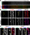
(A) Immunostaining of frozen lens section from 6-week-old Tmod1+/+ mice for α-actinin (green) and F-actin (red). α-Actinin is present in epithelial cells with increased signal in maturing fiber cells. The staining signal is enriched on the short sides of mature fiber cells with a punctate pattern. (B, C) Immunostaining of single mature fiber cells (2D, single optical plane) from 6-week-old Tmod1+/+ and Tmod1−/− lenses for Tmod1 (green), F-actin (red), and α-actinin (blue). Similar to Tmod1 and β2-spectrin, α-actinin is enriched in large puncta in valleys between large paddle domains in the Tmod1+/+ fiber (arrows in [B, C]). In the Tmod1−/− fiber, α-actinin is present along the entire cell membrane. Enlargement shows that α-actinin is enriched near the base of small protrusions in the Tmod1−/− mature fiber cell (arrowheads in [C]). Scale bars: 20 μm (A); 4 μm (B); 2 μm (C).
Figure 6
Figure 6
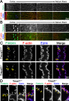
(A) Immunostaining of frozen lens section from 6-week-old Tmod1+/+ mice for ezrin (green) and F-actin (red). Ezrin is present along the membranes of lens fiber cells and enriched at the vertices of cortical fibers and along the short sides of mature fiber cells. (B) Immunostaining of frozen lens section from 6-week-old Tmod1+/+ mice for fimbrin (green) and F-actin (red). Fimbrin is present in epithelial and fiber cells. In fiber cells, fimbrin is distributed along the membrane in differentiating and mature fiber cells. (C, D) Immunostaining of single mature fiber cells (2D, single optical plane) from 6-week-old Tmod1+/+ and Tmod1−/− lenses for fimbrin (green), F-actin (red), and ezrin (blue). As expected, ezrin colocalizes with F-actin along the cell membrane and in interdigitations of Tmod1+/+ and Tmod1−/− fibers. Interestingly, fimbrin is enriched in puncta near the base of small interlocking protrusions in Tmod1+/+ and Tmod1−/− fibers (arrows in [C, D]). Enlargements show ezrin enrichment (arrowheads in [D]) at the base of small protrusions. Scale bars: 20 μm (A, B); 4 μm (C); 2 μm (D).
Figure 7
Figure 7
Immunostaining of single mature fiber cells (2D, single optical plane) from 6-week-old Tmod1+/+ and Tmod1−/− lenses for Arp3 (green), F-actin (red), and N-cadherin (blue). (A) N-cadherin is localized along the cell membrane in Tmod1+/+ and Tmod1−/− fibers and appears enriched along the base of protrusions and valleys between large paddles. Arp3 is enriched in puncta (arrows) near the base of small interlocking protrusions (arrows) in Tmod1+/+ and Tmod1−/− fibers. Arp3 is also found in valleys between large paddles in the Tmod1+/+ fiber (arrowheads). (B) Enlargements show that weak Arp3 staining extends into small protrusions (open triangles) in Tmod1+/+ and Tmod1−/− fibers. Arp3 puncta at the base of small protrusions are often accompanied by enriched N-cadherin staining (arrows). Scale bars: 4 μm (A); 2 μm (B).
Figure 8
Figure 8
(A) During fiber cell maturation, large paddle domains with small interlocking protrusions form along the vertices of hexagonal fiber cells. Tmod1 is required for normal formation of large paddles between mature fiber cells. (B) Tmod1 may stabilize the F-actin–spectrin network as well as α-actinin–crosslinked antiparallel F-actin bundles in valleys between large paddles to maintain their structure. Formation of small protrusions may be facilitated by Arp3-nucleated actin networks, and fimbrin-crosslinked parallel F-actin bundles at the base of protrusions, while ezrin may stabilize actin networks along the entire fiber cell membrane and N-cadherin promotes cell–cell interactions.

Comment in

Similar articles

Cited by

References

    1. Lovicu FJ,, Robinson ML. Development of the Ocular Lens. Cambridge UK, New York: Cambridge University Press; 2004: xv, 398.
    1. Kuszak JR,, Zoltoski RK,, Sivertson C. Fibre cell organization in crystalline lenses. Exp Eye Res. 2004; 78: 673–687. - PubMed
    1. Kuszak JR,, Mazurkiewicz M,, Jison L,, Madurski A,, Ngando A,, Zoltoski RK. Quantitative analysis of animal model lens anatomy: accommodative range is related to fiber structure and organization. Vet Ophthalmol. 2006; 9: 266–280. - PubMed
    1. Piatigorsky J. Lens differentiation in vertebrates. A review of cellular and molecular features. Differentiation. 1981; 19: 134–153. - PubMed
    1. Bassnett S. On the mechanism of organelle degradation in the vertebrate lens. Exp Eye Res. 2009; 88: 133–139. - PMC - PubMed

Publication types

MeSH terms