. 2016 Aug 18:6:31918.
doi: 10.1038/srep31918.

The modulation of MiR-155 and MiR-23a manipulates Klebsiella pneumoniae Adhesion on Human pulmonary Epithelial cells via Integrin α5β1 Signaling

Affiliations

The modulation of MiR-155 and MiR-23a manipulates Klebsiella pneumoniae Adhesion on Human pulmonary Epithelial cells via Integrin α5β1 Signaling

Yan Teng et al. Sci Rep. .

Abstract

Micro-RNAs (miRNAs) critically regulate several host defense mechanisms, but their roles in the bacteria-epithelium interplay remain unclear. Our results displayed that the expression of miR-155 and miR-23a were down-regulated in K. pneumoniae-infected pulmonary epithelial cells. The elevated bacterial adhesion on A549 cells followed the enhancement of the cellular levels of these two miRNAs. Meanwhile, a mechanistic study demonstrated that miR-155 promoted integrin α5β1 function and resulted in the increased actin polymerization. Moreover, a non-histone nuclear protein, high mobility group nucleosomal-binding domain 2 (HMGN2) served as the potential target of miR-155 and miR-23a to regulate the integrin α5β1 expression and K. pneumoniae adhesion. Furthermore, the expression of a known integrin transcription suppressor-Nuclear Factor-I (NFI) was also repressed by miR-155, which paralleled with its chromatin location in the promoter regions of integrin α5 and β1. These results uncover novel links between miRNAs and integrin function to regulate bacterial adhesion, indicating a potential mechanism of host cell autonomous immune response to K. pneumoniae infection.


Figures
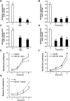
Figure 1
Figure 1. MiR-155 and miR-23a expression were down-regulated in K. pneumoniae infected A549 cells and promoted K. pneumoniae adhesion.
A549 cells were exposed to increasing doses of K. pneumoniae (MOI = 0, 50, 100) for 2 hours, or fixed amount of bacteria (MOI = 100) at indicated time points (0 hr, 2 hr, 4 hr and 6 hr). The expression levels of miR-155 (A,B) and miR-23a (C,D) were examined by RT-qPCR. The Relative expression was normalized to U6 and then converted to the fold change over uninfected. A549 cells were transfected with miR-155 mimic (E), miR-23a mimic (F), miR-155 inhibitor (G) and according negative controls (miR-NC or NC) for 24 hours prior to 100 MOI of bacterial exposure. The relative K. pneumoniae adhesion at indicated time points were determined by colony counts. Relative K. p adhesion was represented after the normalization to 1 hour bacterial adhesion of miR-NC or NC. (Data are the mean ± SD and represent three individual experiments. *p < 0.05, **p < 0.01 compared with K. pneumoniae uninfected, miR-NC or NC).
Figure 2
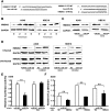
Figure 2. HMGN2 was the potential target of miR-155 and miR-23a to involve in regulating K. pneumoniae adhesion.
(A) Schematic presentation of base pairing between the 3′ UTR of HMGN2 and miR-155 or miR-23a by erect likes. The mutant seed sequence of the HMGN2 3′ UTR matching miR-23a is also presented by dots. (B) Western blot analysis showing the change of HMGN2 protein level in K. pneumoniae infected A549 and HBE16 cells at different infection time (MOI = 100). (C) Western blot analysis showing the effect of miR-23a mimic on the protein expressions of HMGN2 in K. pneumoniae infected A549 or HBE16 cells (MOI = 100 for 2 hours, same as DF). (D) Western blot analysis showing the effect of miR-155 mimic or inhibitor on the protein expressions of HMGN2 in K. pneumoniae infected or uninfected cells. (E) A549 cells were co-transfected with miR-23a mimic or miR-NC along with HMGN2 3′ UTR wild-type (WT) or mutant (MUT) reporter. Luciferase activity was measured 24 hr after transfection. (F) The relative K. pneumoniae adhesion in A549 cells co-transfected with pEx-HMGN2 and miR-155 or miR-23a mimic. (Data are the mean ± SD and represent three individual experiments. **p < 0.01 compared with miR-NC and pEx-NC cotransfection).
Figure 3
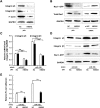
Figure 3. MiR-155 promoted integrin/Rac1 activity during K. pneumoniae infection.
A549 cells were transfected with miR-155 mimic and according negative controls (miR-NC), or miR-155 inhibitor and according negative controls (NC) for 24 hours prior to 100 MOI of K. pneumoniae infection. (A) RT-qPCR analysis showing the mRNA levels of integrin α5 and β1. (B) Western blot analysis showing the expressions of integrin α5, integrin β1. (C) Microscopic images displaying the expressions of integrin α5 and β1 (green fluorescence, 40×). Blue fluorescence represented the nucleus staining by DAPI. (D) Fibronectin (FN)-cell Adhesion assay was performed to evaluate the effect of miR-155 mimic on uninfected A549 cells to associate with fibronectin coated plates. (E) Western blot analysis showing the pull-downed active form of Rac1 (Rac1-GTP) and total Rac1 levels. (F) Microscopic images displaying the membrane ruffles formed by polymerized F-actin (F-actin: red fluorescence, DAPI: blue fluorescence, 100×). (G) Western blot analysis showing the expressions of F-actin. (Data are the mean ± SD and represent three individual experiments. *p < 0.05, **p < 0.01 compared with miR-NC).
Figure 4
Figure 4. HMGN2 was involved in miR-155 mediated Integrin/Rac1 regulation during K. pneumoniae infection.
Western blot analysis showing the expressions of integrin α5, integrin β1 and F-actin (A) or Rac1-GTP and the total Rac1 (B) in A549 cells transfected with pEx-HMGN2 and/or siRNA-HMGN2 prior to K. pneumoniae exposure (MOI = 100 for 2 hours, same as C and D). The relative mRNA level of integrin α5 and β1 (C), the protein level of integrin α5, integrin β1 and Rac1-GTP (D) of A549 cells co-transfected with pEx-HMGN2 and miR-155 mimic prior to K. pneumoniae exposure. (E) The relative FN-cell adhesion of uninfected A549 cells transfected as (C,D). (Data are the mean ± SD and represent three individual experiments. **p < 0.01 normalized with miR-NC and pEx-NC co-transfection).
Figure 5
Figure 5. MiR-155 inhibited a known integrin transcription suppressor NFI during K. pneumoniae infection.
(A) Schematic presentation of base pairing between miR-155 and the 3′ UTR of NFIA or NFIB by erect lines. The mRNA expressions (B) and the protein levels (C) of NFIA and NFIB in A549 cells transfected with miR-155 mimic or inhibitor prior to K. pneumoniae exposure (MOI = 100 for 2 hours). (D) The schematic diagram of NFI binding motifs (TTGGC, GCCAA) on promoters of integrin α5 or β1 (−2000 bp to TSS site ATG). The primers for ChIp assay were designed for about 60–70 bp up-stream or down-stream of NFI binding motifs (−102 to +18 bp on α5 promoter, −1132 and −983 bp on β1 promoter). (E) ChIp assay showingthe NFI recruitment at the desired regions shown in (D) under the same condition as (B,C), the relative occupancy is the ratio of immunoprecipitated NFI to input DNA. (Data are the mean ± SD and represent three individual experiments. **p < 0.01 compared with miR-NC or NC).
Figure 6
Figure 6. Pharmacological inhibition of Integrin/Rac1 and acting polymerization partially blocked miR-155 and miR-23a induced K. pneumoniae adhesion.
K. pneumoniae adhesion was measured on A549 cells transfected with miRNA mimics and then treated with RGD (50 nM, 24 hours) or NSC23766 (NSC, 50 μM, 2 hours) (A) or cytochalasin (B) (CytoB, 10 μM, 2 hours) (C) prior to K. pneumoniae exposure (MOI = 100 for 2 hours). Untreated miR-NC was defined as 100% of relative adhesion. (B) Western blot analysis showing the expressions of F-actin in A549 cells treated with RGD or NSC23766 under the same condition as (A). (Data are the mean ± SD and represent three individual experiments. **p < 0.01). (D) The schematic diagram depicting miR-155 and miR23a pathway: miRNAs promote intergrin α5β1 and Rac1 activities by targeting negative transcriptional regulators of integrin -HMGN2 and NFI, and result in actin polymerization (black solid lines). The exposure of K. pneumoniae causes the active reduction of these two miRNAs, which subsequently de-represses HMGN2 and NFI to inhibit integrin/Rac1 function and slows actin polymerization (green and red arrows).

Similar articles

Cited by

References

    1. Li B., Zhao Y., Liu C., Chen Z. & Zhou D. Molecular pathogenesis of Klebsiella pneumoniae. Future microbiology 9, 1071–1081, 10.2217/fmb.14.48 (2014). - DOI - PubMed
    1. Hsu C. R. et al.. Klebsiella pneumoniae translocates across the intestinal epithelium via Rho GTPase- and phosphatidylinositol 3-kinase/Akt-dependent cell invasion. Infection and immunity 83, 769–779, 10.1128/IAI.02345-14 (2015). - DOI - PMC - PubMed
    1. Hauck C. R., Borisova M. & Muenzner P. Exploitation of integrin function by pathogenic microbes. Current opinion in cell biology 24, 637–644, 10.1016/j.ceb.2012.07.004 (2012). - DOI - PubMed
    1. Hynes R. O. Integrins: bidirectional, allosteric signaling machines. cell 110, 673–687 (2002). - PubMed
    1. DeMali K. A., Wennerberg K. & Burridge K. Integrin signaling to the actin cytoskeleton. Current opinion in cell biology 15, 572–582 (2003). - PubMed

Publication types

LinkOut - more resources