. 2016 Aug 30;113(35):9804-9.
doi: 10.1073/pnas.1605031113. Epub 2016 Aug 15.

Crystal structure of an Fe-S cluster-containing fumarate hydratase enzyme from Leishmania major reveals a unique protein fold

Affiliations

Crystal structure of an Fe-S cluster-containing fumarate hydratase enzyme from Leishmania major reveals a unique protein fold

Patricia R Feliciano et al. Proc Natl Acad Sci U S A. .

Abstract

Fumarate hydratases (FHs) are essential metabolic enzymes grouped into two classes. Here, we present the crystal structure of a class I FH, the cytosolic FH from Leishmania major, which reveals a previously undiscovered protein fold that coordinates a catalytically essential [4Fe-4S] cluster. Our 2.05 Å resolution data further reveal a dimeric architecture for this FH that resembles a heart, with each lobe comprised of two domains that are arranged around the active site. Besides the active site, where the substrate S-malate is bound bidentate to the unique iron of the [4Fe-4S] cluster, other binding pockets are found near the dimeric enzyme interface, some of which are occupied by malonate, shown here to be a weak inhibitor of this enzyme. Taken together, these data provide a framework both for investigations of the class I FH catalytic mechanism and for drug design aimed at fighting neglected tropical diseases.

Keywords: Fe-S cluster; X-ray crystallography; fumarate hydratase; leishmaniases.


Conflict of interest statement

The authors declare no conflict of interest.

Figures

Scheme 1.
Scheme 1.
Fig. 1.
Fig. 1.
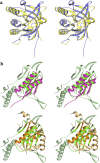
Crystal structure of LmFH-2. (A) Overall structure of the LmFH-2 functional dimer. The Upper and Lower panels represent two orthogonal views of the structure with two domains: N terminal (blue and green) and C terminal (yellow). The Left and Right panels show the cartoon and electrostatic surface potential representation of the LmFH-2 dimer, respectively. The [4Fe-4S] clusters are shown in magenta. (B) Ribbon diagram of LmFH-2 monomer. The N-terminal domain is divided into two nonsequential subdomains 1 (light blue and dark blue) and 2 (green), and is connected to C-terminal domain (yellow) by a linker (black arrow), as indicated in the linear schematic. (C) Ribbon diagram of LmFH-2 N-terminal subdomains 1 and 2. (D) Ribbon diagram of LmFH-2 C-terminal domain.
Fig. 2.
Fig. 2.
LmFH-2 active site. (A) The residues of chains A and B are shown in white and light blue, respectively. The substrate S-malate, [4Fe-4S] cluster and water molecule are shown in green, yellow (S) and orange (Fe), and cyan, respectively. Mesh represents the final 2FoFc electron density map contoured at 1.5 σ level (blue) for S-malate and the [4Fe-4S] cluster. A stereoview is shown in Fig. S4. (B) Interactions between S-malate and the active site residues in LmFH-2. The water molecule, C, N, O, Fe, and S atoms are shown in cyan, black, blue, red, orange, and yellow, respectively. The hydrogen bonds are shown as green dashed lines. The distance between the OH group of Thr-467 to the S-malate C3 carbon is shown as a black dashed line. Image created with LigPlot (30). (C) Proposed mechanism for class I FHs to catalyze the dehydration of S-malate to fumarate. The first step is thought to be deprotonation of S-malate C3 by a catalytic base (B:). The closest residue to C3 is Thr-467 (3.34 Å), which is near a water molecule and two Arg residues (Arg-421 and Arg-471), any of which could accept the proton. The residue Asp-135, and its hydrogen-bonding partner His-334/B, are ideally positioned to play a role as a catalytic acid to protonate the C2 hydroxyl group of S-malate for elimination as H2O and subsequent formation of fumarate.
Fig. S1.
Fig. S1.
Topology diagram of LmFH-2. LmFH-2 is described in two domains: N- and C-terminal domains. The N-terminal domain is divided into subdomains 1 and 2 that are shown in blue and green, respectively. The C-terminal domain is shown in yellow. The topology was performed using PDBsum, a pictorial database of 3D structures in the Protein Data Bank (www.ebi.ac.uk/thornton-srv/databases/cgi-bin/pdbsum/GetPage.pl?pdbcode=index.html).
Fig. S2.
Fig. S2.
Structural comparison of the proteins that share similarity to N- and C-terminal domains of LmFH-2 according to the DALI server. (A) Stereoview of the superposition between LmFH-2 C-terminal domain (residues 385–568) (yellow) and full β-subunit of FH from Archaeoglobus fulgidus (blue; PDB ID code 2ISB), showing a high level of similarity. Orientation is the same as in Fig. 1D. (B) Stereoview of the superposition between subdomain 1 (residues 28–213 and 349–375) of the LmFH-2 N-terminal domain (green and light green), with (Upper) Ni-binding domain (residues 1–68) of HypA from Thermococcus kodakaraensis KOD1 (pink; PDB ID code 3A43) and (Lower) the N-terminal β-domain (residues 2–161) of l-serine dehydratase from Legionella pneumophila (orange and light orange; PDB ID code 4RQO). Secondary structure in light orange does not align with LmFH-2. Orientation is the same as in Fig. 1C. Of the 11 β-strands of the subdomain 1 of the LmFH-2 N-terminal domain, only 3 β-strands (HypA; pink) or 5 β-strands (l-serine dehydratase; orange) are similar, and of 6 helices of the subdomain 1, 2 helices from HypA (pink), and l-serine dehydratase (orange) are similar. No matches to the subdomain 2 of the LmFH-2 N-terminal domain were identified.
Fig. S3.
Fig. S3.
Sequence alignment of class I FHs. LmFH-2 (cytosolic isoform of L. major), LtFH-2 (cytosolic isoform of Leishmania tropica), LdFH-2 (cytosolic isoform of Leishmania donovani), LiFH-2 (cytosolic isoform of Leishmania infantum), LbFH-2 (cytosolic isoform of Leishmania braziliensis), TcFH-2 (cytosolic isoform of Trypanosoma cruzi), TbFH-2 (cytosolic isoform of Trypanosoma brucei), TvFH-2 (cytosolic isoform of Trypanosoma vivax), SpFH (Strongylocentrotus purpuratus), SmFH (Schistosoma mansoni), SjFH (Schistosoma japonicum), AcFH (Acanthamoeba castellanii str. Neff), EcFH-A (fum A of E. coli), SeFH (Salmonella enterica), EnterobacterFH (Enterobacter sp. SST3), SbFH (Shigella boydii), YpFH (Yersinia pseudotuberculosis IP 31758), RhodococcusFH (Rhodococcus sp. P14), LmFH-1 (mitochondrial isoform of L. major), LiFH-1 (mitochondrial isoform of L. infantum), LmxFH-1 (mitochondrial isoform of Leishmania mexicana), LbFH-1 (mitochondrial isoform of L. braziliensis), TbFH-1 (mitochondrial isoform of T. brucei), TcongoFH-1 (mitochondrial isoform of Trypanosoma congolense), TvFH-1 (mitochondrial isoform of T. vivax), TcFH-1 (mitochondrial isoform of T. cruzi). The conserved residues are indicated in the blue boxes. The similar residues are indicated in blue texts. The conserved active site residues among class I FHs are indicated in the pink boxes. The three conserved cysteine residues, which are shown to bind a [4Fe-4S] cluster, are indicated in the yellow boxes. The conserved dimer interface residues among class I FHs are indicated in green circles. The alignment was performed using MULTALIN (31) and graphically displayed using ESPript (32).
Fig. S4.
Fig. S4.
Stereoview of LmFH-2 active site. The residues of chains A and B are shown in white and light blue, respectively. The substrate S-malate, [4Fe-4S] cluster, and water molecule are shown in green, yellow (S) and orange (Fe), and cyan, respectively. Mesh represents the final 2FoFc electron density map contoured at 1.5 σ level (blue) for S-malate and [4Fe-4S] cluster.
Fig. 3.
Fig. 3.
Substrate access to the active site of LmFH-2. (A) Electrostatic surface potential of the substrate-binding pocket showing the access of another S-malate (green) to the active site. The [4Fe-4S] cluster is shown in yellow (S) and orange (Fe). (B) Interactions between the second molecule of S-malate found in the positive cavity and N-terminal domain residues. The hydrogen bonds are shown as dashed lines. Image created with LigPlot (30).
Fig. S5.
Fig. S5.
B-factor versus number of residues of LmFH-2 chain A. The blue and orange bars represent the residues of N- and C-terminal domains, respectively. The graphic was generated by phenix.pdbtools (26).
Fig. 4.
Fig. 4.
LmFH-2 cavities. (A) View of the tunnel (pink), formed between the N-terminal domains, which goes through the entire protein. The ligands polyethylene glycol (orange), glycerol (yellow), and malonate (cyan) are found in this tunnel. (B) View of the dimer surface, showing the entrance of the tunnel (pink) and the binding pockets of malonate (cyan), S-malate (green), and [4Fe-4S] cluster [yellow (S) and orange (Fe)]. (C and D) The interactions between malonate (cyan) and the residues of the binding pocket on the top of protein and in the dimer interface, respectively. Blue mesh represents the final 2FoFc electron density map contoured at 1.5 σ level for residues of LmFH-2. Green mesh represents the FoFc difference electron density map contoured at 3.0 σ level for malonate.
Fig. S6.
Fig. S6.
Determination of IC50 values for malonate. The enzymatic activity was determined under anaerobic conditions in an MBraun glovebox by measuring the fumarate production or consumption at 250 nm, using 0–120 mM malonate in 50 mM Tris, pH 9, 150 mM NaCl, 0.5 mM fumarate (A) or 2 mM S-malate (B). The reaction was initiated by the addition of the LmFH-2 at 10 μg/mL. IC50 values were determined by fitting the data to the equation v = V0/[1 + (I/IC50)], where v is the observed velocity, V0 is the uninhibited velocity, I is the inhibitor concentration, and IC50 is the 50% inhibitory concentration.

Similar articles

Cited by

References

    1. Coustou V, et al. A mitochondrial NADH-dependent fumarate reductase involved in the production of succinate excreted by procyclic Trypanosoma brucei. J Biol Chem. 2005;280(17):16559–16570. - PubMed
    1. Cordeiro AT, Feliciano PR, Pinheiro MP, Nonato MC. Crystal structure of dihydroorotate dehydrogenase from Leishmania major. Biochimie. 2012;94(8):1739–1748. - PubMed
    1. Yogev O, et al. Fumarase: A mitochondrial metabolic enzyme and a cytosolic/nuclear component of the DNA damage response. PLoS Biol. 2010;8(3):e1000328. - PMC - PubMed
    1. Coustou V, et al. Fumarate is an essential intermediary metabolite produced by the procyclic Trypanosoma brucei. J Biol Chem. 2006;281(37):26832–26846. - PubMed
    1. Feliciano PR, et al. Fumarate hydratase isoforms of Leishmania major: Subcellular localization, structural and kinetic properties. Int J Biol Macromol. 2012;51(1-2):25–31. - PubMed

Publication types

MeSH terms

Associated data

LinkOut - more resources