Review
. 2016 Aug;203(4):1563-99.
doi: 10.1534/genetics.112.145243.

The Nuts and Bolts of Transcriptionally Silent Chromatin in Saccharomyces cerevisiae

Affiliations
Review

The Nuts and Bolts of Transcriptionally Silent Chromatin in Saccharomyces cerevisiae

Marc R Gartenberg et al. Genetics. 2016 Aug.

Abstract

Transcriptional silencing in Saccharomyces cerevisiae occurs at several genomic sites including the silent mating-type loci, telomeres, and the ribosomal DNA (rDNA) tandem array. Epigenetic silencing at each of these domains is characterized by the absence of nearly all histone modifications, including most prominently the lack of histone H4 lysine 16 acetylation. In all cases, silencing requires Sir2, a highly-conserved NAD(+)-dependent histone deacetylase. At locations other than the rDNA, silencing also requires additional Sir proteins, Sir1, Sir3, and Sir4 that together form a repressive heterochromatin-like structure termed silent chromatin. The mechanisms of silent chromatin establishment, maintenance, and inheritance have been investigated extensively over the last 25 years, and these studies have revealed numerous paradigms for transcriptional repression, chromatin organization, and epigenetic gene regulation. Studies of Sir2-dependent silencing at the rDNA have also contributed to understanding the mechanisms for maintaining the stability of repetitive DNA and regulating replicative cell aging. The goal of this comprehensive review is to distill a wide array of biochemical, molecular genetic, cell biological, and genomics studies down to the "nuts and bolts" of silent chromatin and the processes that yield transcriptional silencing.

Keywords: Sir2; chromatin; histone deacetylation; silencing; yeast.


Figures

Figure 1
Figure 1
Representative silent chromatin domains of budding yeast. (A) The HMR locus. Silent chromatin is shown in pink and cis-acting silencer elements, E and I, are shown in orange. At HMR, silent chromatin silences the a mating-type genes. (B) Discontinuous silent chromatin domains at telomere VIIIR silence the subtelomeric IMD2 gene. (C) A ribosomal rDNA repeat element. Silent chromatin domains spanning the intergenic sequences, IGS1 and IGS2, suppress Pol II transcription. rDNA silencing requires Sir2 but not Sir1, Sir3, or Sir4. Loci are not drawn to scale.
Figure 2
Figure 2
Local and long-range interactions of silent chromatin. (A) The arrangements of components within a typical locus of silent chromatin. Sir2 (2), Sir3 (3) and Sir4 (4) form the Sir2/3/4 complex that binds histones throughout the silenced domain. Histones within the domain lack post-translational modifications with the exception of H2AS129 phosphorylation and some H4K12 acetylation. ORC, Rap1 (R), Abf1 (A) and Sir1 (1) bind to cis-acting silencer elements and interact with proteins of the Sir2/3/4 complex. Specific interactions between these components are documented in subsequent figures. Ac, acetylation. (B) The folded-back structure of silent chromatin at a telomere. (C) The long-range interactions between the silent chromatin domains at HML and HMR cause chromosome III to fold back upon itself. (D) Interactions between the silent chromatin domains of different telomeres and interactions between silent chromatin and docking sites at the nuclear membrane cause chromosome ends to cluster at the nuclear periphery.
Figure 3
Figure 3
DNA binding sites within silencers and proto-silencers. Direct DNA binding by Sum1 contributes to the function of the HML-E silencer. The X and Y′ subtelomeric repeat elements of telomere VIIIR are shown.
Figure 4
Figure 4
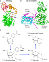
Sir2 structure and enzymatic activity. (A) Crystal structure of the Archaeoglobus fulgidus homolog Sir2Af2. The nucleotide-binding Rossmann fold is shown in green, zinc-binding (ribbon) in red, and small helical domain in orange. Zn2+ ion is shown coordinated with four cysteine residues (yellow). The positions of NAM and NAD+ binding are indicated. Protein Data Bank (PDB) accession number 1S7G. (B) Crystal structure of S. cerevisiae Sir2 core (green) and N-terminally extended (cyan) domains are shown in contact with the SID of Sir4 (magenta). PDB accession number 4IAO. (C) Chemical reaction of lysine deacetylation by Sir2 and other sirtuins.
Figure 5
Figure 5
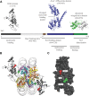
Domain structure and nucleosome binding of Sir3. (A) The structural and functional domains of Sir3. The BAH domain from PDB accession number 3TU4, the AAA+ ATPase-like domain from 3TE6, and the winged-helix domain from 3ZCO. (B) Crystal structure of the Sir3 BAH domain bound to the nucleosome core particle. The BAH domain is shown with a dark gray ribbon. The H4K16 and H3K79 histone residues critical for Sir3 binding are shown in red and green space-filling spheres, respectively. PDB accession number 3TU4. (C) The positions of H4K16 and H3K79 on the nucleosome-binding surface of the Sir3 BAH domain.
Figure 6
Figure 6
The structural and functional domains of Sir4. Each of the amino acids highlighted in the coiled-coil structure, M1307, E1310, I1311, and K1324 disrupts Sir3 binding when mutated. PDB accession number 1NYH.
Figure 7
Figure 7
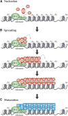
Nucleation, spreading, and maturation of silent chromatin. (A) Nucleation. The known network of interactions between Sir proteins and silencer-bound factors is shown. (B) Spreading. Deacetylation of neighboring histones by Sir2 creates additional binding sites for Sir3 and Sir4. Successive rounds of histone deacetylation and Sir2/3/4 binding expands the silent chromatin domain until a barrier is reached. (C) Maturation. Numerous conditions have been found where Sir2/3/4 spreading does not produce transcriptional repression. These circumstances suggest that nascent silent chromatin may undergo a final maturation step (e.g., removal of H3K79 methylation) to yield transcriptional silencing.
Figure 8
Figure 8
Antisilencing and barriers to silent chromatin spreading. (A) Discontinuities in chromatin created by highly-dynamic or displaced nucleosomes disfavor silent chromatin spreading. A barrier created by the nucleosome-depleted tRNA gene next to HMR is shown. (B) At silent chromatin borders, the Sas2 acetyltransferase and Sir2 histone deacetylase compete to determine the acetylation state of H4K16. The histone tail is then bound by effector proteins that demarcate the silent chromatin boundary. In the case of deacetylation, the Sir2/3/4 complex binds to extend the silent chromatin domain and in the case of acetylation, barrier proteins Bdf1 and Yta7 bind to create a boundary. H4K16ac also favors incorporation of histone variant H2A.Z, another barrier to silent chromatin spreading. (C) Loss of genome-wide antisilencing. Global histone modifications that disfavor Sir2/3/4 complex binding increase the available pool of Sir proteins available for binding at telomeres. When these histone modifications are lost, as shown in the figure, Sir proteins are titrated from bona fide silent chromatin sites by nonspecific binding elsewhere.
Figure 9
Figure 9
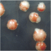
Epigenetic inheritance of the silent state. Genetically identical colonies with a telomeric ADE2 reporter were plated on low adenine media. The red sectors contain lineages of cells with ADE2 in the silenced state. The white sectors contain cell lineages with the ADE2 gene in the nonsilent state. Red and white sectors intermingle within a given colony, indicating that cells periodically switch between silent and nonsilent states.
Figure 10
Figure 10
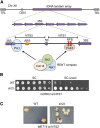
Sir2-dependent silencing in the rDNA locus. (A) Schematic representation of the rDNA repeat organization on chromosome XII. The array consists of ∼150 repeats of 9.1 kb each. Purple arrows represent individual Pol I transcription units of the 35S precursor rRNA. In between are the IGS consisting of NTS1 and NTS2, divided by the Pol III-transcribed 5S gene. Pol I sits at the rDNA promoter region in NTS2 and Fob1 on the RFB site in NTS1. The RENT complex is recruited to NTS2 and NTS1 via interactions with Pol I and Fob1, respectively. (B) Example of Sir2-dependent rDNA silencing phenotype using the mURA3 reporter gene integrated at NTS1. (C) Example of rDNA silencing using MET15 at NTS2 as a colorimetric reporter on lead nitrate-containing plates. White color indicates loss of silencing. Dark brown sectors indicate loss of the marker due to rDNA recombination. CEN, centromere; Chr, chromosome; TEL, telomere; WT, wild type.
Figure 11
Figure 11
Spreading of rDNA silencing. Schematic representation of chromosome XII organization at the interface between the leftmost (centromere-proximal) rDNA gene and unique sequence. NTS1 sequence of the leftmost repeat (thick purple arrow) is truncated at the middle of the Ter1 RFB site. Sir2 overexpression results in spreading of silencing ∼2.7 kb, up to tRNAGln gene, tQ(UUG)L, which acts as a boundary element.
Figure 12
Figure 12
Silencing and endogenous noncoding RNAs and cohibin function in the rDNA. (A) X-ray crystal structure of the cohibin complex consisting of Csm1 (full length 190 amino acids) and an N-terminal portion of Lrs4 (amino acids 1–102), although only a small N-terminal α-helical portion of Lrs4 (purple, amino acids 3–33) is visible in structure. PDB accession number 3N7M. (B) Model for cohibin function at the rDNA where it bridges an interaction between Fob1, RENT, and Tof2 bound to the RFB sites, with the inner nuclear membrane CLIP complex. Cohesin and condensin then associate to align rDNA repeats and stabilize rDNA array structure. (C) Schematic diagram indicating sites of noncoding RNA transcription emanating from NTS1 and NTS2 (green dashed arrows), including bidirectional transcription from E-pro. CAR indicates a cohesin-associated region in NTS2. The rDNA ARS in NTS2 is also indicated.
Figure 13
Figure 13
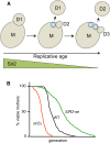
Replicative aging in S. cerevisiae. (A) Mother cells (M) produce daughter cells (D) that leave behind chitinous bud scars (blue circles). Older mother cells therefore harbor more bud scars, which is commonly used as an indicator of average replicative age of a population. (B) Example of a typical survival curve showing short life span of a sir2∆ strain and extended life span when Sir2 is modestly overexpressed (Sir2oe).

Similar articles

Cited by

References

    1. Abraham J., Nasmyth K., Strathern J. N., Klar A. J. S., Hicks J. B., 1984. Regulation of mating-type information in yeast. J. Mol. Biol. 176: 307–331. - PubMed
    1. Ai W., Bertram P. G., Tsang C. K., Chan T.-F., Zheng X. F. S., 2002. Regulation of subtelomeric silencing during stress response. Mol. Cell 10: 1295–1305. - PubMed
    1. Altaf M., Utley R. T., Lacoste N., Tan S., Briggs S. D., et al. , 2007. Interplay of chromatin modifiers on a short basic patch of histone H4 tail defines the boundary of telomeric heterochromatin. Mol. Cell 28: 1002–1014. - PMC - PubMed
    1. Anderson R. M., Bitterman K. J., Wood J. G., Medvedik O., Cohen H., et al. , 2002. Manipulation of a nuclear NAD+ salvage pathway delays aging without altering steady-state NAD+ levels. J. Biol. Chem. 277: 18881–18890. - PubMed
    1. Anderson R. M., Bitterman K. J., Wood J. G., Medvedik O., Sinclair D. A., 2003a Nicotinamide and PNC1 govern lifespan extension by calorie restriction in Saccharomyces cerevisiae. Nature 423: 181–185. - PMC - PubMed

MeSH terms

LinkOut - more resources