

Human umbilical cord-derived mesenchymal stem cells protect against experimental colitis via CD5(+) B regulatory cells
- PMID: 27515534
- PMCID: PMC4981968
- DOI: 10.1186/s13287-016-0376-2
Human umbilical cord-derived mesenchymal stem cells protect against experimental colitis via CD5(+) B regulatory cells
Erratum in
-
Correction to: Human umbilical cord-derived mesenchymal stem cells protect against experimental colitis via CD5+ B regulatory cells.Stem Cell Res Ther. 2019 Jan 21;10(1):33. doi: 10.1186/s13287-019-1132-1. Stem Cell Res Ther. 2019. PMID: 30665468 Free PMC article.
Abstract
Background: To clarify the effect of human umbilical cord-derived mesenchymal stem cell (hUC-MSCs) treatment on colitis and to explore the role of CD5(+) B cells in MSC therapy.
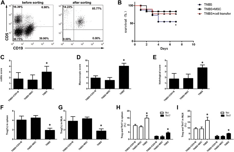
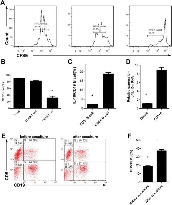
Methods: The trinitrobenzenesulfonic acid (TNBS)-induced colitis mouse model was used. HUC-MSCs were transferred peritoneally. Survival rates, colitis symptoms, and macroscopic and histologic scores were evaluated. CD4(+) T helper (Th) cell subgroups and CD5(+) regulatory B cell (Bregs) in lymphocytes were quantitated by flow cytometry. Cytokine levels were detected by ELISA and Bio-plex. CD5(+) B cells were isolated for in vitro co-culture and adaptive transfer.
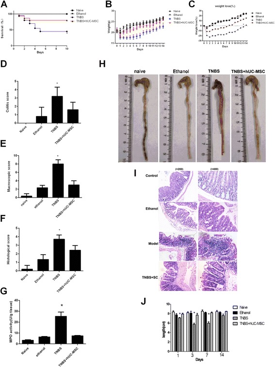
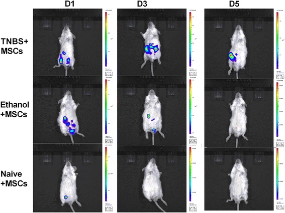
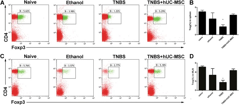
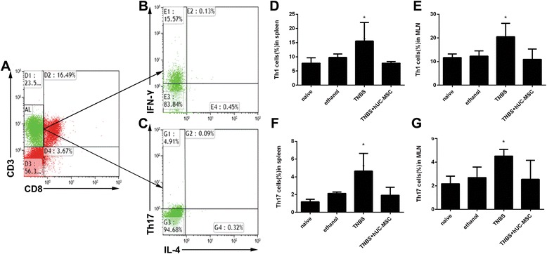
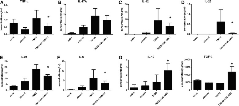
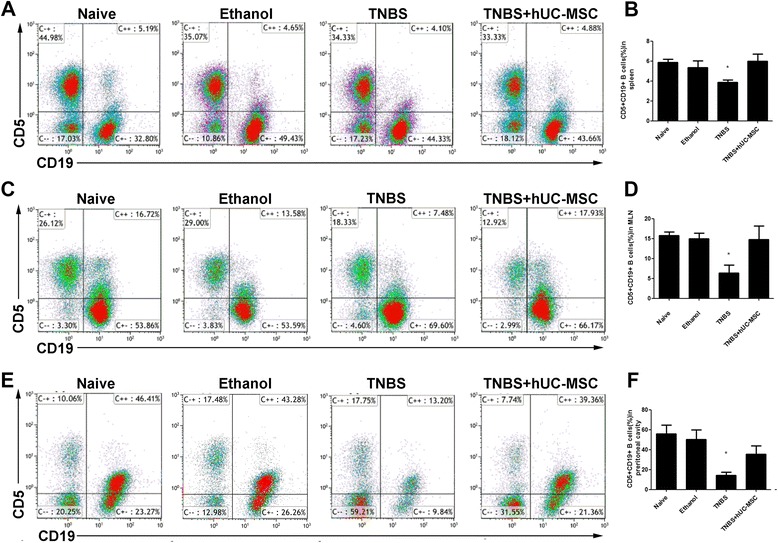
Results: HUC-MSC treatment alleviated TNBS-induced colitis by increasing survival rates, relieving symptoms, and improving macroscopic and histologic scores. Labeled hUC-MSCs were located in the inflamed areas of colitis mice. Increases in regulatory T cells (Tregs) and CD5(+) B cells and decreases in Th1 cells, Th17 cells, and several pro-inflammatory cytokines were observed with hUC-MSC treatment. After adaptive transfer, CD5(+) B cells, which were located mainly in the peritoneal lavage fluid, improved TNBS-induced colitis by correcting Treg/Th1/Th17 imbalances. CD5(+) B cells also inhibited T-cell proliferation and produced interleukin (IL)-10.
Conclusions: HUC-MSCs protected against experimental colitis by boosting the numbers of CD5(+) B cells and IL-10-producing CD5(+) Bregs, and correcting Treg/Th17/Th1 imbalances.
Keywords: B regulatory cell; Colitis; Crohn’s disease; Mesenchymal stem cells; T helper cell.
Figures
Similar articles
-
TLR3 preconditioning enhances the therapeutic efficacy of umbilical cord mesenchymal stem cells in TNBS-induced colitis via the TLR3-Jagged-1-Notch-1 pathway.Mucosal Immunol. 2017 May;10(3):727-742. doi: 10.1038/mi.2016.78. Epub 2016 Sep 21. Mucosal Immunol. 2017. PMID: 27649928
-
Human umbilical cord-derived mesenchymal stem cells downregulate inflammatory responses by shifting the Treg/Th17 profile in experimental colitis.Pharmacology. 2013;92(5-6):257-64. doi: 10.1159/000354883. Epub 2013 Nov 20. Pharmacology. 2013. PMID: 24280970
-
Human umbilical cord mesenchymal stem cells ameliorate mice trinitrobenzene sulfonic acid (TNBS)-induced colitis.Cell Transplant. 2011;20(9):1395-408. doi: 10.3727/096368910X557245. Epub 2011 Mar 9. Cell Transplant. 2011. PMID: 21396175
-
Mesenchymal stem cell effects on T-cell effector pathways.Stem Cell Res Ther. 2011 Aug 11;2(4):34. doi: 10.1186/scrt75. Stem Cell Res Ther. 2011. PMID: 21861858 Free PMC article. Review.
-
The therapeutic potential of stem cell-derived exosomes in the ulcerative colitis and colorectal cancer.Stem Cell Res Ther. 2022 Apr 1;13(1):138. doi: 10.1186/s13287-022-02811-5. Stem Cell Res Ther. 2022. PMID: 35365226 Free PMC article. Review.
Cited by
-
Human Mesenchymal Stem Cell-Treated Regulatory CD23+CD43+ B Cells Alleviate Intestinal Inflammation.Theranostics. 2019 Jun 24;9(16):4633-4647. doi: 10.7150/thno.32260. eCollection 2019. Theranostics. 2019. PMID: 31367246 Free PMC article.
-
Transplantation of mesenchymal stem cells ameliorates systemic lupus erythematosus and upregulates B10 cells through TGF-β1.Stem Cell Res Ther. 2021 Sep 25;12(1):512. doi: 10.1186/s13287-021-02586-1. Stem Cell Res Ther. 2021. PMID: 34563233 Free PMC article.
-
Biological Characteristics of Umbilical Cord Mesenchymal Stem Cells and Its Therapeutic Potential for Hematological Disorders.Front Cell Dev Biol. 2021 May 3;9:570179. doi: 10.3389/fcell.2021.570179. eCollection 2021. Front Cell Dev Biol. 2021. PMID: 34012958 Free PMC article. Review.
-
Intravenous infusion of small umbilical cord mesenchymal stem cells could enhance safety and delay retinal degeneration in RCS rats.BMC Ophthalmol. 2022 Feb 11;22(1):67. doi: 10.1186/s12886-021-02171-3. BMC Ophthalmol. 2022. PMID: 35144581 Free PMC article.
-
Recent advances in pre-conditioned mesenchymal stem/stromal cell (MSCs) therapy in organ failure; a comprehensive review of preclinical studies.Stem Cell Res Ther. 2023 Jun 7;14(1):155. doi: 10.1186/s13287-023-03374-9. Stem Cell Res Ther. 2023. PMID: 37287066 Free PMC article. Review.
References
MeSH terms
Substances
LinkOut - more resources
Full Text Sources
Other Literature Sources
Medical
Research Materials
