

Identification and validation of multiple cell surface markers of clinical-grade adipose-derived mesenchymal stromal cells as novel release criteria for good manufacturing practice-compliant production
- PMID: 27515308
- PMCID: PMC4982273
- DOI: 10.1186/s13287-016-0370-8
Identification and validation of multiple cell surface markers of clinical-grade adipose-derived mesenchymal stromal cells as novel release criteria for good manufacturing practice-compliant production
Abstract
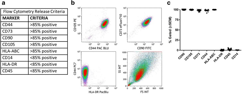
Background: Clinical translation of mesenchymal stromal cells (MSCs) necessitates basic characterization of the cell product since variability in biological source and processing of MSCs may impact therapeutic outcomes. Although expression of classical cell surface markers (e.g., CD90, CD73, CD105, and CD44) is used to define MSCs, identification of functionally relevant cell surface markers would provide more robust release criteria and options for quality control. In addition, cell surface expression may distinguish between MSCs from different sources, including bone marrow-derived MSCs and clinical-grade adipose-derived MSCs (AMSCs) grown in human platelet lysate (hPL).
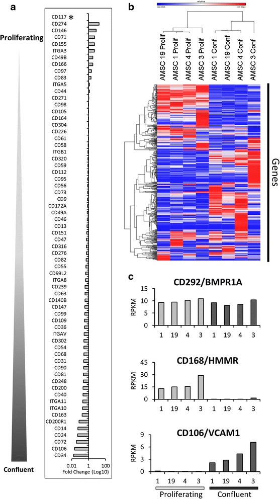
Methods: In this work we utilized quantitative PCR, flow cytometry, and RNA-sequencing to characterize AMSCs grown in hPL and validated non-classical markers in 15 clinical-grade donors.
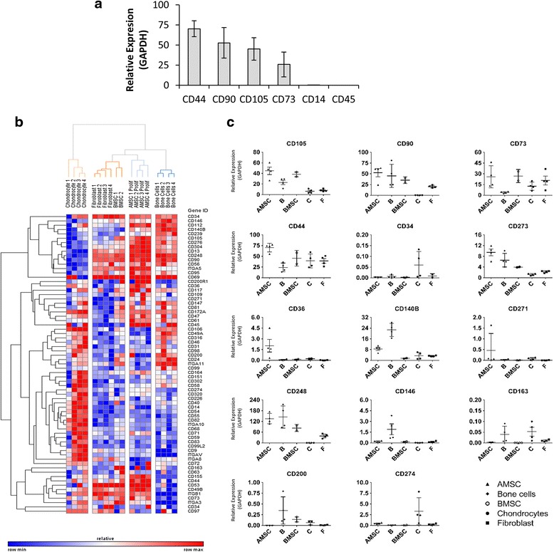
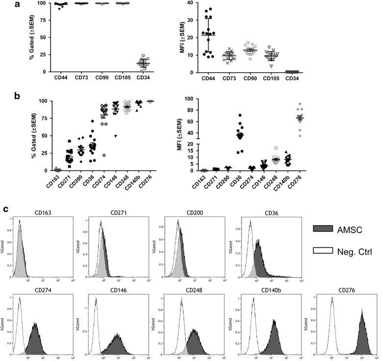
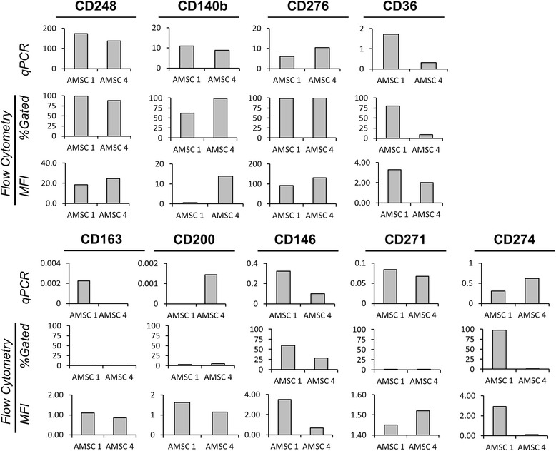
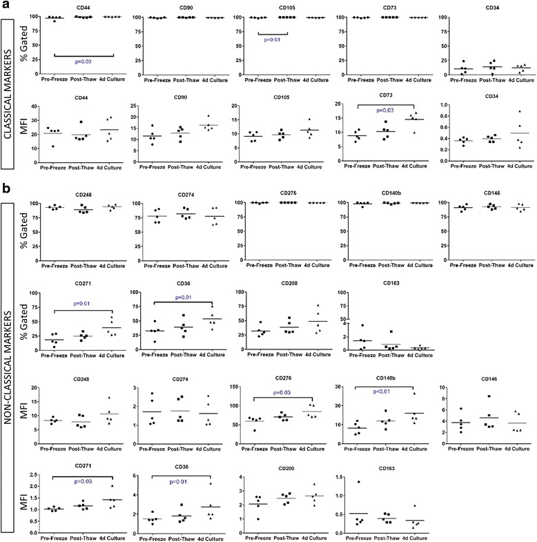
Results: We characterized the surface marker transcriptome of AMSCs, validated the expression of classical markers, and identified nine non-classical markers (i.e., CD36, CD163, CD271, CD200, CD273, CD274, CD146, CD248, and CD140B) that may potentially discriminate AMSCs from other cell types. More importantly, these markers exhibit variability in cell surface expression among different cell isolates from a diverse cohort of donors, including freshly prepared, previously frozen, or proliferative state AMSCs and may be informative when manufacturing cells.
Conclusions: Our study establishes that clinical-grade AMSCs expanded in hPL represent a homogeneous cell culture population according to classical markers,. Additionally, we validated new biomarkers for further AMSC characterization that may provide novel information guiding the development of new release criteria.
Clinical trials: Use of Autologous Bone Marrow Aspirate Concentrate in Painful Knee Osteoarthritis (BMAC): Clinicaltrials.gov NCT01931007 . Registered August 26, 2013. MSC for Occlusive Disease of the Kidney: Clinicaltrials.gov NCT01840540 . Registered April 23, 2013. Mesenchymal Stem Cell Therapy in Multiple System Atrophy: Clinicaltrials.gov NCT02315027 . Registered October 31, 2014. Efficacy and Safety of Adult Human Mesenchymal Stem Cells to Treat Steroid Refractory Acute Graft Versus Host Disease. Clinicaltrials.gov NCT00366145 . Registered August 17, 2006. A Dose-escalation Safety Trial for Intrathecal Autologous Mesenchymal Stem Cell Therapy in Amyotrophic Lateral Sclerosis. Clinicaltrials.gov NCT01609283 . Registered May 18, 2012.
Keywords: Adipose-derived mesenchymal stromal cells; CD markers; Flow cytometry; Human platelet lysate; Manufacturing; RNA-sequencing; Release criteria.
Figures
Similar articles
-
Regulatory-compliant conditions during cell product manufacturing enhance in vitro immunomodulatory properties of infrapatellar fat pad-derived mesenchymal stem/stromal cells.Cytotherapy. 2020 Nov;22(11):677-689. doi: 10.1016/j.jcyt.2020.06.007. Epub 2020 Jul 26. Cytotherapy. 2020. PMID: 32723596
-
Isolation, characterization, and in vitro proliferation of canine mesenchymal stem cells derived from bone marrow, adipose tissue, muscle, and periosteum.Am J Vet Res. 2012 Aug;73(8):1305-17. doi: 10.2460/ajvr.73.8.1305. Am J Vet Res. 2012. PMID: 22849692
-
Comparative analysis of human mesenchymal stem cells from bone marrow and adipose tissue under xeno-free conditions for cell therapy.Stem Cell Res Ther. 2015 Apr 13;6(1):55. doi: 10.1186/s13287-015-0066-5. Stem Cell Res Ther. 2015. PMID: 25884704 Free PMC article.
-
Isolation and characterization of primary bone marrow mesenchymal stromal cells.Ann N Y Acad Sci. 2016 Apr;1370(1):109-18. doi: 10.1111/nyas.13102. Ann N Y Acad Sci. 2016. PMID: 27270495 Review.
-
Single-Cell Profiles and Clinically Useful Properties of Human Mesenchymal Stem Cells of Adipose and Bone Marrow Origin.Am J Sports Med. 2019 Jun;47(7):1722-1733. doi: 10.1177/0363546519848678. Epub 2019 May 17. Am J Sports Med. 2019. PMID: 31100005 Review.
Cited by
-
The Myofibroblast Fate of Therapeutic Mesenchymal Stromal Cells: Regeneration, Repair, or Despair?Int J Mol Sci. 2024 Aug 9;25(16):8712. doi: 10.3390/ijms25168712. Int J Mol Sci. 2024. PMID: 39201399 Free PMC article. Review.
-
Umbilical cord blood derived cell expansion: a potential neuroprotective therapy.Stem Cell Res Ther. 2024 Jul 29;15(1):234. doi: 10.1186/s13287-024-03830-0. Stem Cell Res Ther. 2024. PMID: 39075614 Free PMC article. Review.
-
New Perspectives to Improve Mesenchymal Stem Cell Therapies for Drug-Induced Liver Injury.Int J Mol Sci. 2022 Feb 28;23(5):2669. doi: 10.3390/ijms23052669. Int J Mol Sci. 2022. PMID: 35269830 Free PMC article. Review.
-
Adipose-Derived Mesenchymal Stem Cells Combined With Extracellular Vesicles May Improve Amyotrophic Lateral Sclerosis.Front Aging Neurosci. 2022 May 18;14:830346. doi: 10.3389/fnagi.2022.830346. eCollection 2022. Front Aging Neurosci. 2022. PMID: 35663577 Free PMC article. Review.
-
The Therapeutic Potential of Mesenchymal Stromal Cells for Regenerative Medicine: Current Knowledge and Future Understandings.Front Cell Dev Biol. 2021 Aug 18;9:661532. doi: 10.3389/fcell.2021.661532. eCollection 2021. Front Cell Dev Biol. 2021. PMID: 34490235 Free PMC article. Review.
References
-
- Use of Autologous Bone Marrow Aspirate Concentrate in Painful Knee Osteoarthritis (BMAC) [database on the Internet]. ClinicalTrials.gov [Internet]. Bethesda (MD): National Library of Medicine (US). Available from: https://clinicaltrials.gov/ct2/show/NCT01931007. Accessed: 16 Nov 2015.
-
- MSC for Occlusive Disease of the Kidney [database on the Internet]. ClinicalTrials.gov [Internet]. Bethesda (MD): National Library of Medicine (US). Available from: https://clinicaltrials.gov/ct2/show/NCT01840540. Accessed: 16 Nov 2015.
-
- Mesenchymal Stem Cell Therapy in Multiple System Atrophy. [database on the Internet]. ClinicalTrials.gov [Internet]. Bethesda (MD): National Library of Medicine (US). Available from: https://clinicaltrials.gov/ct2/show/NCT02315027. Accessed: 16 Nov 2015.
MeSH terms
Substances
Associated data
Grants and funding
LinkOut - more resources
Full Text Sources
Other Literature Sources
Molecular Biology Databases
Research Materials
Miscellaneous
