. 2016:2016:5151843.
doi: 10.1155/2016/5151843. Epub 2016 Jul 18.

Mystixin-7 Peptide Protects Ionotropic Glutamatergic Mechanisms against Glutamate-Induced Excitotoxicity In Vitro

Affiliations

Mystixin-7 Peptide Protects Ionotropic Glutamatergic Mechanisms against Glutamate-Induced Excitotoxicity In Vitro

Anatoly A Mokrushin. Int J Pept. 2016.

Abstract

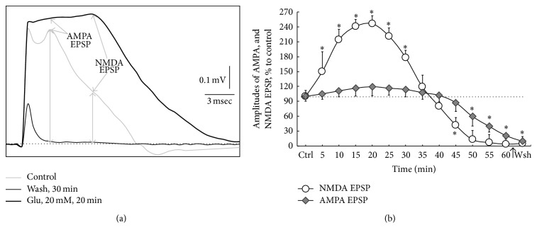
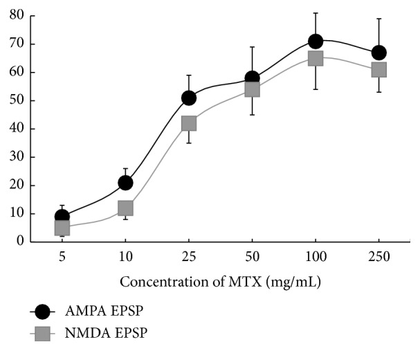
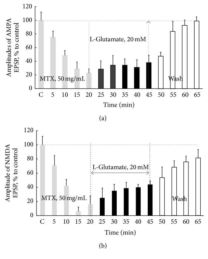
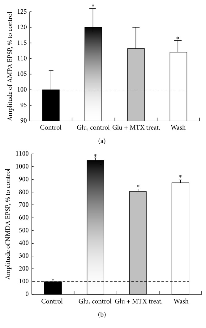
Hyperactivation of the N-methyl-D-aspartic acid type glutamate receptors (NMDARs) causes glutamate excitotoxicity, a process potentially important for many neurological diseases. This study aims to investigate protective effects of the synthetic corticotrophin-releasing factor-like peptide, mystixin-7 (MTX), on model glutamate-induced excitotoxicity in vitro. The technique online monitoring of electrophysiological parameters (excitatory glutamatergic alpha-amino-3-hydroxy-5-methyl-4-isoxazolepropionic (AMPAR) and NMDAR-dependent postsynaptic mechanisms) in the olfactory cortex slices was used. Application of L-glutamate in toxic concentration (20 mM) on slices evoked hyperactivation of NMDARs and weaker activation of the AMPARs. Upon further action agonist, the excessive activation of glutamate receptors was replaced by their irreversible blockade. Pretreatment of the slices using MTX in different concentrations (50 and 100 mg/mL) protected both NMDARs and AMPARs from glutamate-induced damage. An enzymatic treatment of MTX reduced hyperactivation of both NMDARs and AMPARs. The present study demonstrated that MTX minipeptide protected the functioning of both NMDARs and AMPARs against glutamate-induced damage. The MTX peptide is a prospective candidate for elaborated medication in treatment of neurological diseases.


Figures

Figure 1
Figure 1
The model of glutamate excitotoxicity in the olfactory cortex slices. (a) Effects application of L-glutamate in toxic concentration (20 mM) on profiles FPs. Representative FPs were recorded in time points 1, 2, and 3, respectively, as indicated in (b). FPs are integral averaging potentials generated by neurons in slices processing by special computer program at four independent experiments performed in triplicate in different slices (n = 12 per treatment condition). The dotted line indicates isoline. Arrows indicate AMPA and NMDA components of the EPSP. The vertical arrows from isoline to the peaks of the AMPA and NMDA EPSP show as conducted the measurements of their amplitudes. The captions about the conditions exposures to slices (legend beneath (a)) indicate time points of the registration FPs, corresponding to the points in (b), namely, (1) “Cntr,” (2) “Glu, 20 min,” and (3) “Wsh, 30 min.” Calibration as is indicated. At the FPs registration, the electronic device (Pavlov Institute of Physiology, RAS) for artefact-rejection was used. (b) Change of the AMPA and NMDA EPSP amplitudes at imitation glutamate excitotoxicity obtained at application of L-glutamate (20 mM) on the olfactory cortex slices. The x-axis—“Ctrl”: control values of AMPA and NMDA EPSP (without the glutamate), 15 min; thick arrow and “Wsh” indicate washout, 30 min. The dotted line indicates the control level corresponding to 100%. The duration application of L-glutamate on the slices for modeling of the glutamate excitotoxicity was 60 min. The results are expressed as percentage of control condition and represent means ± SEM of 4 independent experiments performed in triplicate in different slices and analyzed statistically by U-test, Wilcoxon-Mann-Whitney matched pairs signed-rank test. p ≤ 0.05, significantly different from control. n = 12, number of slices per time point performed in the repeated at least four independent experiments. At the FPs registration, the electronic device (Pavlov Institute of Physiology, RAS) for artefact-rejection was used. Note that L-glutamate induces the most significant hyperactivity of NMDARs with a maximum value at 20 min. Then the amplitudes of these components FPs progressively decreased and to 60th min were irreversibly blocked. The increase in AMPARs activity at action agonist was smaller, but with longer duration than NMDARs and after 40 min there was a decrease and irreversible blockade of these receptors' activity.
Figure 2
Figure 2
Protective effects of different concentrations MTX on the degree of preservation of activities of both AMPARs and NMDARs after exposure to glutamate in toxic concentration (20 mM). y-axis: amplitudes of AMPA and NMDA EPSP (mV) after pretreatment slices by MTX and further glutamate exposure (20 mM). x-axis: irregular. n = 7 for every point. Note that suppression of hyperactivation of AMPARs and NMDARs induced of L-glutamate was dependent upon the concentration of MTX at pretreatment of slices with this peptide.
Figure 3
Figure 3
Neuroprotective effects pretreatment of slices by MTX at concentration 50 mg/mL on AMPARs (a) and NMDARs (b) activities at the action of L-glutamate in toxic concentration (20 mM). The left parts of the schedule (in (a) and (b)) show changes in the averaged amplitudes of AMPA and NMDA EPSP under the action MTX in concentration 50 mg/mL. Black columns indicate the beginning and the termination of L-glutamate action. Duration of L-glutamate action was 20 min. This time range has been used by us because according to the data presented in Figure 1(b) the maximum activating effects of glutamate in a toxic concentration of 20 mM on both NMDA and AMPA EPSP were at this duration of agonist exposure. “Wash, 20 min”: washing slices by aCSF during 20 min. Horizontal dotted line (in (a) and (b)) means the control values of the NMDA and AMPA EPSP amplitudes (without MTX and glutamate) before MTX application, and on the x-axis it is marked for “C, control” and the subsequent action of L-glutamate as well as at washing process. The data are presented as percentage of control condition and represent means ± SEM of five independent experiments performed in different slices and analyzed statistically by U-test, Wilcoxon-Mann-Whitney matched pairs signed-rank test. n = 16, number of slices per time point. p ≤ 0.05, significantly different from control. Note that suppression of hyperactivation of AMPARs and NMDARs induced L-glutamate at pretreatment of slices with this peptide. Recovery of the AMPARs and NMDARs activities at washout reached the control level.
Figure 4
Figure 4
An enzymatic treatment of MTX (100 mg/mL) by trypsin resulted in a reduction of the peptide-mediated neuroprotection of AMPARs and NMDARs in slices of rat olfactory cortex. The dashed lines in (a) and (b) show control level of the AMPA and NMDA EPSP amplitudes. On the abscissa: control: the averaged values of the AMPA and NMDA EPSP amplitudes performed during 15 min; “Glu, ctrl”: effect of L-glutamate in toxic concentration 20 mM during 20 min; “Glu + MTX treat.”: effects of L-glutamate in toxic concentration 20 mM and subsequent action of the MTX pretreated during 25 min; and “wash”: washout, 20 min. The data are expressed as percentage of control value and represent means ± SEM analyzed statistically by U-test, Wilcoxon-Mann-Whitney matched pairs signed-rank test. p ≤ 0.05, significantly different from control. n = 12, number of slices per column. Statistically significant differences as compared to control are indicated by asterisks; U-test, p ≤ 0.05. n = 12 for every point. Note that the enzymatic pretreatment of MTX resulted in a decrease (statistically insignificant) in the amplitudes of AMPA EPSP but does not completely block theirs. The same values of responses persisted after washout. Hyperactivity of NMDARs to glutamate action and subsequent treatment of slices by MTX significantly decreased but remained increased compared with the control level.

Similar articles

References

    1. Hudspith M. J. Glutamate: a role in normal brain function, anaesthesia, analgesia and CNS injury. British Journal of Anaesthesia. 1997;78(6):731–747. doi: 10.1093/bja/78.6.731. - DOI - PubMed
    1. Meldrum B. S. Glutamate as a neurotransmitter in the brain: review of physiology and pathology. The Journal of Nutrition. 2000;130(4):1007S–1015S. - PubMed
    1. Segovia G., Porras A., Del Arco A., Mora F. Glutamatergic neurotransmission in aging: a critical perspective. Mechanisms of Ageing and Development. 2001;122(1):1–29. doi: 10.1016/s0047-6374(00)00225-6. - DOI - PubMed
    1. Obrenovitch T. P., Urenjak J. Altered glutamatergic transmission in neurological disorders: from high extracellular glutamate to excessive synaptic efficacy. Progress in Neurobiology. 1997;51(1):39–87. doi: 10.1016/s0301-0082(96)00049-4. - DOI - PubMed
    1. Choi D. W. Glutamate neurotoxicity and diseases of the nervous system. Neuron. 1988;1(8):623–634. doi: 10.1016/0896-6273(88)90162-6. - DOI - PubMed

LinkOut - more resources