. 2016 Sep 15;197(6):2409-20.
doi: 10.4049/jimmunol.1502702. Epub 2016 Aug 8.

TLR2 Activation Limits Rhinovirus-Stimulated CXCL-10 by Attenuating IRAK-1-Dependent IL-33 Receptor Signaling in Human Bronchial Epithelial Cells

Affiliations

TLR2 Activation Limits Rhinovirus-Stimulated CXCL-10 by Attenuating IRAK-1-Dependent IL-33 Receptor Signaling in Human Bronchial Epithelial Cells

Shyamala Ganesan et al. J Immunol. .

Abstract

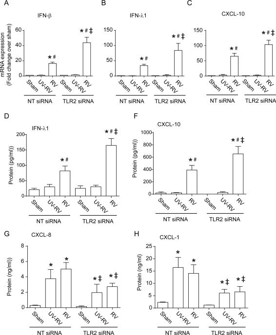
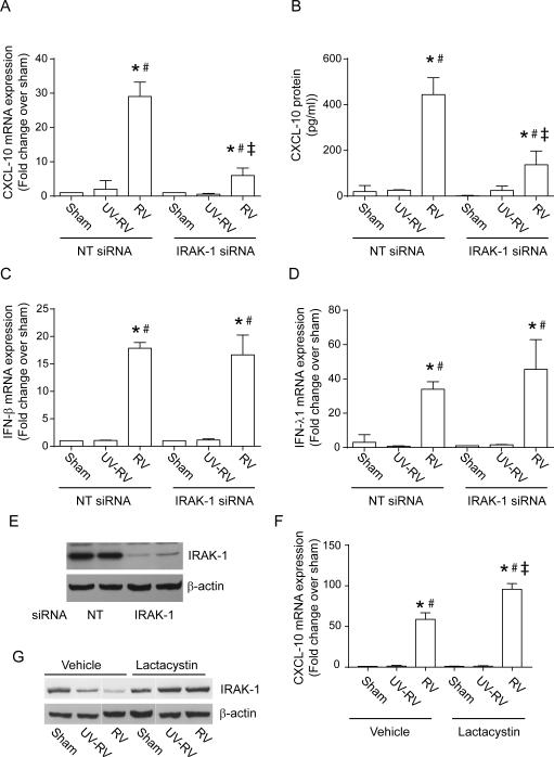
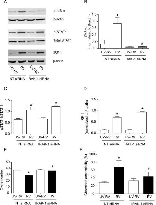
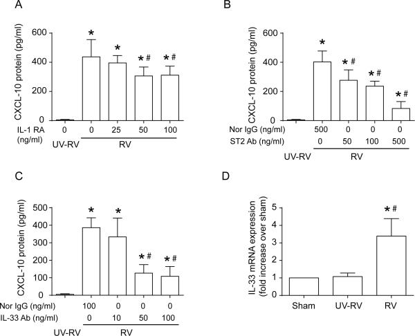
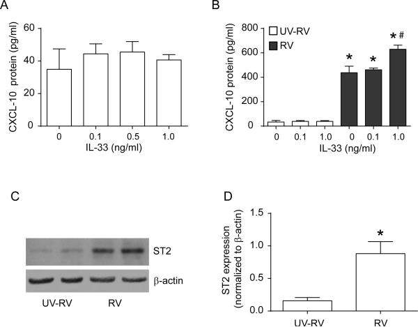
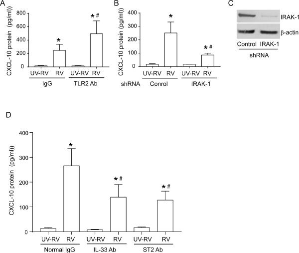
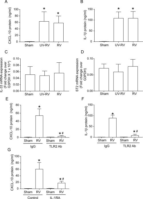
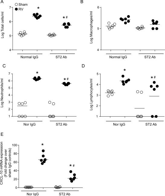
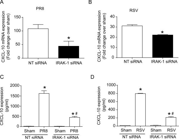
Airway epithelial cells are the major target for rhinovirus (RV) infection and express proinflammatory chemokines and antiviral cytokines that play a role in innate immunity. Previously, we demonstrated that RV interaction with TLR2 causes ILR-associated kinase-1 (IRAK-1) depletion in both airway epithelial cells and macrophages. Further, IRAK-1 degradation caused by TLR2 activation was shown to inhibit ssRNA-induced IFN expression in dendritic cells. Therefore, in this study, we examined the role of TLR2 and IRAK-1 in RV-induced IFN-β, IFN-λ1, and CXCL-10, which require signaling by viral RNA. In airway epithelial cells, blocking TLR2 enhanced RV-induced expression of IFNs and CXCL-10. By contrast, IRAK-1 inhibition abrogated RV-induced expression of CXCL-10, but not IFNs in these cells. Neutralization of IL-33 or its receptor, ST2, which requires IRAK-1 for signaling, inhibited RV-stimulated CXCL-10 expression. In addition, RV induced expression of both ST2 and IL-33 in airway epithelial cells. In macrophages, however, RV-stimulated CXCL-10 expression was primarily dependent on TLR2/IL-1R. Interestingly, in a mouse model of RV infection, blocking ST2 not only attenuated RV-induced CXCL-10, but also lung inflammation. Finally, influenza- and respiratory syncytial virus-induced CXCL-10 was also found to be partially dependent on IL-33/ST2/IRAK-1 signaling in airway epithelial cells. Together, our results indicate that RV stimulates CXCL-10 expression via the IL-33/ST2 signaling axis, and that TLR2 signaling limits RV-induced CXCL-10 via IRAK-1 depletion at least in airway epithelial cells. To our knowledge, this is the first report to demonstrate the role of respiratory virus-induced IL-33 in the induction of CXCL-10 in airway epithelial cells.


Figures

Figure 1
Figure 1
TLR2 alters RV-induced cytokine expression. BEAS-2B cells were transfected with either NT or TLR2 siRNA. Cells were then infected with sham, UV-RV or RV and incubated for 16h. Total RNA was isolated, subjected to qPCR and results were normalized to house-keeping gene, G3PDH and expressed as fold increase over sham controls (A to C). Media was used to determine the protein levels of cytokines by ELISA (D to H). Data represent mean ± SEM calculated from at least 3 independent experiments done in duplicate (* p≤0.05, ANOVA, different from sham; # p≤0.05, ANOVA, different from UV-RV-infected cells; ‡ p≤0.05, ANOVA, different from NT siRNA-transfected cells).
Figure 2
Figure 2
Genetic inhibition of IRAK-1 inhibits RV-stimulated expression of CXCL-10, but not IFNs. BEAS-2B cells were transfected with either NT- or IRAK siRNA and then infected with sham, UV-RV and RV and incubated for 16h. Total RNA was isolated and subjected to qPCR, Results were normalized to house-keeping gene, G3PDH and expressed as fold increase over sham controls (A, C and D). Media was used to determine CXCL-10 protein levels by ELISA (B). From identical experiments total cell lysates were subjected to Western blot analysis with IRAK-1 antibody to confirm the knockdown of IRAK-1 by gene-specific siRNA (E). BEAS-2B cells were infected with sham, UV-RV or RV and incubated in the presence or absence of 5 μM lactacystin for 16 h. CXCL-10 mRNA expression was assessed by qPCR and expressed as fold increase over sham control (F) and IRAK-1 expression was determined by Western blot analysis (G). Line in image G represents lanes from different parts of the same gel. Data in panels A-D and F represent mean ± SEM calculated from at least 3 independent experiments done in duplicates. (* p≤0.05, ANOVA, different from respective sham and UV-RV- infected cells; # ANOVA, different from UV-RV-infected cells; # p≤0.05, ANOVA, different from NT siRNA-transfected; ‡ p≤0.05, ANOVA, different from NT siRNA-transfected or media treated control cells). Images in E and G are representative of 3 and 10 independent experiments respectively.
Figure 3
Figure 3
IRAK-1 is required for RV-stimulated phosphorylation of IκB-α and to enhance binding of NF-κB to CXCL-10 promoter. BEAS-2B cells were transfected with either NT or IRAK siRNA and then infected with UV-RV or RV and incubated for 4 h. Cell lysates were subjected to Western blot analysis (A). Images are representative of 3 independent experiments. Phosphorylated IκB-α, p-STAT-1 and IRF-1 was quantified by densitometry and normalized to β-actin, total STAT-1 and β-actin respectively (B-D). From identical experiments, Chip assay was performed to examine the binding of NF-κB to the promoter region of CXCL-10 (E). From another set of experiments, total DNA was isolated and used for determination of chromatin accessibility in the promoter region of CXCL-10 (F). Data represent mean and SEM calculated from 3 independent experiments Data in panels B to F represent mean ± SEM calculated from at least 3 independent experiments done in duplicates (* p≤0.05, ANOVA, different from respective UV-RV- infected cells; # p≤0.05, ANOVA, different from NT siRNA-transected or media treated control cells).
Figure 4
Figure 4
IL-1 and IL-33 receptor contribute to RV-induced CXCL-10. BEAS-2B cells were infected with UV-RV or RV and incubated in the presence of IL-1RA (A), neutralizing antibody to ST2 (B) or IL-33 (C). CXCL-10 protein was estimated in the cell culture media by ELISA (A to C). BEAS-2B cells were infected with sham, UV-RV or RV and expression of IL-33 was determined by q-PCR (D). Data represent mean ± SEM calculated from 3 to 4 independent experiments done in triplicates (* p≤0.05, ANOVA, different from respective UV-RV- infected cells).
Figure 5
Figure 5
Recombinant IL-33 enhances rhinovirus-stimulated expression of CXCL-10, and induces ST2 receptor expression. BEAS-2B cells were treated with recombinant IL-33 (A) or infected with UV-RV or RV and incubated in the presence of recombinant IL-33 for 16 h. CXCL-10 protein in media was estimated by ELISA. BEAS-2B cells were infected with UV-RV or RV and incubated for 16 h. The expression of ST2 was determined by Western blot analysis (C). Band intensities were quantified by image J and expressed as fold control over β-actin (D). Data in A, B and D) represent mean and SEM calculated from 3-4 independent experiments performed in triplicates (* p≤0.05, ANOVA (B) or t test (D), different from UV-RV; # p≤0.05, ANOVA, different from RV alone infected cells). Image in C is representative of 4 independent experiments.
Figure 6
Figure 6
RV stimulates CXCL-10 expression in primary airway epithelial cells, which is partially dependent on IL-33 and ST2. Mucociliary differentiated primary airway epithelial cells were preincubated with normal IgG or TLR2 antibody (0.5 μg/ml), infected apically with UV-RV or RV and incubated in the presence of 0.5 μg/ml normal IgG, or TLR2 antibody for 16 h and CXCL-10 was measured in the basolateral medium by ELISA (A). Control or IRAK-1 shRNA-transduced primary airway epithelial cells were infected with UV-RV or RV and CXCL-10 in the medium was measured after 16 h (B). Knockdown of IRAK-1 was confirmed by Western blot analysis (C). Primary airway epithelial cell cultures infected with UV-RV or RV were incubated in the presence of 1 μg/ml of normal IgG, or antibodies to IL-33 or ST2 and CXCL-10 in the medium was assessed after 16 h (D). Data in A, B, and D represent mean ± SEM calculated from 3-4 independent experiments done in duplicate or triplicates (* p≤0.05, ANOVA, different from UV-RV-infected cells; # p≤0.05, ANOVA, different from RV-infected IgG treated cells). Image in C is representative of 4 independent experiments.
Figure 7
Figure 7
RV stimulated CXCL-10 is independent of IL-33/ST2 in macrophages: PMDMs were infected with sham, UV-RV or RV and incubated for 16 h. Protein expression of CXCL-10 and IL-1β in the spent medium was assessed by ELISA (A and B). Expression of IL-33 and ST2 was assessed by qPCR (C and D). PMDMs were preincubated for 1 h with 0.5 μg/ml of normal IgG or antibody to TLR2 (E and F), or with 100 ng/ml IL-1RA (G), infected with sham or RV and incubation continued for another 16 h, and the expression of IL-1β or CXCL-10 was determined by ELISA. Data represent mean ± SEM calculated from 3 independent experiments done in duplicate or triplicates (* p≤0.05, ANOVA, different from UV-RV-infected cells; # p≤0.05, ANOVA, different from RV-infected cells treated with normal IgG or media alone).
Figure 8
Figure 8
BALB/C (6-8 weeks old) mice were infected with sham or RV by intranasal route and 100 μl of endotoxin free PBS containing 5 μg/ml normal IgG or ST2 antibody was injected by intraperitoneal route on the day of infection and again after 24 h post-infection. Mice were sacrificed 48 h post-infection, BAL was performed to assess the total and differential cell counts (A to D) and lungs were processed for determination of CXCL-10 by qPCR (E). Data represent geomean with range from two independent experiments with three replicates (* p≤0.05, ANOVA on ranks, different from UV-RV-infected cells; # p≤0.05, ANOVA on ranks, different from RV-infected cells treated with normal IgG or media alone).
Figure 9
Figure 9
IRAK-1 also contributes to RSV and influenza virus induced CXCL-10. BEAS-2B cells were transfected with either NT or IRAK siRNA and then infected with sham, RSV at MOI of 1 or influenza virus at MOI of 0.1 and incubated for 16h. Expression of CXCL-10 mRNA (A and B) and CXCL-10 protein in the media (C and D) was determined by q-PCR and ELISA respectively. Data represents mean ± SEM calculated from at least 3 independent experiments done in duplicates (* p≤0.05, t test, different from sham; # p≤0.05, ANOVA, different from respective NT siRNA-transfected cells).
Figure 10
Figure 10
Neutralization of ST2 inhibits RSV and influenza virus-stimulated CXCL-10. BEAS-2B cells were infected with sham, influenza virus (PR8) or RSV and the expression of IL-33 mRNA was determined by q-PCR (A and B) and protein was determined by Western blot analysis (C and D). BEAS-2B cells were infected with sham, influenza virus or RSV and incubated in the presence of 500μg/ml normal IgG or antibody to ST2. CXCL-10 protein in the media was quantified by ELISA (E and F). Data presented in panels A, B, D, E and F are mean and SEM calculated from 3 independent experiments (* p≤0.05, t test). Image in panel C is representative of 4 independent experiments.

Similar articles

Cited by

References

    1. Contoli M, Marku B, Conti V, Saturni S, Caramori G, Papi A. Viral infections in exacerbations of asthma and chronic obstructive pulmonary disease. Minerva Med. 2009;100:467–478. - PubMed
    1. Kim WK, Gern JE. Updates in the relationship between human rhinovirus and asthma. Allergy, asthma & immunology research. 2012;4:116–121. - PMC - PubMed
    1. Wat D, Gelder C, Hibbitts S, Cafferty F, Bowler I, Pierrepoint M, Evans R, Doull I. The role of respiratory viruses in cystic fibrosis. J Cyst Fibros. 2008;7:320–328. - PMC - PubMed
    1. Schneider D, Ganesan S, Comstock AT, Meldrum CA, Mahidhara R, Goldsmith AM, Curtis JL, Martinez FJ, Hershenson MB, Sajjan U. Increased cytokine response of rhinovirus-infected airway epithelial cells in chronic obstructive pulmonary disease. Am J Respir Crit Care Med. 2010;182:332–340. - PMC - PubMed
    1. Unger BL, Faris AN, Ganesan S, Comstock AT, Hershenson MB, Sajjan US. Rhinovirus Attenuates Non-typeable Hemophilus influenzae-stimulated IL-8 Responses via TLR2-dependent Degradation of IRAK-1. PLoS Pathog. 2012;8:e1002969. - PMC - PubMed

Publication types

MeSH terms