. 2016 Nov;64(11):1987-2004.
doi: 10.1002/glia.23037. Epub 2016 Jul 29.

Epidermal growth factor preserves myelin and promotes astrogliosis after intraventricular hemorrhage

Affiliations

Epidermal growth factor preserves myelin and promotes astrogliosis after intraventricular hemorrhage

Govindaiah Vinukonda et al. Glia. 2016 Nov.

Abstract

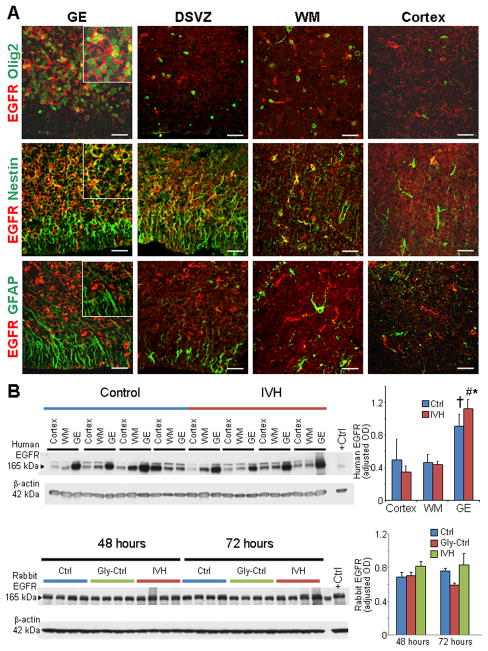
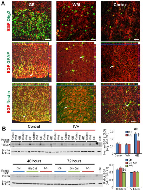
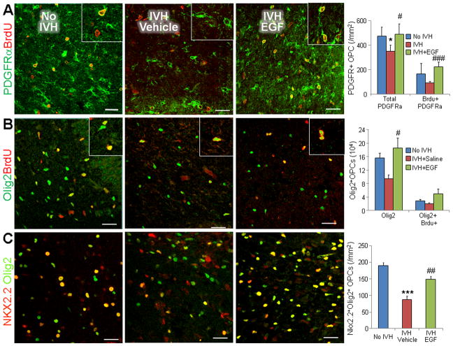
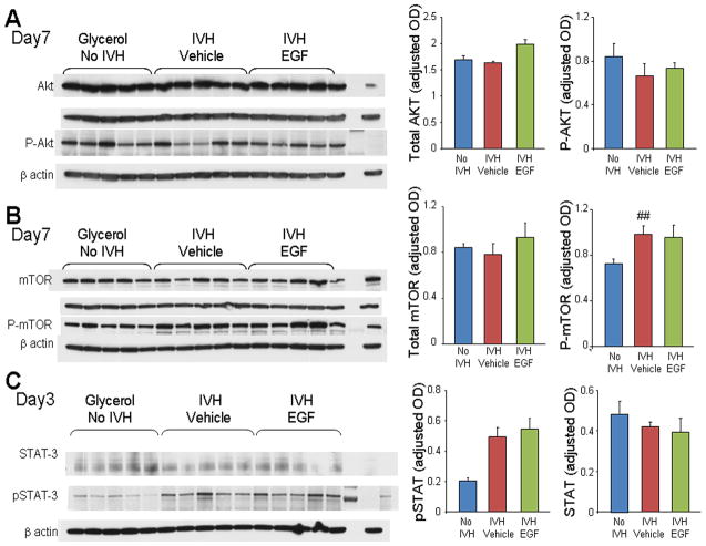
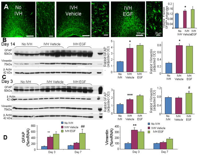
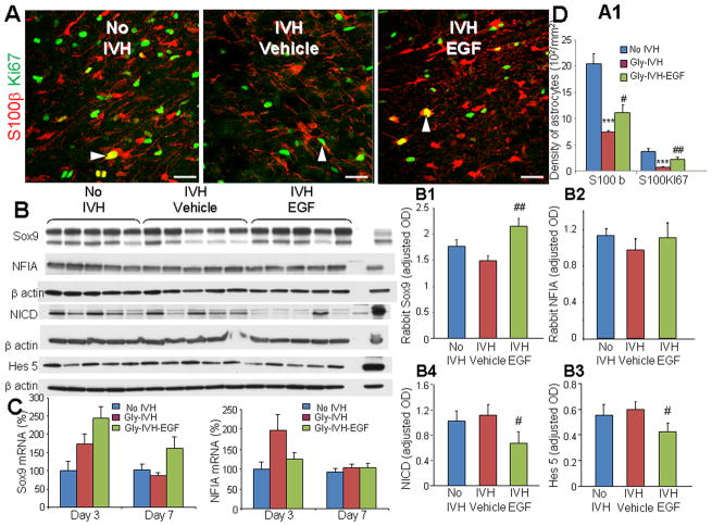
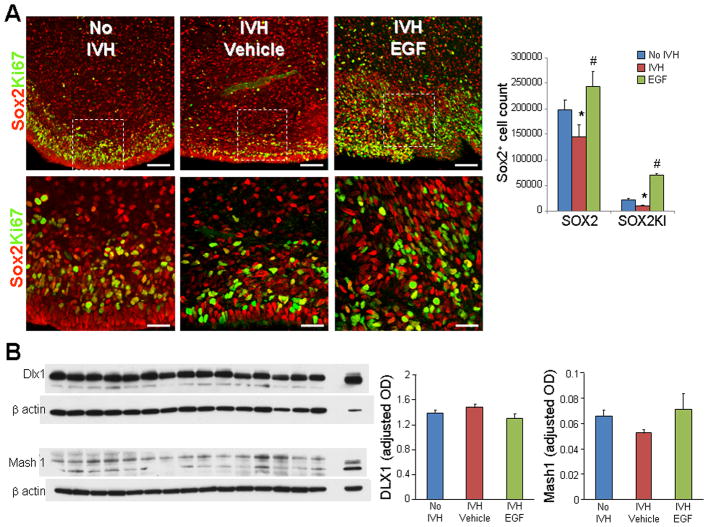
Intraventricular hemorrhage (IVH) leads to reduced myelination and astrogliosis of the white matter in premature infants. No therapeutic strategy exists to minimize white matter injury in survivors with IVH. Epidermal growth factor (EGF) enhances myelination, astrogliosis, and neurologic recovery in animal models of white matter injury. Here, we hypothesized that recombinant human (rh) EGF treatment would enhance oligodendrocyte precursor cell (OPC) maturation, myelination, and neurological recovery in preterm rabbits with IVH. In addition, rhEGF would promote astrogliosis by inducing astroglial progenitor proliferation and GFAP transcription. We tested these hypotheses in a preterm rabbit model of IVH and evaluated autopsy samples from human preterm infants. We found that EGF and EGFR expression were more abundant in the ganglionic eminence relative to the cortical plate and white matter of human infants and that the development of IVH reduced EGF levels, but not EGFR expression. Accordingly, rhEGF treatment promoted proliferation and maturation of OPCs, preserved myelin in the white matter, and enhanced neurological recovery in rabbits with IVH. rhEGF treatment inhibited Notch signaling, which conceivably contributed to OPC maturation. rhEGF treatment contributed to astrogliosis by increasing astroglial proliferation and upregulating GFAP as well as Sox9 expression. Hence, IVH results in a decline in EGF expression; and rhEGF treatment preserves myelin, restores neurological recovery, and exacerbates astrogliosis by inducing proliferation of astrocytes and enhancing transcription of GFAP and Sox9 in pups with IVH. rhEGF treatment might improve the neurological outcome of premature infants with IVH. GLIA 2016;64:1987-2004.

Keywords: astrogliosis; epidermal growth factor; intraventricular hemorrhage; myelination; oligodendrocyte progenitor cells; premature infants.


Conflict of interest statement

Nothing to report.

Figures

Fig. 1
Fig. 1. Human ganglionic eminence exhibits abundance of EGFR; and IVH does not affect EGFR expression
A) Representative immunofluorescence of cryosections from 23 gw infant, which was labeled using EGFR with Olig2, (upper panel), nestin (middle panel), or GFAP (lower panel) specific antibody. EGFR was abundantly expressed on Olig2+ and nestin+ precursor cells of the medial GE and white matter. GFAP+ radial glia in the GE and astrocytes in the embryonic whiter matter (WM) weakly expressed EGFR. Scale bar, 20 μm. B) Western blot analyses were performed for EGFR on homogenates made from tissues taken from the cortical plate (cortex), WM, and GE of preterm infants with and without IVH, as indicated. The bar charts are mean ± s.e.m. (n = 6 each). Values were normalized to β actin levels. EGFR expression was significantly higher in the GE compared to the cortical plate and WM of infants with IVH. In infants without IVH, EGFR levels were elevated in the GE compared to the white matter, but not to the cortex. EGFR expression was comparable between infants with and without IVH for the three brain regions. **P<0.01 GE vs. WM, # P<0.01 GE vs. cortex in infants with IVH; †P<0.05 GE vs. WM in infants without IVH. C) Western blot analyses for EGFR was performed on lysates made from the forebrain slices of rabbit pups with and without IVH at 48 and 72 h age. Bar chart shows mean ± s.e.m. (n=5 each group). EGFR protein levels were comparable between pups with and without IVH. DSVZ, dorsal cortical subventricular zone.
Fig. 2
Fig. 2. EGF expression was reduced in both rabbits and humans with IVH
A) Representative immunofluorescence of cryosections from 23 gw infant double labeled with EGF and Olig2 (upper panel), GFAP (middle panel), or nestin (lower panel) antibodies. For middle and lower panel, above and to the right of images are orthogonal views in x–z and y–z planes of a composite z-stalk of a series of confocal images taken 0.4 μm apart. Arrow shows co-localization of EGF and GFAP/nestin expression in the GE. Note that EGF is abundantly expressed on GFAP+ and nestin+ radial glia of the medial GE and dorsal cortical SVZ. Expression of EGF was weak-to-absent on Olig2+ cells. EGF immunoreactivity is more intense in the GE relative to the WM. Scale bar, 20 μm. B) Representative Western blot analyses for EGF on homogenates made from tissues taken from cortex, WM, and GE of preterm infants with and without IVH, as indicated. The bar charts are mean ± s.e.m. (n = 6 each). Values were normalized to β actin levels. Note higher EGF expression (15kDa) in the GE relative to the cortex and WM in infants without IVH and reduced EGF levels in the GE of infants with IVH compared to controls without IVH. C) Western blot analyses for EGF were performed on lysates made from forebrain slices of rabbit pups with IVH and without IVH at 48 and 72 h age. Bar chart shows mean ± s.e.m. (n=5 each group). EGF protein levels were lower in pups with IVH compared to pups without IVH at 48h. *P<0.01 GE vs. WM; # P<0.01 GE vs. cortex; †P<0.05 IVH vs. no IVH in the GE; ‡P<0.05 for IVH vs. untreated no IVH controls; ϕP<0.01 for IVH vs. glycerol treated controls.
Fig. 3
Fig. 3. EGF treatment preserves myelin in rabbits with IVH
A) Coronal brain slice from frontal lobe of E29 rabbit pup show normal slit-like ventricle (left panel), moderate hemorrhage in the ventricle (middle panel) and severe hemorrhage (right panel) resulting in fusion of the two ventricles (bottom panel). Scale bar, 1 cm. B, B′) Representative immunofluorescence of myelin basic protein (MBP) in the corona radiata of D14 pups. Data are mean ± s.e.m. (n = 5 each group). Volume fractions of MBP were higher in the corpus callosum and corona radiata of EGF-treated pups compared with vehicle controls with IVH. Scale bar, 200 μm. V, ventricular side. C, Cand C″) Representative Western blot analysis for MBP and MAG in the forebrain of premature rabbit pups, as indicated, at D14. Adult rat brain was used as positive control. Each lane represents lysate from a whole coronal slice taken at the level of midseptal nucleus of one brain. Bar chart shows mean ± s.e.m. (n=5 each group). MBP and MAG expression were higher in EGF treated pups compared with vehicle treated controls. D, D′) Typical electron micrograph from rabbit pups with and without IVH, and pups with IVH treated with rhEGF at D 14. Note that myelinated axons were fewer in pups with IVH compared to controls without IVH and that rhEGF treatment significantly increased the number of myelinated axons in pups with IVH. Scale bar, 1 μm. *P<0.05,**<0.01, ***P<0.001 pups with vs. without IVH. #P<0.05, ###P<0.001 vehicle vs. rhEGF treated pups with IVH.
Fig. 4
Fig. 4. EGF administration promotes OPC proliferation and maturation
A) Representative immunofluorescence of cryosections from a 3 d old pup double-labeled with PDGFRα and BrdU specific antibodies. Graphs show mean ± SEM (n = 5 each). Cycling PDGFRα+ cells (arrowhead) were reduced in pups with IVH, and rhEGF treatment increased their density. B) Representative immunofluorescence of cryosections from a 3 d old pup double-labeled with Olig2 and BrdU specific antibodies. Bar-charts are mean±SEM (n= 5 each). Note total and cycling Olig2+ cells show a trend toward reduction in pups with IVH (compared to no IVH controls) and an increase in rhEGF treated pups relative to vehicle controls. Arrowhead indicated Olig2+BrdU+ cells. C) Cryosections were double labeled with Nkx2.2 and Olig2 antibodies. Graphs show mean±SEM (n=5 each). Note reduced number of cells co-labeled with Nkx2.2+Olig2+ (arrowhead) in pups with IVH compared to vehicle controls, and rhEGF treatment increased the number in this subset of cells. *P<0.05, ***P<0.001 pups with vs. without IVH. #P<0.05, ##P<0.01, ###P<0.001 vehicle vs. rhEGF treated pups with IVH. Scale bar, 20μm.
Fig. 5
Fig. 5. RhEGF treatment does not affect mTOR, Akt, or STAT-3 signaling
Representative Western blot analyses performed on forebrain homogenates (1 cm slice at mid-septal nucleus) of pups at D3 or 7, as indicated. Adult rat brain was positive-control. Graph shows mean ± s.e.m. (n = 5 each). Values were normalized to β-actin. A, B, C) mTOR, Akt, phospho-mTOR, phosphor-Akt and STAT-3 were comparable between controls without IVH, vehicle- and rhEGF-treated pups with IVH. However, P-STAT-3 levels were higher in pups with IVH compared to controls without IVH and rhEGF treatment did not make any difference. **P < 0.01 for no IVH vs. IVH.
Fig. 6
Fig. 6. rhEGF treatment promotes astrogliosis
A) Representative immunofluorescence of cryosections from pups (D14) labeled with GFAP antibody are shown, as indicated. ‘V’ indicates ventricular side. Rectangular panels are high power view of the adjacent image. Note abundant hypertrophic astrocytes in vehicle and rhEGF treated pups with IVH. Scale bar, 200 μm and 20 μm, as indicated. Graph shows mean ± s.e.m. (n = 5 each). rhEGF treatment did not affect volume fraction of astroglial fibers compared to vehicle controls on stereological-analyses. B) Western blot analyses for GFAP and vimentin were performed in forebrain homogenates of pups (D 14). Adult rat brain was positive-control. Graph shows mean ± s.e.m. (n = 5 each). Values were normalized to β-actin. rhEGF treatment did not affect GFAP or vimentin expression in pups with IVH. C) Western blot analyses for GFAP and vimentin were performed in forebrain homogenates of pups (D 3). Adult rat brain was positive-control. Graph shows mean ± s.e.m. (n = 5 each). Values were normalized to β-actin. rhEGF treatment increased both GFAP and vimentin expression in pups with IVH. D) Data are mean ± s.e.m. GFAP mRNA expression increased with rhEGF treatment at D7, not D3. However, vimentin mRNA expression was not affected by rhEGF treatment at D 3 or 7. *P < 0.05, **P < 0.01, ***P < 0.001 for no IVH vs. IVH. #P < 0.05, ##P < 0.01for vehicle treated vs. rhEGF treated pups with IVH.
Fig. 7
Fig. 7. rhEGF increases astrocytic proliferation, upregulates Sox9 and blocks Notch signaling
A, A1) Representative immunofluorescence of cryosections from pups (D3) labeled with S100β and Ki67 antibodies. Note reduced density of total and proliferating (arrowhead) S100β in pups with IVH and increase in their density on rhEGF treatment. Data are mean ± s.e.m. Scale Bar, 20μm. B–B4) Representative Western blot analyses of Sox9, NFIA, NICD, and Hes5 performed in forebrain homogenates of pups at D3. Adult rat brain was positive-control. Graph shows mean ± s.e.m. (n = 5 each). Values were normalized to β-actin. rhEGF treatment increased Sox9 protein levels, reduced NICD and Hes5 expression, and did not change NFIA levels. C) Bar Chart shows mean ± s.e.m. (n = 5 each). rhEGF treatment increased Sox9 mRNA accumulation in pups with IVH, but did not affect NFIA mRNA. P < 0.001 for no IVH vs. IVH. #P < 0.05, ##P < 0.01 for vehicle treated vs. rhEGF treated pups with IVH.
Fig. 8
Fig. 8. rhEGF treatment induces proliferation of Sox2+ cells and does not affect Dlx1, Mash1 proteins
A) Representative immunofluorescence of cryosections from pups (D3) labeled with Sox2 and Ki67 antibodies. Lower panels are high magnification images of the boxed area in the upper panel. Note reduced density of total and proliferating (arrowhead) Sox2 in pups with IVH and increase in their density on rhEGF treatment. Data are mean ± s.e.m. Scale Bar, 50 μm (upper panel), 20μm (lower panel). B) A typical Western blot analyses of Dlx1, and Mash1 performed on forebrain homogenates of pups at D3. Adult rat brain was positive-control. Graph shows mean ± s.e.m. (n = 5 each). Values were normalized to β-actin. rhEGF treatment did not affect Dlx1 and Mash1 levels. *P < 0.05 for no IVH vs. IVH. #P < 0.05, for vehicle treated vs. rhEGF treated pups with IVH.

Similar articles

Cited by

References

    1. Aguirre A, Dupree JL, Mangin JM, Gallo V. A functional role for EGFR signaling in myelination and remyelination. Nat Neurosci. 2007;10:990–1002. - PubMed
    1. Aguirre A, Gallo V. Reduced EGFR signaling in progenitor cells of the adult subventricular zone attenuates oligodendrogenesis after demyelination. Neuron Glia Biol. 2007;3:209–20. - PMC - PubMed
    1. Aguirre A, Rizvi TA, Ratner N, Gallo V. Overexpression of the epidermal growth factor receptor confers migratory properties to nonmigratory postnatal neural progenitors. J Neurosci. 2005;25:11092–106. - PMC - PubMed
    1. Amir-Levy Y, Mausner-Fainberg K, Karni A. Treatment with Anti-EGF Ab Ameliorates Experimental Autoimmune Encephalomyelitis via Induction of Neurogenesis and Oligodendrogenesis. Mult Scler Int. 2014;2014:926134. - PMC - PubMed
    1. Ballabh P. Pathogenesis and prevention of intraventricular hemorrhage. Clin Perinatol. 2014;41:47–67. - PMC - PubMed

Publication types

MeSH terms

Substances