. 2016 Jul 28;7(7):e2308.
doi: 10.1038/cddis.2016.115.

PML nuclear body disruption impairs DNA double-strand break sensing and repair in APL

Affiliations

PML nuclear body disruption impairs DNA double-strand break sensing and repair in APL

A di Masi et al. Cell Death Dis. .

Abstract

Proteins involved in DNA double-strand break (DSB) repair localize within the promyelocytic leukemia nuclear bodies (PML-NBs), whose disruption is at the root of the acute promyelocytic leukemia (APL) pathogenesis. All-trans-retinoic acid (RA) treatment induces PML-RARα degradation, restores PML-NB functions, and causes terminal cell differentiation of APL blasts. However, the precise role of the APL-associated PML-RARα oncoprotein and PML-NB integrity in the DSB response in APL leukemogenesis and tumor suppression is still lacking. Primary leukemia blasts isolated from APL patients showed high phosphorylation levels of H2AX (γ-H2AX), an initial DSBs sensor. By addressing the consequences of ionizing radiation (IR)-induced DSB response in primary APL blasts and RA-responsive and -resistant myeloid cell lines carrying endogenous or ectopically expressed PML-RARα, before and after treatment with RA, we found that the disruption of PML-NBs is associated with delayed DSB response, as revealed by the impaired kinetic of disappearance of γ-H2AX and 53BP1 foci and activation of ATM and of its substrates H2AX, NBN, and CHK2. The disruption of PML-NB integrity by PML-RARα also affects the IR-induced DSB response in a preleukemic mouse model of APL in vivo. We propose the oncoprotein-dependent PML-NB disruption and DDR impairment as relevant early events in APL tumorigenesis.


Figures

Figure 1
Figure 1
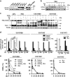
PML-NB integrity and γ-H2AX dephosphorylation kinetics. (a) Representative images of the double immunofluorescence analysis of γ-H2AX (Alexa Fluor 488, green fluorophore) and PML (Alexa Fluor 610, red fluorophore) foci in APL blasts untreated (Ctrl) and exposed to 1 Gy of X-rays and fixed after 0.5, 3, and 24 h (cell image: bright field; confocal microscopy images, magnification × 63), and NB4 cells treated or not with 1 μM RA for 72 h, and then exposed to IR and fixed after 0.5, 3, 24, and 48 h (counterstain: DAPI; confocal microscopy images, magnification × 63). (b) Quantification of the mean number of γ-H2AX foci/cell and (c) analysis of the rejoining capability measured as percentage of DSBs in untreated and irradiated APL blasts derived from 3 different individuals, and NB4 and NB4-MR4 cells untreated or treated with 1 μM RA for 72 h and then irradiated with 1 Gy. For NB4 and NB4-MR4 DSB rejoining graphs; * and ** indicate significant differences of RA-treated NB4 cells with respect to RA-untreated NB4 cells and NB4-MR4 cells. Mean values were derived from the analysis of 100 cells in 3 independent experiments±S.D. For DSBs, the mean number of γ-H2AX foci/cell measurable at 0.5 h after IR was taken as 100%. *P<0.05; **P<0.01. Confocal analysis was performed using the LCS Leica confocal microscope (Leica Microsystems, Heidelberg, Germany)
Figure 2
Figure 2
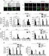
(a) Representative immunoblot analysis of H2AX and H2AX phosphorylation at the Ser139 residue in untreated human CD34− and CD34+ cells isolated from the peripheral blood of normal donors, in three APL patients, in NB4 and NB4-MR4 cells. (b) Representative immunoblot analysis of H2AX phosphorylation in NB4 cells treated or not with 1 μM RA for 72 h and then irradiated with 1 Gy of X-rays and lysed after 0.5, 3, and 24 h. (c) Representative immunoblot analysis of RARα and PML-RARα expression levels in APL blasts, NB4, U937/PR9, and U937/MT cells exposed to IR and lysed after 0.5 h; before irradiation, NB4 cells were treated or not with 1 μM RA for 72 h, whereas U937/PR9 cells were treated or not with 100 μM ZnSO4 for 8 h and then with 1 μM RA for 72 h, as indicated; filters were probed with anti-RARα antibody, and tubulin was used as loading control. (d) Quantification of the mean number of γ-H2AX foci/cell and (e) analysis of the rejoining capability measured as percentage of DSBs in U937/PR9, U937/MT, and U937/WT cells treated or not with 100 μM ZnSO4 for 8 h and/or with 1 μM RA for 72 h, as indicated; cells were then exposed to IR and fixed at the indicated times. Mean values were derived from the analysis of 100 cells in three independent experiments±S.D. For DSBs, the mean number of γ-H2AX foci/cell measurable at 0.5 h after IR was taken as 100%. **P<0.01. For U937/PR9 DSB rejoining graph; **indicates significant differences of ZnSO4-treated U937/PR9 cells with respect to U937/PR9 control cells. Confocal analysis was performed using the LCS Leica confocal microscope (Leica Microsystems)
Figure 3
Figure 3
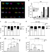
PML-NB integrity and 53BP1 recruitment to the DSBs. (a) Representative images of 53BP1 foci disappearance in APL blasts untreated (Ctrl) and exposed to 1 Gy and fixed after 0.5, 3, and 24 h, and in RA-untreated (NB4) and RA-treated (NB4+RA) cells non-irradiated (Ctrl) and exposed to IR and fixed after 0.5 h (cell image: bright field; confocal microscopy images, magnification × 63; 53BP1 (Alexa Fluor 488, green fluorophore); PML (Alexa Fluor 610, red fluorophore)). (b) Quantification of 53BP1 foci/cell, reported as the mean value of 53BP1 foci in APL blasts, NB4 and NB4-MR4 cells untreated or treated with 1 μM RA for 72 h, and U937/PR9 cells either untreated or exposed for 8 h to 100 μM ZnSO4 and then treated or not with 1 μM RA for 72 h. After ZnSO4 and/or RA treatment, cells were irradiated with 1 Gy and fixed at the indicated time points. (c) Quantification of 53BP1 foci association events with PML per nucleus in APL blasts, NB4, NB4-MR4, and U937/PR9 cells. (d) Quantification of 53BP1 foci/cell and 53BP1 foci association events with PML per nucleus in U937/MT cells. Before irradiation, U937/MT cells were either untreated or exposed for 8 h to 100 μM ZnSO4 and then treated or not with 1 μM RA for 72 h. Mean values were derived from the analysis of 100 cells in three independent experiments±S.D. *P<0.05; **P<0.01. Confocal analysis was performed using the LCS Leica confocal microscope (Leica Microsystems)
Figure 4
Figure 4
Chromosomal damage, cell cycle distribution, and apoptosis in PML-RARα-expressing cells. (a) Representative image of an mFISH-stained NB4 karyotype. Karyotype was established considering conserved translocations that appear in >90% of the cells analyzed in controls. Metaphases were captured with the Axio Imager M1 microscope (Carl Zeiss, Oberkochen, Germany). Karyotyping and cytogenetic analysis of each single chromosome was performed by the ISIS software. (b) Frequency per cell of chromosomal aberrations (i.e., fragments, exchanges, and breaks) after irradiation with 1 Gy in NB4 cells untreated or exposed to 1 μM RA for 72 h. (c) Cell cycle distribution of APL blasts and of NB4 cells untreated or treated with 1 μM RA for 72 h and then irradiated and fixed after 24 h. (d) Cell cycle distribution of U937/PR9 cells untreated or treated with 100 μM ZnSO4 for 8 h to induce PML-RARα expression and then irradiated and fixed after 24 h. (e) Sub-G1 population analyzed by flow cytometry in APL blasts and in NB4 cells untreated or treated with 1 μM RA for 72 h, and then irradiated and fixed after 24 h. (f) Sub- G1 population analyzed by flow cytometry in U937/PR9 cells untreated or treated with 100 μM ZnSO4 for 8 h, and then irradiated and fixed after 24 h. Mean values were derived from three independent experiments±S.D. **P<0.01
Figure 5
Figure 5
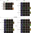
PML-NB integrity and ATM activation. Double immunofluorescence of pSer1981-ATM (Alexa Fluor 488, green fluorophore) and PML (Alexa Fluor 610, red fluorophore) foci performed in cells untreated or irradiated with 1 Gy and fixed after 0.5 and 3 h. Representative images of the analysis performed in (a) APL blasts (cell image: bright field), (b) NB4 cells untreated or treated with 1 μM RA for 72 h previous to IR (counterstain: DAPI), and (c) U937/PR9 cells either untreated or exposed for 8 h to 100 μM ZnSO4 and then untreated or treated with 1 μM RA for 72 h previous to IR (counterstain: DAPI). Some colocalization signals have been highlighted and marked within the cell by a white square. Confocal microscopy images, magnification × 63; LCS Leica confocal microscope (Leica Microsystems)
Figure 6
Figure 6
PML-NB integrity and ATM-dependent NBN phosphorylation. Double immunofluorescence of pSer1981-ATM (Alexa Fluor 488, green fluorophore) and pSer343-NBN (Alexa Fluor 610, red fluorophore) foci performed in cells untreated or irradiated with 1 Gy and fixed after 0.5 and 3 h. Representative images of the analysis performed in (a) APL blasts (cell image: bright field), (b) NB4 cells untreated or treated with 1 μM RA for 72 h previous to IR (counterstain: DAPI), and (c) U937/PR9 cells either untreated or exposed for 8 h to 100 μM ZnSO4 and then treated or not with 1 μM RA for 72 h previous to IR (counterstain: DAPI). Some colocalization signals have been highlighted and marked within the cell by a white square. Confocal microscopy images, magnification × 63; LCS Leica confocal microscope (Leica Microsystems)
Figure 7
Figure 7
Phosphorylation of ATM kinase and its substrates in PML-RARα-expressing cells. (a) Immunoblot analysis of pSer1981-ATM, pSer343-NBN, and pThr68-CHK2 phosphorylation in APL blasts, NB4, NB4-MR4, and U937/PR9 cells. Previous to IR, NB4 and NB4-MR4 cells were either untreated or exposed to 1 μM RA for 72 h, whereas U937/PR9 cells were either untreated or exposed for 8 h to 100 μM ZnSO4, and then treated or not with 1 μM RA for 72 h. Cells were then exposed to 1 Gy of X-rays and lysed after 0.5, 3, and 24 h. (b) Immunoblot analysis of ATM phosphorylation at the Ser1981 residue after 24 h from IR in NB4 and U937/PR9 cells. (c) U937/PR9 cells were first treated with 100 μM of ZnSO4, then exposed to 10 μg/ml cycloheximide, and finally irradiated with 1 Gy and lysed after 0.5 h. Immunoblots were performed using anti-γ-H2AX, anti-pSer343-NBN, anti-NBN, anti-pThr68-CHK2, and anti-CHK2 antibodies. Blots presented are exemplificative of the results obtained from two independent experiments
Figure 8
Figure 8
In vivo validation of the DDR in the preleukemic mouse model of APL. WT and preleukemic mice knock-in for PML-RARα (PR) were irradiated with 5.5 Gy of X-rays and sacrificied after 0.5, 3, 6, and 24 h. Lin− cells were isolated from the bone marrow of three pooled mice. (a) Immunoblot analysis of RARα and PML-RARα expression, and of H2AX phosphorylation at Ser139 residue, in untreated and irradiated WT and PR mice. (b) Representative images of the double immunofluorescence analysis of γ-H2AX (Alexa Fluor 488, green fluorophore) and PML (Alexa Fluor 610, red fluorophore) foci in untreated and irradiated WT and PR mice. (c) Representative images of the 53BP1 foci in untreated and irradiated WT and PR mice. (d) The DSBs rejoining analysis was reported as the mean value of γ-H2AX foci/cell and as the percentage of residual DSBs in untreated and irradiated WT and PR mice. The DSBS repair was also analyzed by counting the number of 53BP1 foci/cell in untreated and irradiated WT and PR mice. Mean values were derived from the analysis of 100 cells from three independent experiments±S.D. *P<0.05, **P<0.01. (e) Representative images of the double immunofluorescence analysis of pSer1981-ATM (Alexa Fluor 488, green fluorophore) and pSer343-NBN (Alexa Fluor 610, red fluorophore) foci in WT and PR mice. (f) Immunoblot analysis of ATM phosphorylation at the Ser1981 residue, NBN phosphorylation at Ser343, CHK2 phosphorylation at Thr68, DNA-PK phosphorylation at Ser2056, and RAD51 expression. Cell image: bright field; confocal microscopy images, magnification × 63; LCS Leica confocal microscope (Leica Microsystems)

Similar articles

Cited by

References

    1. Madhusudan S, Middleton MR. The emerging role of DNA repair proteins as predictive, prognostic and therapeutic targets in cancer. Cancer Treat Rev 2005; 31: 603–617. - PubMed
    1. Pardo B, Gomez-Gonzalez B, Aguilera A. DNA repair in mammalian cells: DNA double-strand break repair: how to fix a broken relationship. Cell Mol Life Sci 2009; 66: 1039–1056. - PMC - PubMed
    1. Rogakou EP, Pilch DR, Orr AH, Ivanova VS, Bonner WM. DNA double-stranded breaks induce histone H2AX phosphorylation on serine 139. J Biol Chem 1998; 273: 5858–5868. - PubMed
    1. Rogakou EP, Boon C, Redon C, Bonner WM. Megabase chromatin domains involved in DNA double-strand breaks in vivo. J Cell Biol 1999; 146: 905–916. - PMC - PubMed
    1. di Masi A, Viganotti M, Polticelli F, Ascenzi P, Tanzarella C, Antoccia A. The R215W mutation in NBS1 impairs gamma-H2AX binding and affects DNA repair: molecular bases for the severe phenotype of 657del5/R215W Nijmegen breakage syndrome patients. Biochem Biophys Res Commun 2008; 369: 835–840. - PubMed

MeSH terms